Link do ustawienia hasła został wysłany na adres W przyszłości dostęp do zakupów będzie można uzyskać po podaniu hasła.
W ramach bezpłatnego okresu próbnego uzyskasz 3 wideo lub 25 obrazów
Dodano nowe filtry
Sortuj wg

133 wyniki wyszukiwania „dimer” w kategorii: wideo

Czy chodziło Ci o: zimer
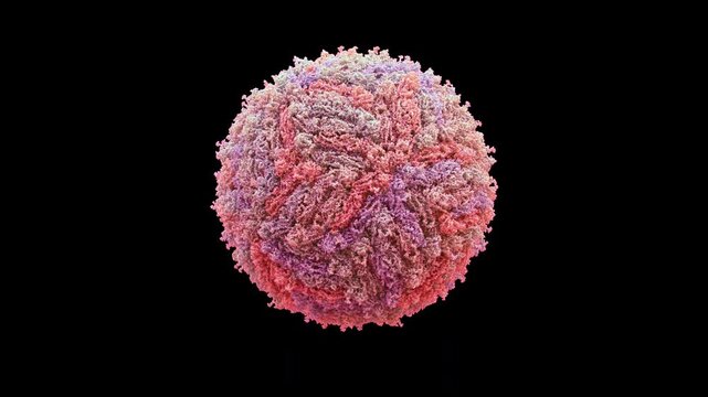
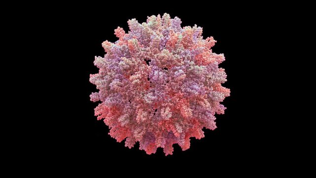
3d structure of cryo-EM zika virus capsid and envelope, icosahedral +ssrna flavivirus, pathogenic and self-replicating, primarily for microcephaly and neurotropic infection, seamless loop animation.
00:08
HD
Wild-type P53 dimer from human cancer cells 3D molecule 4K spinning
00:48
HD
Enzyme or protein in motion
00:16
HD
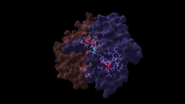
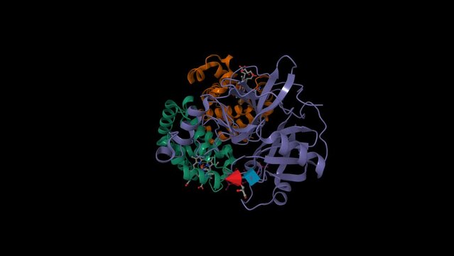
Epidermal growth factor receptor dimer in complex with allosteric inhibitor (red) and AMP (cyan). Animated 3D cartoon and Gaussian surface models, chain id color scheme, PDB 6p8q, black background
00:32
HD
Medical 3D animation of an insulin molecule docking with its receptor, triggering a clear glucose uptake cascade for healthy cellular balance
00:07
HD
Molecular Activation: ABA-Induced Protein Dimerization
00:05
HD
Non-coding RNA structures. Complex RNA assemblies forming helices and cage-like architectures involved in gene regulation and cellular function.
00:09
HD
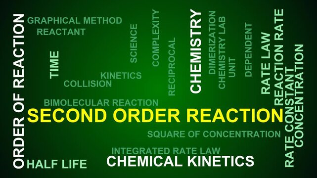
Second Order Reaction Word Cloud
00:11
HD
High Contrast Grayscale Close Up Of Two Fused Cells.
00:06
HD
Dinitrogen tetroxide rotating molecular structure, 3d ball-and-stick model, looped video oxidizing agent
00:15
HD
A dedicated scientist examines samples with a microscope, focusing on D Dimer test results for crucial data
00:18
HD
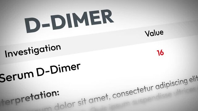
D-dimer test results design in medical report with highlighted value and interpretation text, 4k footage
00:10
HD
Molecular Signaling: Receptor Tyrosine Kinase Activation Cascade
00:05
HD
Ultraviolet radiation damaging skin cells at molecular level causing DNA breaks cellular self destruction and peeling tissue response
00:08
HD
Detailed animated coagulation panel report template: interactive D-dimer and INR test results with elevated clotting values flagged
00:07
HD
Cancer immunotherapy: Therapeutic antibodies block HER2 on cancer cells
00:15
HD
UV C Ultraviolet Light Damaging Viral RNA and Inactivating Pathogen During Microscopic Disinfection Process
00:08
HD
Doctor holding in hand Enzyme Replacement Therapy
00:29
HD
female hand twists the focus adjustment ring of the microscope. doctor's fingers twist the dimer in the hospital. close-up
00:05
HD
Hyponitrous acid (Diazenediol, Hyponitrous acid dimer) is a chemical compound with formula H2N2O2. 3D render. Seamless loop. Chemical structure model: Ball and Stick.
00:12
HD
3D structure Dengue Virus Serotype 2 DENV-2 comparison, compact 28 celsius breathing dynamics, expanded 37 celsius states primarily  for ADE Antibody-Dependent enhancement epitope exposure research.
00:08
HD
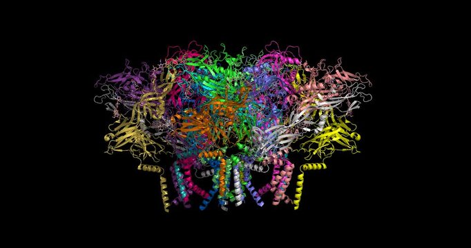
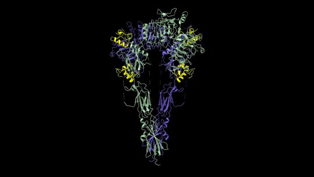
Andes virus (Hantavirus)  glycoprotein dimer of tetramers in conformation I, 9P3X cryo-EM 3D structure 4K spinning
00:16
HD
Proteins with lymphocytes , t cells or cancer cells
00:16
HD
Structure of native human insulin oligomer. Animated 3D cartoon and Gaussian surface models, chain id color scheme,  PDB 4f1b, black background
00:53
HD
Rotating cartoon models of single-strand binding protein. Wrapped DNA shown as ball and stick. Seamless loop
00:15
HD
enzyme linked receptor, ligand, ADP, ATP, cell membrane, Enzyme-linked receptors, cell surface proteins that activate intracellular pathways upon ligand binding.
00:11
HD
Zika virus concept / 3D animation of Zika virus (ZIKV) showing distinct 3-like organisation of surface dimers of genus Flavivirus
00:12
HD
A macro shot of an audio console with buttons that pressed by fingers. radio station, Israel.
00:17
HD
Molecule Structure of Ebola Virus VP40 Dimer protein
00:15
HD
3d illustration proteins or enzymes
00:16
HD
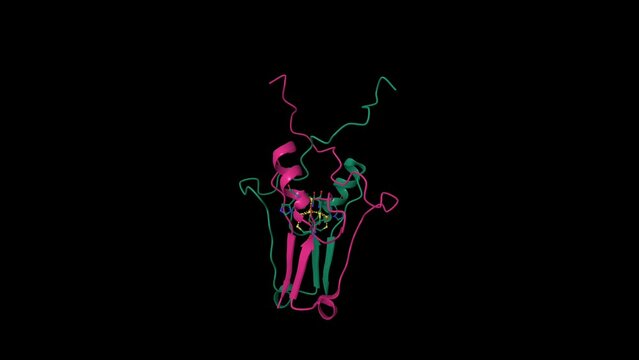
Heat-shock protein 90 dimer (pink-blue)-HSP90 co-chaperone Cdc37 (yellow)-cyclin-dependent kinase 4 (green) complex and ATP (red). Animated 3D cartoon and Gaussian surface models, PDB 5fwk
00:32
HD
Enzymes or proteins in motion
00:16
HD
Estrogen receptor beta dimer in complex with estradiol (orange red), animated 3D cartoon and Gaussian surface models, chain id color scheme, based on PDB 5toa, black background
00:20
HD
Structure of human Interleukin-5 homodimer in complex with its alpha receptor, 3D cartoon and Gaussian surface models, black background
00:32
HD
Structure of lac repressor dimer of E. coli bound to operator DNA. Animated 3D cartoon and Gaussian surface models, chain id color scheme, PDB 1efa, black background
00:28
HD
3d illustration proteins with lymphocytes , t cells or cancer cells
00:10
HD
Small circular RNA dimer replication intermediate from rolling circle synthesis, 3D cryo-EM model 4K spinning
00:16
HD
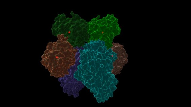
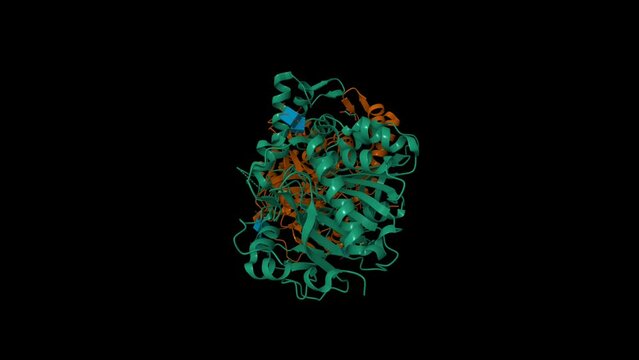
Native structure of soluble human primary amine oxidase AOC3. Animated 3D cartoon and Gaussian surface models, chain id color scheme, PDB 2y73, black background
00:35
HD
Structure of the dimeric extracellular domain of human carbonic anhydrase XII with inhibitor. Animated 3D cartoon and molecular surface models, chain id color scheme, pdb 1jd0, black background
00:30
HD
Crystal structure of a dimeric caspase-9. Animated 3D cartoon and molecular surface models, chain id color scheme, PDB 2ar9, black background
00:29
HD
Hexafluoropropylene oxide dimer acid, persistent environmental PFAS, 3D molecule spinning, 4K
00:16
HD
KaiCBA cyanobacterial circadian clock backbone model, 6 KaiA dimers (blue), 6 KaiB monomers (orange), KaiC hexamer (green), based on PDB 5n8y, 3D cartoon and Gaussian surface models, black background
00:18
HD
Crystal structure of human CYP7A1 dimer. Animated 3D cartoon and Gaussian surface models, PDB 3dax, black background.
00:22
HD
Structure of human cytosolic 5'-nucleotidase II in complex with adenosine. Animated 3D cartoon model, chain id color scheme, PDB 2jc9, black background
00:20
HD
Human alpha-galactosidase substrate bound. Animated 3D cartoon and Gaussian surface models, PDB 3hg3, black background
00:34
HD
Structure of human complement C3. Animated 3D cartoon and molecular surface models, chain id color scheme, pdb 2a73, black background
00:30
HD
Anthrax toxin lethal factor. Animated 3D cartoon and Gaussian surface models, PDB 1j7n, chain id color scheme, black background
00:35
HD
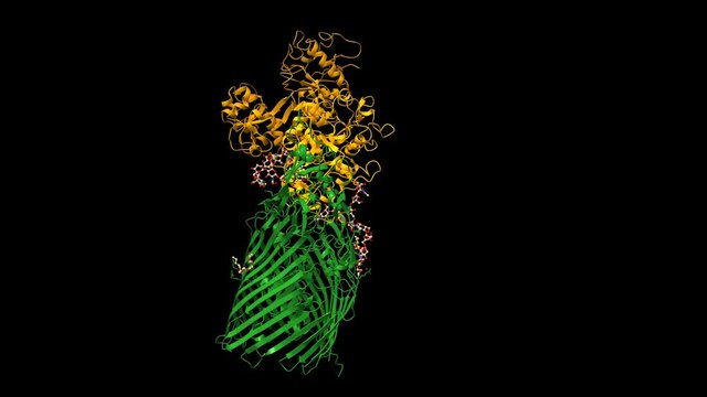
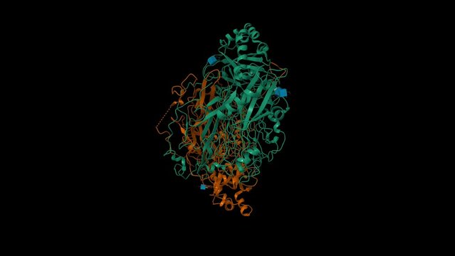
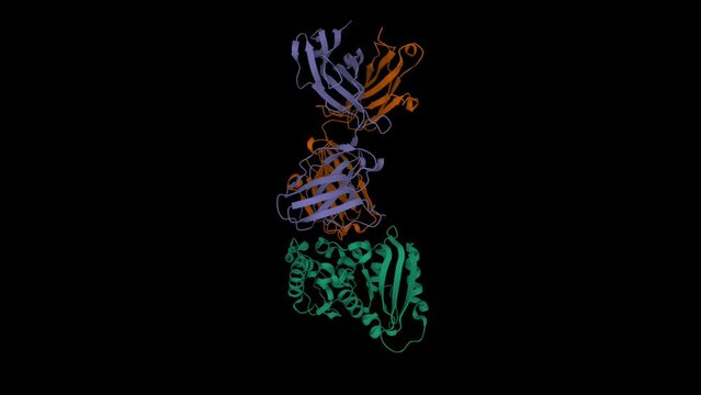
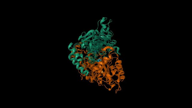
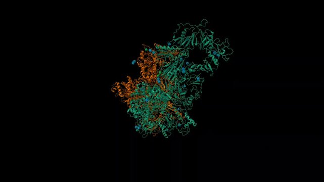
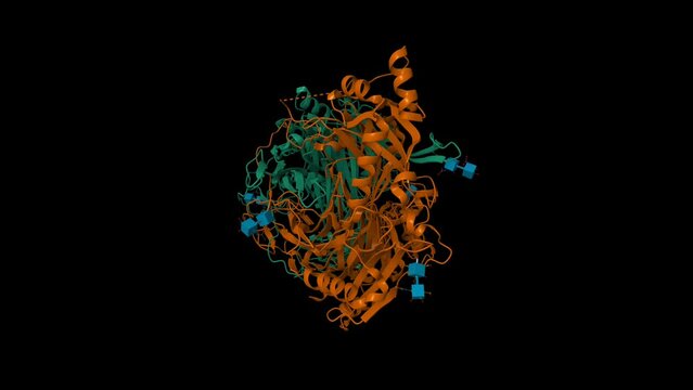
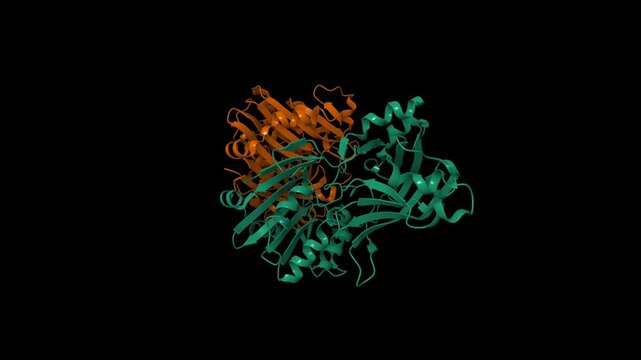
Structure of transferrin binding protein A (green) from Neisseria meningitidis complexed with full length human transferrin (orange). Animated 3D cartoon and Gaussian surface models, PDB 3v8x
00:28
HD
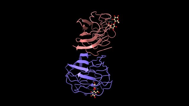
X-ray crystal structure of the human dimeric s-lac lectin in complex with lactose.  Animated 3D cartoon and Gaussian surface model, chain id color scheme, PDB 1hlc, black background.
00:28
HD
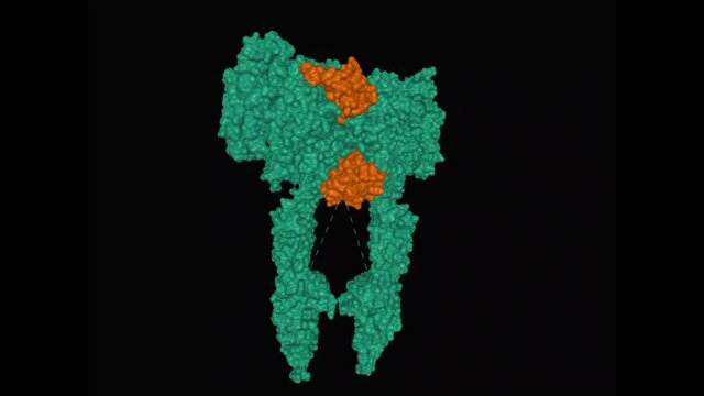
Structure of full-length insulin receptor (green) bound to four insulin molecules (brown), animated rotating 3D surface model, black background
00:20
HD
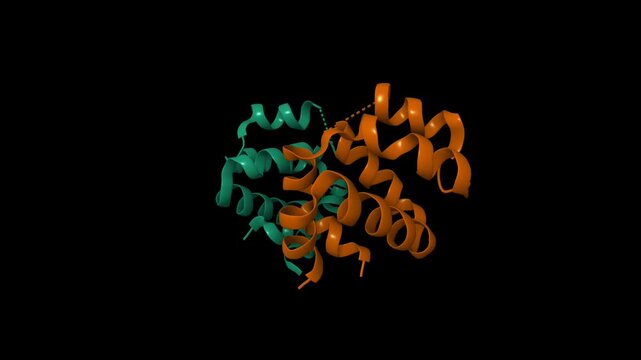
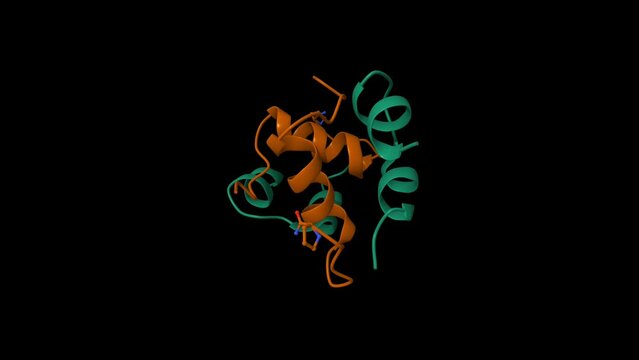
Dimeric structure of CARMA1 CARD. Animated 3D cartoon and molecular surface models, PDB 4jup, chain id color scheme, black background
00:29
HD
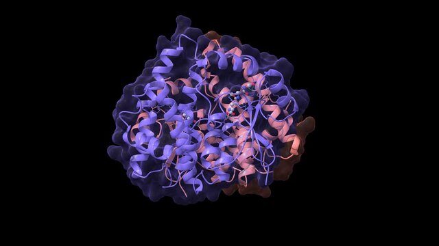
Structure of human interleukin-7 (blue) in complex with its alpha receptor (pink), animated 3D cartoon and Gaussian surface models, black background
00:15
HD
Crystal structure of human wild type GlyRS dimer bound with tRNAGly. Animated 3D cartoon and Gaussian surface model, PDB 5e6m, black background.
00:32
HD
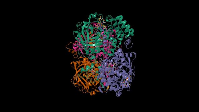
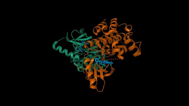
Human CYP11A1 dimer in complex with adrenodoxin (orange) and cholesterol (red). The protoporphyrin is shown in green. Animated 3D cartoon and Gaussian surface models, PDB 3n9y, black background.
00:29
HD
Human transferrin receptor (blue)-transferrin (green-red) complex. Animated 3D cartoon and Gaussian surface models, chain id color scheme (identical chains are colored similarly), PDB 1suv.
00:25
HD
Crystal structure of human vascular adhesion protein-1. Animated 3D cartoon and Gaussian surface models, chain id color scheme,  PDB 1us1, black background
00:47
HD
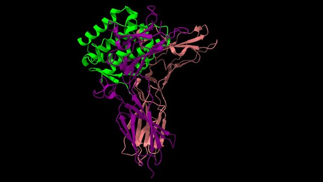
Staphylocoagulase (green) complexed with human thrombin heterodimer. Animated 3D cartoon and Gaussian surface models in two purpendicular projections, PDB 1nu7, black background
00:23
HD
Structure of full-length insulin receptor (blue and grey) bound to four insulin molecules (yellow). Animated rotating 3D cartoon and surface models, PDB 6pxv, black background
00:28
HD
Crystal structure of CDK4 (pink) in complex with cyclin D1 (blue) and P27 inhibitor (green). Animated 3D cartoon and Gaussian surface models, PDB 6p8e, black background
00:14
HD
Crystal structure of cathepsin A, apo-structure. Animated 3D cartoon and Gaussian surface models, chain id color scheme, PDB 4ci9, black background
00:34
HD
Structure of natriuretic peptide receptor-C dimer complexed with atrial natriuretic peptide (blue). Animated 3D cartoon and Gaussian surface models, PDB 1yk0, chain id color scheme, black background
00:34
HD
Crystal structure of c-raf (raf-1),  a mitogen-activated protein kinase dimer. Animated 3D cartoon and Gaussian surface models, chain id color scheme, PDB 3omv, black background
00:32
HD
Crystal structure of CD38 (green) with a novel CD38-targeting antibody SAR650984 (isatuximab). Animated 3D cartoon and Gaussian surface models, PDB 4cmh, black background
00:35
HD
Structure of antibiotic vancomycin, animated 3D ball-and-stick and Gaussian surface models, PDB 1aa5, black background
00:26
HD
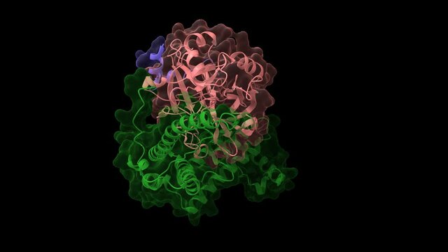
Structure of cyclin-dependent kinase 2 (CDK2, blue) in complex with cyclin E (pink). Animated 3D cartoon and Gaussian surface models, PDB 1w98, black background
00:14
HD
Structure of human erythrocyte catalase. Animated 3D cartoon and Gaussian surface models, chain id color scheme,  PDB 1dgg, black background
00:43
HD
Structure of the human proto-oncogene ALK GRD bound to inhibitor. Animated 3D cartoon and molecular surface models, pdb 7ls0, chain id color scheme, black background.
00:31
HD
Structure of human haptoglobin(violet)-haemoglobin(green-brown) complex. Animated 3D cartoon and Gaussian surface models, chain id color scheme,  PDB 4x0l, black background
00:44
HD
Crystal structure of human ligand-free mature caspase-6. Animated 3D cartoon and molecular surface models, PDB 3k7e, chain id color scheme, black background
00:29
HD
Structure of signaling thrombopoietin(green)-MPL receptor(violet-brown) complex. Animated 3D cartoon and molecular surface models, pdb 8g04, chain id color scheme, black background
00:31
HD
Betaine-homocysteine S-methyltransferase in complex with S-(delta-carboxybutyl)-L-homocysteine. Animated 3D cartoon and molecular surface models, pdb 1lt8, chain id color scheme, black background.
00:31
HD
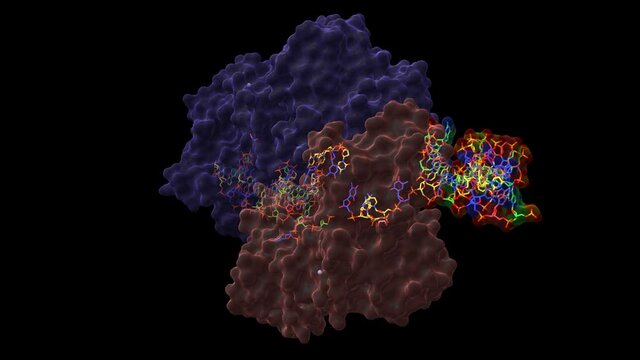
Cas1(green)-Cas2(brown)-prespacer binary complex. Animated 3D cartoon and Gaussian surface models, chain entity color scheme, PDB 5xvn, black background
00:35
HD
Crystal structure of human nicotinamide phosphoribosyltransferase (NMPRTase) complexed with nicotinamide (blue). Animated 3D cartoon and Gaussian surface models, PDB 2e5d, black background
00:52
HD
Structure of bone morphogenetic protein 3 homodimer, animated 3D cartoon and Gaussian surface model, black background
00:34
HD
Structure of the trimeric globular domain of adiponectin. Animated 3D cartoon and molecular surface models isolated, PDB 6u66, chain id color scheme, black background
00:29
HD
Structure of human glucose-6-phosphate isomerase, animated 3D cartoon and Gaussian surface models, chain ID color scheme, based on PDB 1jlh, black background
00:20
HD
Structure of KRAS4B-GDP homodimer on a lipid bilayer nanodisc, animated 3D cartoon model isolated, black background
00:23
HD
Complex of diphtheria toxin (green) and heparin-binding epidermal growth factor (brown). Animated 3D cartoon and Gaussian surface model, PDB 1xdt, black background
00:23
HD
Crystal structure of a hemagglutinin component (HA1) from type C Clostridium botulinum. Animated 3D cartoon and Gaussian surface models. PDB 1qxm, black background.
00:29
HD
Human topoisomerase 2 beta in complex with DNA and anticancer drug mitoxantrone (magenta). Animated 3D model in two perpendicular projections, PDB 4g0v, black background.
00:32
HD
Crystal structure of human glutathione transferase (GST) A1-1 in complex with glutathione, animated 3D cartoon and Gaussian surface models, chain id color scheme, based on PDB 1pkw, black background
00:44
HD
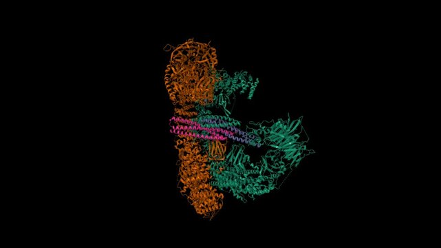
Cryo-EM structure of human BIRC6 (brown-green) in complex with SMAC (pink-violet). Animated 3D cartoon and molecular surface models, PDB 8auw, chain id color scheme, black background.
00:29
HD
Semi-synthetic highly active analogue of human insulin D-ProB26-DTI- NH2. Animated 3D cartoon and Gaussian surface models, chain id color scheme,  PDB 2wrw, black background
00:50
HD
Cryo-EM structure of human hormone thyroglobulin. Animated 3D cartoon and molecular surface models, PDB 7b75, black background
00:29
HD
Hsp90-Sba1/p23 closed chaperone complex, 3D biological assembly 4K spinning
00:16
HD
B-Raf Kinase V600E oncogenic mutant dimer in complex with Dabrafenib. Animated 3D cartoon and molecular surface models, PDB 4xv2, chain id color scheme, black background
00:30
HD
3D structure human hepatitis B virus HBV core particle hbcag capsid, t=4 and t=3 icosahedral shell four-helix bundle spikes, replication-defective vlp pathogenic virus, primarily for vaccine platform.
00:08
HD
Crystal structure of TAP01 in complex with amyloid beta peptide (green). Animated 3D cartoon and molecular surface models, chain id color scheme, pdb 7ow1, black background
00:29
HD
Structure of streptococcal pyrogenic exotoxin A1, animated 3D cartoon and Gaussian surface models, chain id color scheme, based on PDB 1b1z, black background.
00:26
HD
Structure of SARS-Cov-2 main protease dimer interacting with antiviral drug, narlaprevir, animated 3D cartoon and Gaussian surface models, black background
00:18
HD
Crystal structure of mitoNEET. Animated 3D cartoon and Gaussian surface models, chain id color scheme,  PDB 3ree, black background
00:47
HD
Ternary structure of human interleukin-20 (green) in complex with its receptor (pink and violet), animated 3D cartoon and Gaussian surface models, black background
00:15
HD
Structure of c-Cbl-ZAP-70 peptide complex. Animated 3D cartoon and molecular surface models, pdb 2y1n, black background.
00:29
HD
DNA-binding domain of human SETMAR in complex with Hsmar1 terminal inverted repeat (TIR) DNA. Animated 3D cartoon and Gaussian surface models, PDB 7s03, black background
00:35
HD
Structure of human RECQ-like helicase in complex with DNA. Animated 3D cartoon and Gaussian surface models in two perpendicular projections, PDB 2wwy, black background
00:32
HD
Crystal structure of human diamine oxidase. Animated 3D cartoon and Gaussian surface models, chain id color scheme, PDB 3hi7, black background
00:34
HD
Human nucleoside diphosphate kinase 4. Animated 3d cartoon and Gaussian surface models, chain id color scheme, PDB 1ehw, black background
00:35
HD
Crystal structure of TANK-binding kinase 1 (TBK1) with inhibitor bound. Animated 3D cartoon and molecular surface models, chain id color scheme, pdb 4iwp, black background
00:29
HD
Structure of the BfiI restriction endonuclease. Animated 3D cartoon and molecular surface models, chain id color scheme, pdb 2c1l, black background
00:31
HD
Structure of human immunoglobulin, animated 3D cartoon and Gaussian surface models, black background
00:14
HD