Et link til indstilling af din adgangskode er sendt til: For at få adgang til dine køb i fremtiden har du brug for en adgangskode.
Få 3 videoer eller 25 billeder med en gratis prøveversion
Nye filtre tilføjet
Sortér efter

33.043 resultater for microscopy i videoer

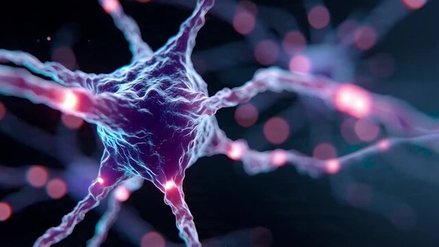
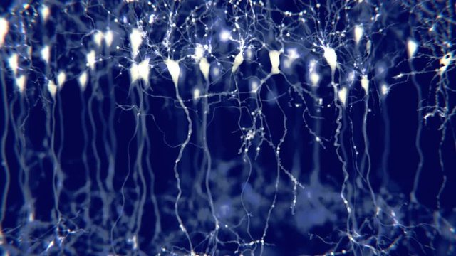
An intricate view of neurons showcasing synaptic connections and electrical activity, highlighting the beauty and complexity of the nervous system in amazing detail.
00:05
Scientific analysis of food samples in a laboratory with detailed observations and microscopy
00:13
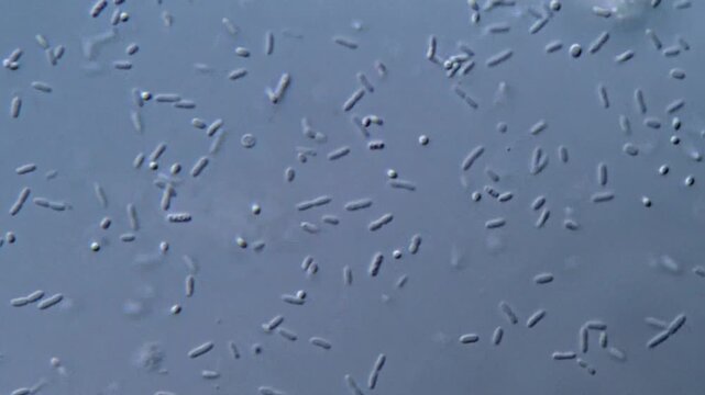
High resolution microscopy of bacteria colonies
00:44
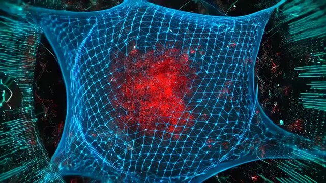
isolated neuron with red nucleus and blue mesh envelope, slow approach revealing membrane tension and surrounding microfilaments, scientific visualization for microscopy and virology
00:10
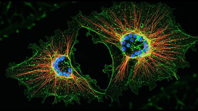
Fluorescence microscopy of actin filaments v1
00:10
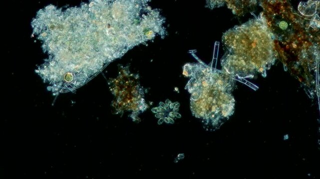
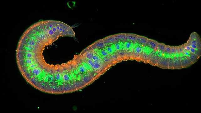
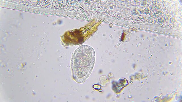
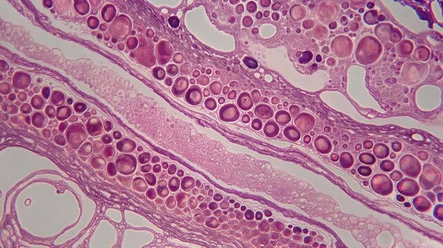
Annelids moving with rotifer colony in background under 40x magnification darkfield microscopy. 4K scientific microscopy footage.
00:27
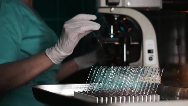
Abstract microscopic view of cells with glowing nucleus and soft texture. Concept for biology, life, medicine, science, health, and innovation
00:06
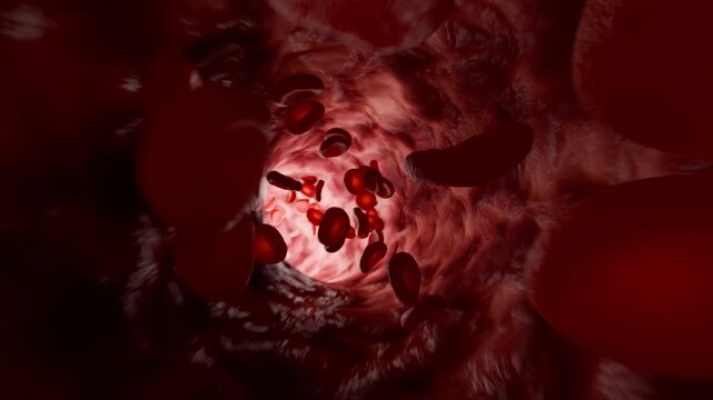
Red blood cells move in human blood stream during a close-up view under a microscope showing various shapes and sizes of the cells in a dark background
00:10
Fibroblast cell neuron network microscopic biology glowing membrane organic structure molecular tissue science research connective extracellular matrix scientific visualization cellular anatomy
00:05
Electron Microscopy Enables Precise Scientific Examination of Glowing Biological Cellular Material
00:10
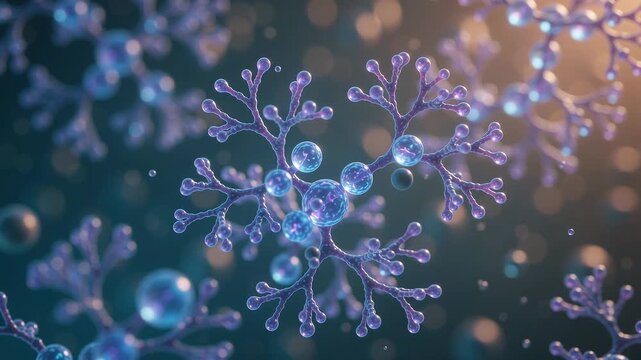
Molecule chemistry science cell biotechnology molecular nad longevity research laboratory featuring glowing molecular structure near cell surface and membrane DNA microscopy genetics medical biology
00:08
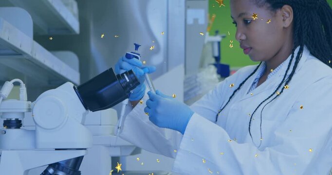
Female lab tech picking up pipette, aspirating sample for microscopy with gold stars overlaying
00:10
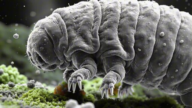
Tardigrade water bear microscopy macro, DNA protection, concept for radiation resistance
00:10
A microscopy image of a thyroid follicle showing the unique shape of thyroid cells resembling tiny sacs filled with hormoneproducing machinery.
00:07
Bright laboratory-style abstract 3D microscopy visualization, multiple types of viruses and bacteria in vivid contrasting colors, fine structural details on surfaces, floating dyna
00:10
Macro cinematic movement through plant spores, scientific discovery concept
00:05
Highdetail depiction of mitochondria in cellular energy production
00:10
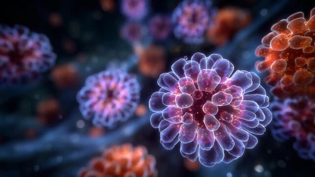
Pathogens infecting and replicating within human cells, representing an abstract biological concept of viral infection, disease, and medical research in microscopy and science
00:05
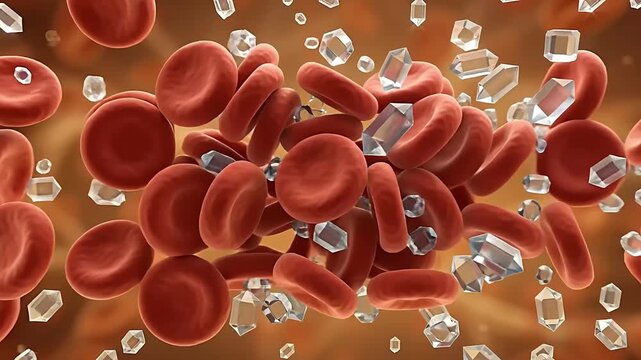
Dynamic blood cells with glucose molecules — vivid biomedical visualization of sugar–blood interaction
00:05
Healthy human liver organ anatomy with translucent glowing bile duct and branching vascular vessel tree transparent liver cross section illustrating illuminated bile duct network, microscopic
00:08
Abstract scientific background showing isolated stem cell under magnification, surrounded by soft blue microscopy textures
00:10
Human cerebellar cortex, light microscopy showing Purkinje cells, granular cells, and molecular layer involved in motor coordination and balance.
00:16
Movement of freshwater Amoebae under microscopy
00:33
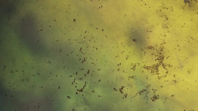
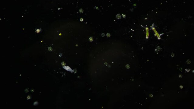
Darkfield Microscopy of Live Microorganisms Moving Fast across Slide with Stationary Algae for Microbiological Research and Exploration of Hidden Ecosystems in Water Samples and Environmental Study
00:18
3D animation: Green glowing plankton, algae, and diatoms float in dark water. Microscopic aquatic life, darkfield microscopy style. Science, biology visualization.
00:10
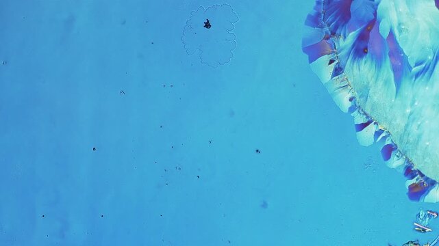
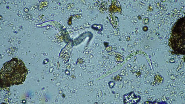
Nematodes actively moving in blue water droplet under 100x magnification brightfield microscopy. 4K scientific microscopy footage.
00:27
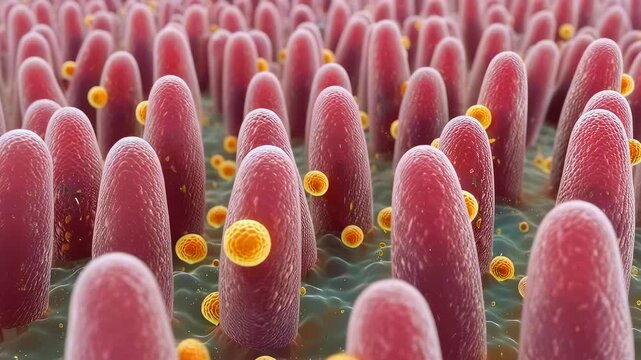
Microscopic intestinal villi covered in bacteria, showing nutrient absorption on mucosal surface, yellow spherical microbes attaching to pink villi, closeup microscopy style, highdetail
00:10
Microscopic Scanning Electron Microscopy View of Stratified Epithelial Cells
00:05
Neuron Network Green Light Patterns.
00:10
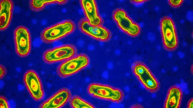
Bacterial infection Yersinia pestis microscopy fluorescent microscopic animation
00:10
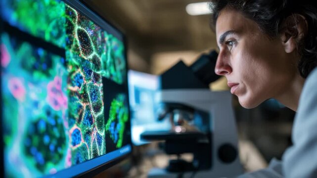
A dedicated researcher conducts scientific analysis using advanced microscopy technology in a professional lab environment. The scene highlights innovation, precision, and scientific discovery
00:10
Woman lifting pipette and drawing into tube in lab, molecules animating on pipette for microscopy
00:10
Molecule network technology for skincare science, nanotechnology chemistry, glowing blue molecular structure with connected nodes, serum delivery innovation, cellular repair research
00:10
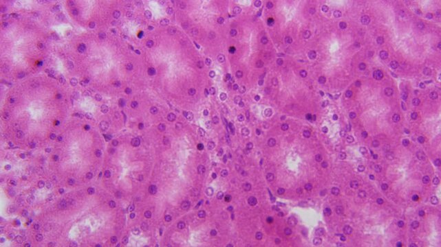
Histology Microscope Video of Mammal Cuboidal Epithelium in Kidney Medulla Tubules (Mouse, 400x)
00:11
Scientist in protective gear working with advanced microscopy equipment
00:10
Macro Close-up of Emerald Green Rippling Liquid Texture with Air Bubble
00:37
Microscopic view of paramecium and amoeba actively swimming in a drop of pond water. Dark-field microscopy lighting makes the microorganisms glow vividly against a deep black background.
00:05
Laboratory research activities scientific lab image series professional setting close-up view microscopy techniques for education
00:09
Female Doctor Engaged In Precise Clinical Research Within Hightech Automated Laboratory Environment
00:10
Microscopic animation of glowing particles within cell-like structures
00:10
Stunning Digital Representation of DNA Helix in Futuristic Blue Tones
00:05
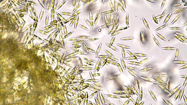
Microscopic diatoms with translucent silica shells scattered across slide, varied shapes and sizes, delicate frustules and algae cells visible, brightfield microscopy for taxonomy and environmental
00:38
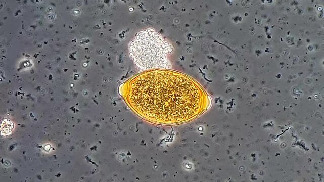
Stool sample under phase microscopy view, Trichuris egg
00:10
Science Research: Advanced microscopy technology performs 3D molecular protein analysis in a modern laboratory.
00:08
Fluorescent microscopy of Caenorhabditis elegans, with glowing neurons and transparent body structure under lab lighting
00:10
Large nematodes aggregation actively moving in blue water droplet under 40x magnification brightfield microscopy. 4K scientific microscopy footage.
00:26
Microbial green chemistry microscopy of algae and microorganism culture water. glass vial and petri showing diverse single celled life soft warm
00:05
Close-up shot of sample slide and microscope with metal lens at laboratory.
00:08
Biomedical animation of clostridium botulinum.
00:15
A Laboratory Scientist Uses Electron Microscopy to Inspect High-Contrast Biological Cell Structures
00:10
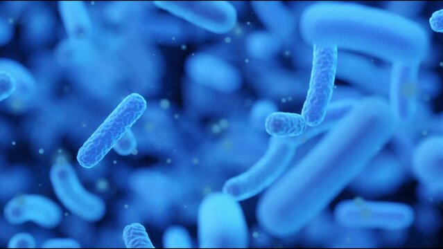
A close up of a bunch of blue bacteria. Resistance to antibacterial agents, growth of bacteria in the intestines, microscopy of bacteria.
00:10
Fluorescent microscopy animation of an activated B lymphocyte surrounded by a glowing ring of antigens.
00:08
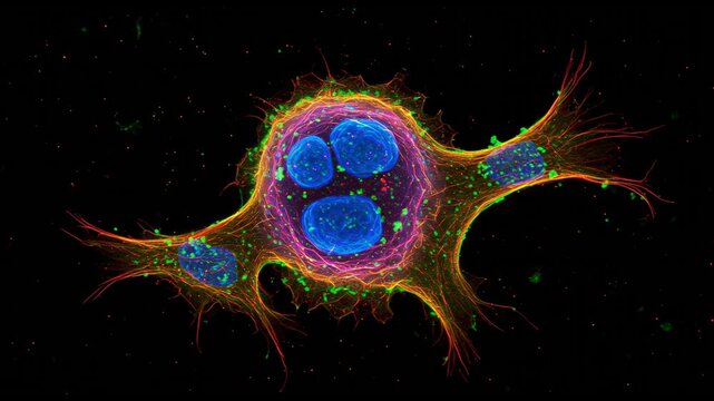
Fibroblast cell microscopy tissue regeneration healing anatomy biology medical research fibroblast cell microscopic anatomy glowing membrane extracellular matrix nucleus filament network dynamic
00:05
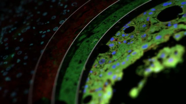
immunostained tissue (mouse liver), split up into single fluorescence channels, confocal microscopy
00:30
Amoeba and many other microorganisms in the frame. Also in the frame are colonies of unicellular algae and bacteria. Combination of oblique illumination microscopy and darkfield microscopy methods
00:15
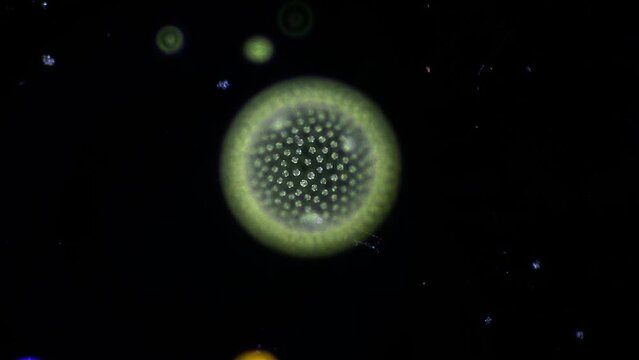
Colony of ball-shaped green algae called Gonium, which assemble with astounding simmetry. They have beating flagella for locomotion. 400x magnification, phase contrast microscopy
00:26
Atomic Surface Patterns via Quantum Tunneling Microscopy Generative AI
00:10
Visual representation of mitochondria highlighting structure and function in cellular energy production during biological processes
00:10
Scientist using microscopy glass setup in laboratory
00:07
Two scientists in protective gear using microscope and advanced holographic interface to analyze DNA data in a high-tech laboratory.
00:16
Looping animated abstract background. 4K Green wave wallpaper for VJ or screensaver.
00:08
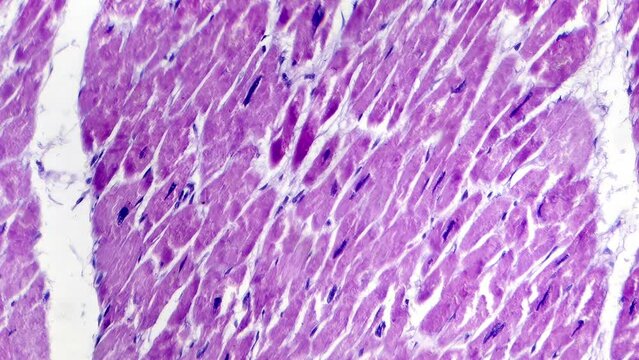
Lobar pneumonia during the hemorrhagic edema period, light microscopy displaying lung tissue inflammation with hemorrhagic changes.
00:16
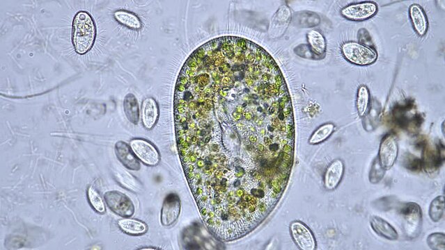
Ciliates Protozoa activity recorded with microscopy
00:28
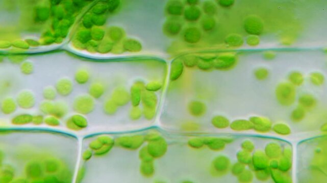
Plant stem cross section microscopy
00:08
Fluorescence microscopy of actin filaments v3
00:10
Nematodes moving slowly in blue water droplet under 400x magnification brightfield microscopy. 4K scientific microscopy footage.
00:17
Resistance to antibacterial agents, growth of bacteria in the intestines, microscopy of bacteria
00:10
Female scientist prepare sample test by drop on microscope slides for microscopy on cells and bacteria in laboratory for research on pharmacy medical for protect disease and virus
00:15
Immune cell T cell antigen receptor activation synapse microvilli membrane biology microscopy micrograph lymphocyte defense immunology
00:05
Lassa virus microscopy video showing cellular degeneration vacuoles and membrane rupture
00:10
Scanning Electron Microscope (SEM) Microstructure Image, Laboratory Analysis
00:36
An infrared microscopy image of mitochondria capturing the heat signatures of these energyproducing organelles within the cell.
00:08
3D rendering of the actin cytoskeleton is essential for maintaining the shape and structure of cells
00:16
immunostained tissue (mouse liver), split up into single fluorescence channels, confocal microscopy
00:30
Amoeba and many other microorganisms in the frame. Also in the frame are colonies of unicellular algae and bacteria. Combination of oblique illumination microscopy and darkfield microscopy methods
00:15
Beautiful and very active single-cell marine microalgae found in the bucket of a fisherman. Microscope footage, time lapse, 200x magnification, bright field microscopy
00:43
digital microscopy
00:08
Ultra-High Resolution Imaging of Ebola Virus Filamentous Morphology, Contagious Agent of Hemorrhagic Fever. Advanced microscopy captures the elongated filamentous structures characteristic of Ebola
00:08
Close-up of a euglena colony, focusing on the greenish hue and flagella, through differential interference contrast microscopy
00:05
Microscopic isolated rotifer revealing internal organelles and ciliary corona, centered on clear background, high contrast brightfield imaging ideal for anatomy and microscopy demonstrations
00:13
Close-up shot of sample slide and microscope with metal lens at laboratory.
00:18
Pyramidale Neuronen in der Großhirnrinde. Die synaptische Signale sind deutlich erkennbar.
00:12
Blepharisma ciliate under microscopy view 400x magnification
00:21
Circular chemistry materials green laboratory molecule polymer reaction solution beaker flask glass molecular network research microscopy scientific
00:05
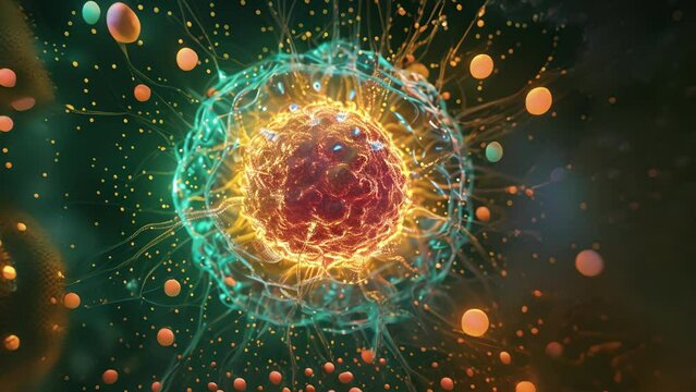
3D animation: Green filamentous algae with smaller protists. Brightfield microscopy style. Aquatic life, biology, science visualization.
00:10
Nematodes actively moving in blue water droplet under 400x magnification brightfield microscopy. 4K scientific microscopy footage.
00:20
Microscopic view of an amoeba with cellular structures.
00:08
check micro electronic chip under a microscope
00:28
Caenorhabditis elegans
01:21
Bacteria and Protozoa in waste water under the microscope.
00:19
Laboratory gel poured into container for biomaterial microscopy research
00:07
A fluorescence microscopy image of liver cells marked with fluorescent stains to visualize the process of detoxification and identify the specific toxins being removed.
00:08
Blood erythrocyte movement in blood vessels capillaries pigment cells microscopy in tadpole tail
00:14
(Daphnia magna, Crustacea, Cladocera), small planktonic crustacean
00:20
Red Blood Cells Moving in FOV 600x Microscope
00:29
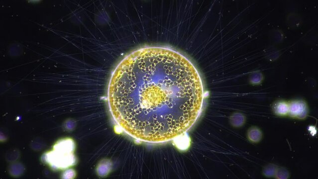
fresh water micro organism, Heliozoa, with its beautiful filaments spreading out like sun rays. 400x magnification, dark field microscopy
00:49
Amoeba and Vorticella hunt among unicellular algae. The incredible and amazing life of microorganisms under the microscope. Bright-field microscopy method
00:29
A scientist places a slide on the specimen stage of a laboratory microscope and then makes adjustments to the focus.
00:19
Diatom under the optical microscope
00:35
Highdetail rotifer portrait showing ciliary corona, mastax and interior structures, sharp focus on translucent body, brightfield microscopy image for highresolution educational content and scientific
00:29