Et link til indstilling af din adgangskode er sendt til: For at få adgang til dine køb i fremtiden har du brug for en adgangskode.
Få 3 videoer eller 25 billeder med en gratis prøveversion
Nye filtre tilføjet
Sortér efter

3.256 resultater for microscopic section i videoer

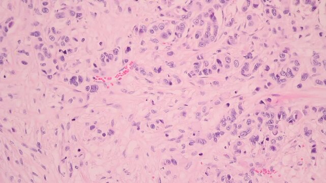
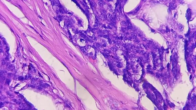
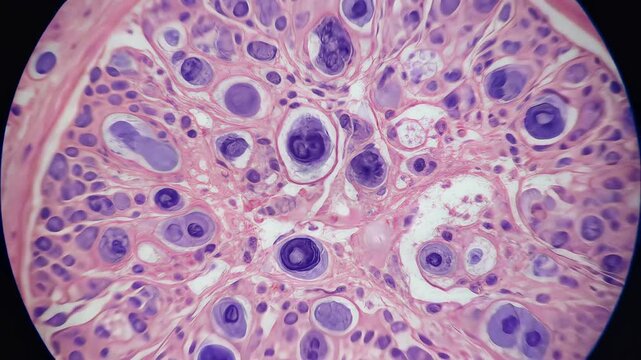
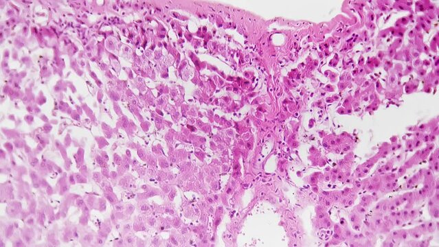
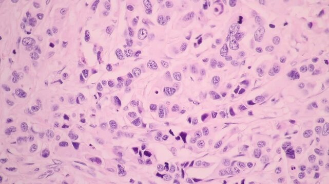
View in microscopic of glandular epithelial cell, adenonocarcinoma from human lung cancer, tissue section by H and E stain.Pathology diagnosis.Medical concept. Under microscope, magnification 600X
00:27
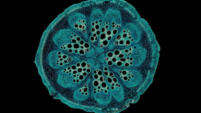
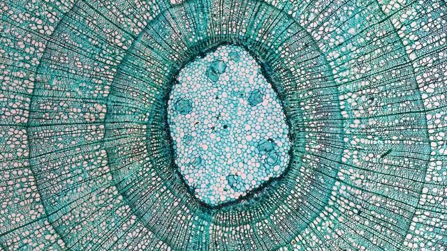
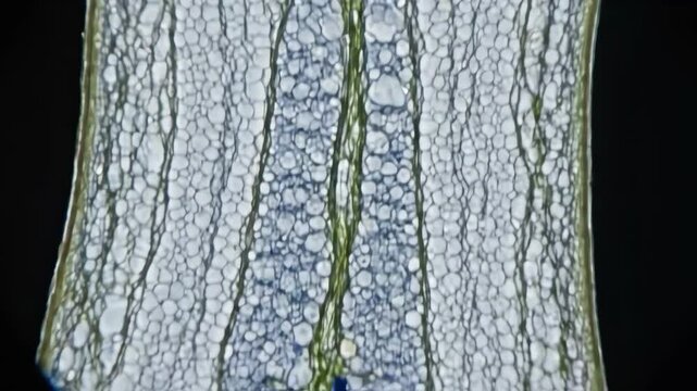
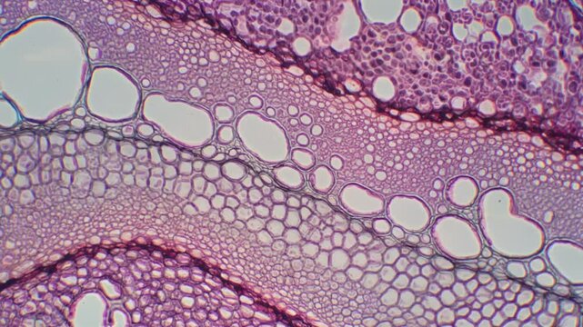
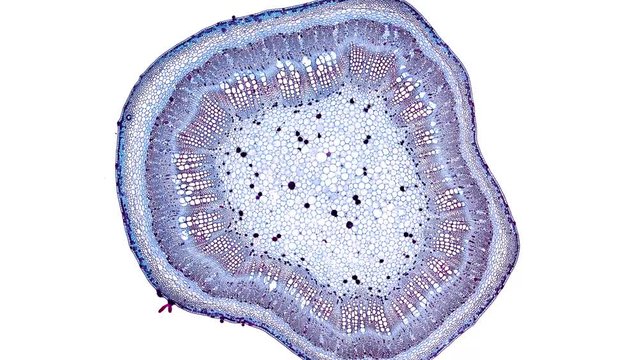
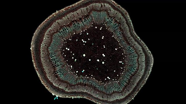
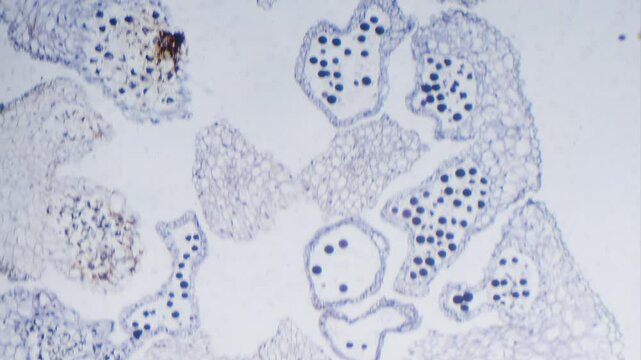
Bryonia, stem with sieve plates - microscopic cross section cut of a plant stem - dark field
00:30
Embryonic mitosis stem cells colony tissue section magnified in microscope. Cellular therapy research, disease treatment 3D animation. Pathology diagnosis. Biology and human body medicine concept. 4K

00:10
Explore human skin layers showing hydration with a close up look at skin cross section and glowing details in different frames
00:06
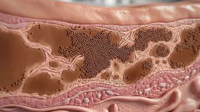
Detailed cross section of human skin layers showing melanin deposits and cellular structure under microscopic view with soft lighting
00:08
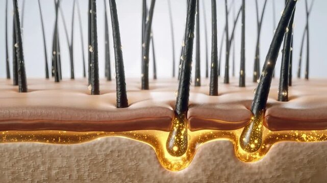
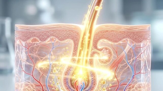
Microscopic view of healthy hair follicles under the skin with nourishing serum close-up cross-section view of roots and beneath the skin a golden nutrient-rich
00:05
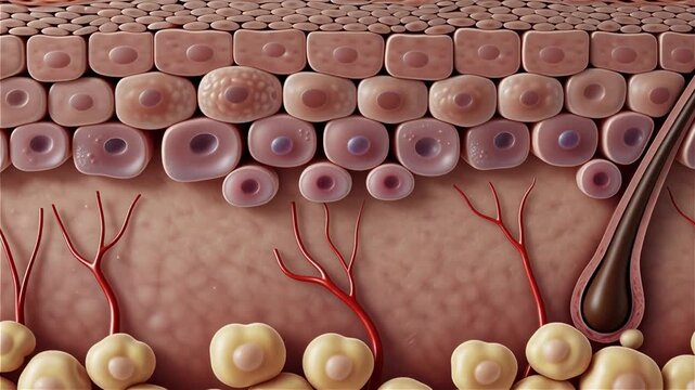
Detailed Illustration of Human Skin Anatomy and Structure
00:05
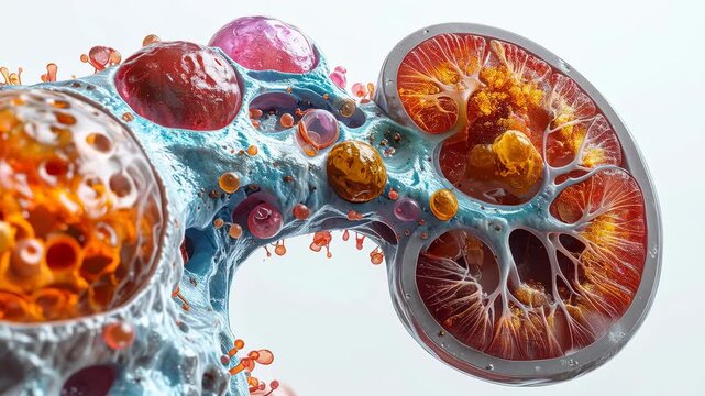
Medical anatomy illustration of human kidney showing serious health problem, with detailed cross section revealing painful stones and polycystic disease for nephrology
00:05
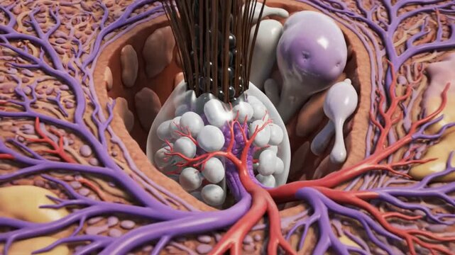
Hair follicle skin anatomy cross section showing dermis epidermis cuticle pore, intricate structure with sebaceous gland, blood vessel, nerve, glowing medical science detail in skin layers
00:08
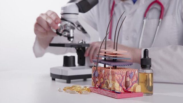
Medical skin cross section model with hair follicles and microscope in laboratory for dermatology education, clinical trichologist.
00:37
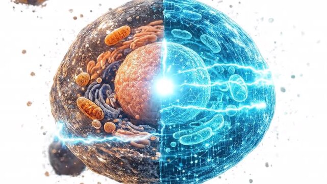
Cross section of cell showing internal structure, biology concept, scientific research, medical study.
00:07
Animation of mathematical equations on mid section of scientist performing experiment at laboratory
00:15
Microscopic view of colorful cells and vessels in a human body tissue section
00:08
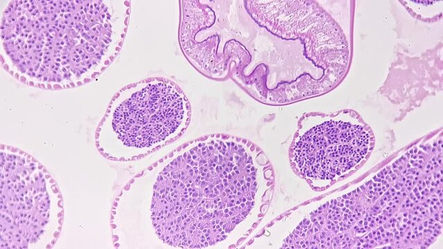
Cross section of ascaris magnified under microscope in 200 times filmed on bright field background. Macro view of parasitic worm living in human intestine and causing disease. Research of pests theme
00:28
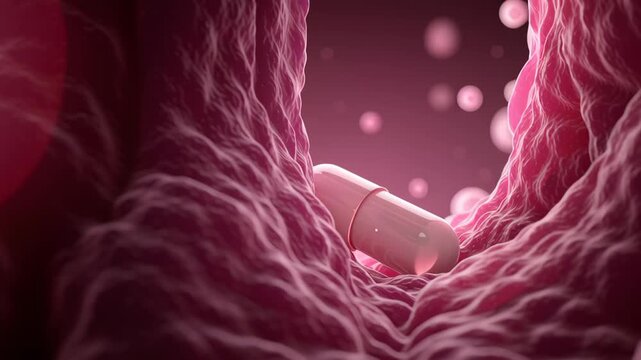
Pink capsule pill inside blood vessel with red blood cells and soft tissue texture, medical anatomy concept for health, drug delivery, and treatment
00:05
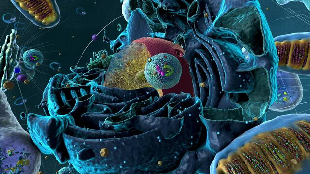
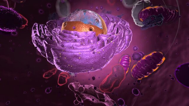
Organelles inside an Eukaryote or eukaryotic cell with focus on a lysosome, component of the cell - 3d illustration
00:30
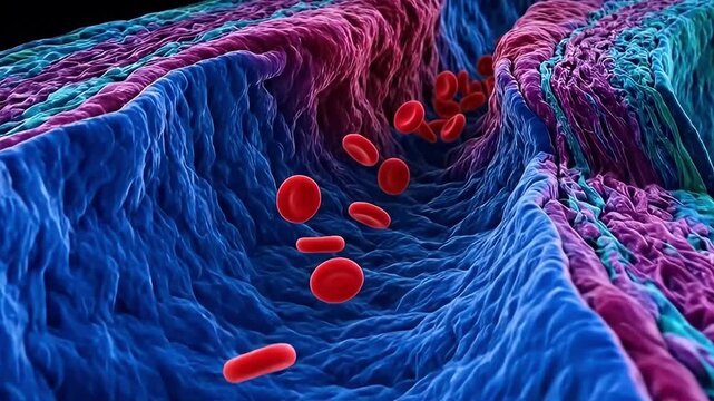
Microscope view 3d render artery blood vessel red blood cell circulation biology anatomy tissue science colorful artery cross section wall microscopic texture vivid medical background
00:08
Histopathology of right breast lump. Infiltrating ductal carcinoma (nst), well differentiated G-1. Section of breast tumour reveal features of an infiltrating ductal carcinoma.
00:12
Magnified section of gallium nitride ribbon reveals microscopic textures and structures, enhancing conductivity with light reflecting off the edges
00:06
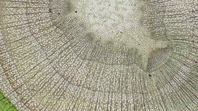
High resolution light photomicrograph of tilia stem cross section seen through a microscope
00:10
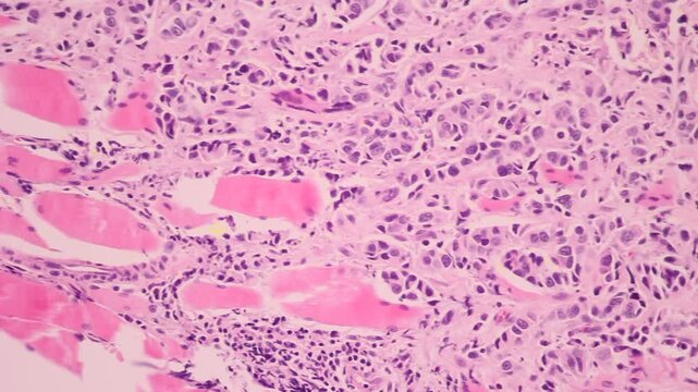
View in microscopic of glandular epithelial cell, adenonocarcinoma from human lung cancer, tissue section by H and E stain.Pathology diagnosis.Medical concept. Under microscope, magnification 600X
00:17
cross section cut of a stem under the microscope
00:30
renal glomerulus histology micrograph showing nephron tissue under microscope
00:08
3D animation of the structure of the hair shaft. The keratin cuticle, cortex, and medulla are shown in layers
00:10
Detailed microscopic cross section of human skin tissue and cells, animated medical website banner
00:05
An educational animation displaying sustainable agriculture data through a digital cross section of soil showing root growth and microscopic nutrient movement in a scientific simulation
00:06
Microscopic cross section of lycopodium stem revealing vascular cells
00:10
Macro cross section of an ATP synthase molecular motor in a mitochondrial membrane
00:10
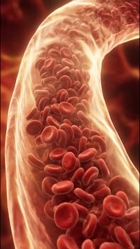
Blood flow veins artery cross section illustration shows red cell movement through vessel wall structure microscope medical anatomy concept for circulation health care blood cell blood flow red cell
00:08
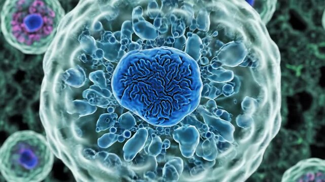
Blue cell nucleus surrounded by cytoplasm and membrane microscopic biology concept showing organelle detail fluorescent plant cells green fluorescent tissue cross section chloroplast stained sample
00:08
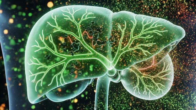
Healthy human liver organ anatomy with translucent glowing bile duct and branching vascular vessel tree transparent liver cross section illustrating illuminated bile duct network, microscopic
00:08
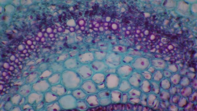
Plant stem cross section microscopic view revealing detailed cellular structure and vascular tissue
00:08
White bean Phaseolus vulgaris, rotating seed coat testa and hilum morphology in botanical section anatomy with microscopic detail, macro laboratory shot
00:16
Microscopic view of plant stem tissue cross section showing cellular structure and vascular bundle development for botanical biology research
00:08
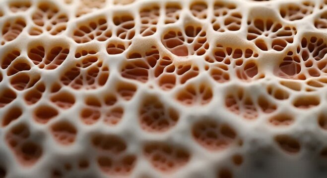
The microscopic view of a bone cross section reveals a seamless trabecular network.
00:15
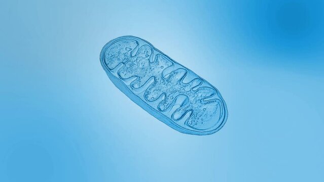
Mitochondria animation, cellular organelles, produce energy, Cell energy and Cellular respiration, DNA, 3D rendering animation
00:30
Stained stem of monocotyledon transverse section under microscope bright field view
00:14
Minimalist outline animation of a human blood vessel section with flowing cells drawing on and fading out. Features a clean pop up style in 3 color setups.
00:12
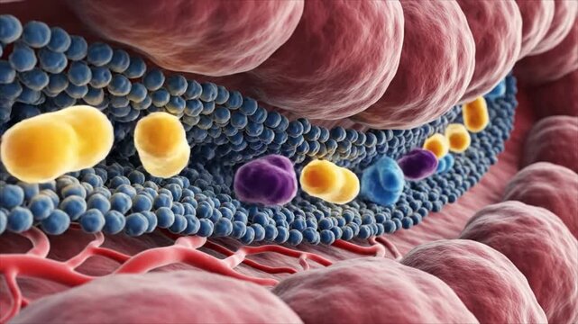
Close-up microscopic view of healthy skin layers with nutrients, Cross-section of radiant skin with hexagonal cells and nourishing particles
00:08
View in microscopic of glandular epithelial cell, adenonocarcinoma from human lung cancer, tissue section by H and E stain.Pathology diagnosis.Medical concept. Under microscope, magnification 600X
00:13
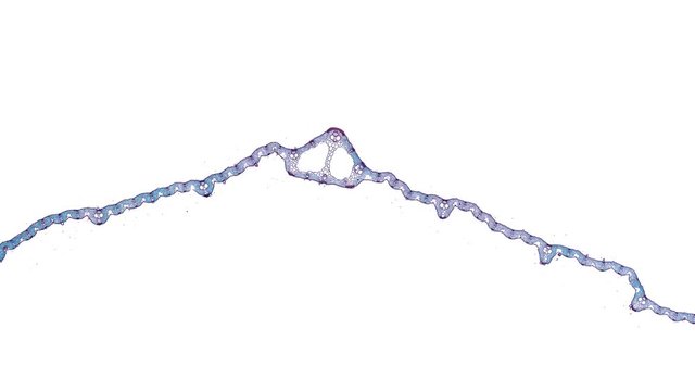
paddy leaf - cross section cut under the microscope – microscopic view of plant cells for botanic education
00:30
Cross section of mitochondrion and cell nucleus in space
00:08
Cross section of a diseased vein with blood pooling. 4K Video
00:05
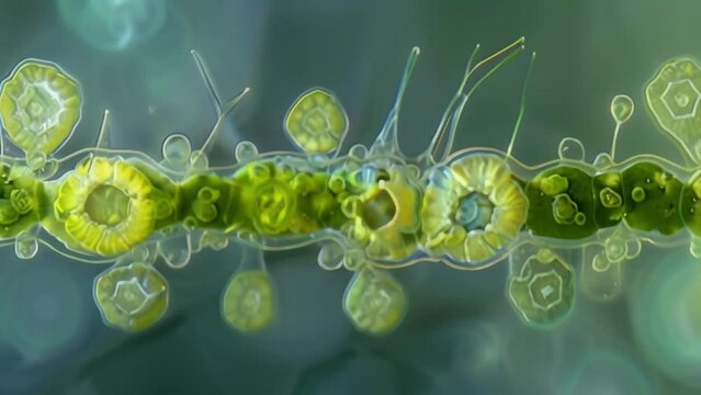
A cross section of a cyanobacteria filament revealing the presence of specialized cells called heterocysts which are responsible for . AI generation.
00:06
Detailed 3D animation showing hair follicle cross section, structure, and associated anatomy
00:08
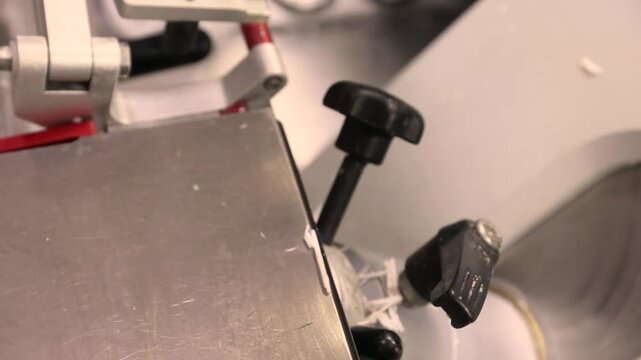
Cyclic motion of a cryostat microtome cutting frozen tissue for rapid intraoperative histology. Preparation of ultrathin sections for microscopic examination.
00:11
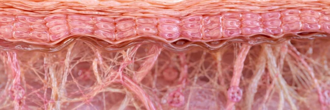
Microscopic cross section of human skin tissue and collagen fibers, dermatology website banner
00:05
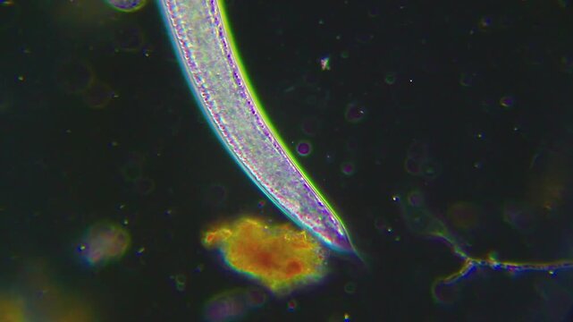
Microscopic view of nematode worm tail section in motion under darkfield 400x magnification
00:06
Macro footage of liver vessel injection in section under microscope 200x on bright field background. Isolated slide of organ serving for detoxification and metabolism. Tissue prepared for examination
00:13
Glowing futuristic nerve root cross section floating above platform microscopic neuron and axon tissue detail with electric sparks clinical laboratory visualization of layered neural tissue cross
00:08
Organelles inside an Eukaryote or eukaryotic cell showing component of the cell
01:00
White bean Phaseolus vulgaris, seed coat testa and hilum morphology in botanical section anatomy with microscopic detail, macro laboratory shot
00:14
Microscopic cross section view of vascular plant stem tissue anatomy.
00:05
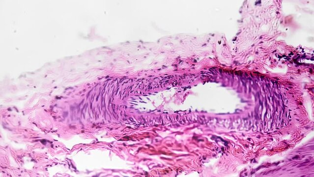
Blood vessel cross section microscopic image anatomy
00:05
Microscopic view of blackhead extraction animation with serum action dissolving sebum plug in skin pore cross section cellular level microscopic
00:08
Macro Soil Cross Section Showing Plant Roots Absorbing Nutrients with Blue Glowing Particles
00:05
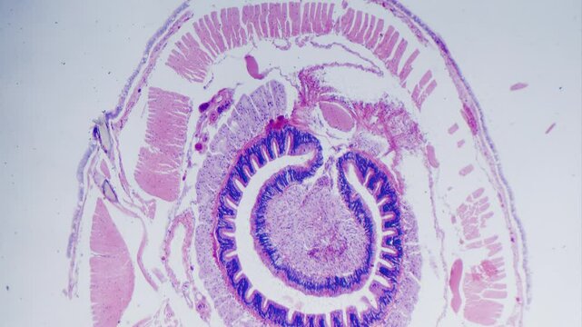
Earthworm transverse section under microscope bright field view, stained sample.
00:10
3D Cross Section of Human Skin with Light Rays Penetrating Layers
00:05
Stamen or Anther under microscope. Cross section of a Lilium stamen, with four locules surrounded by the tapetum. Sexual reproduction in plants. 100x times magnificatiion
00:21
View in microscopic of ductal cell carcinoma, adenonocarcinoma from human breast cancer, tissue section by H and E stain.Pathology diagnosis.Medical concept. Under microscope, magnification 600X
00:28
cotton stem - cross section cut under the microscope – microscopic view of plant cells for botanic education
00:30
3D rendering of a cross section of multiple layers of skin tissue
00:05
Cross section of human blood vessel with glowing inflammation. 4K Video
00:05
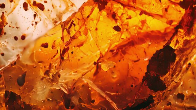
A microscope view of a thin section of fossilized amber with scientists using trace element ysis to determine the age of the amber and any fossils contained within it.
00:07
Macro view of soil section revealing intricate root network beneath the surface
00:05
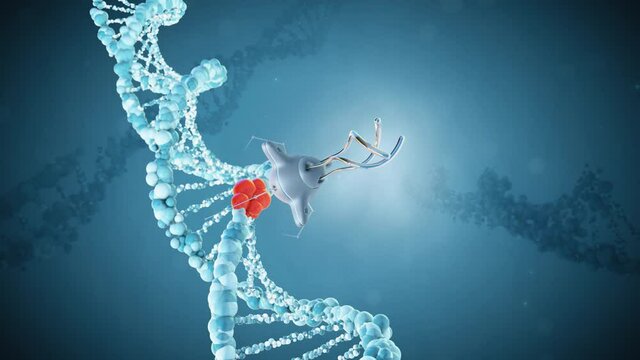
Medical nanobots repair a damaged section of DNA. The concept of the future. Genetic engineering and treatment. 3D render.
00:18
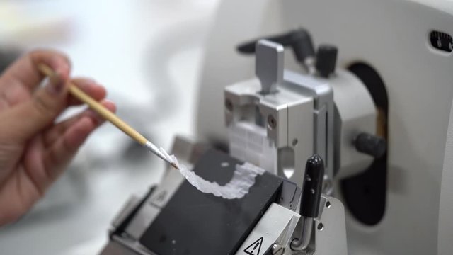
Rotary Microtome Section for diagnosis in pathology make microscope slide histology. Human tissue equipment.
00:08
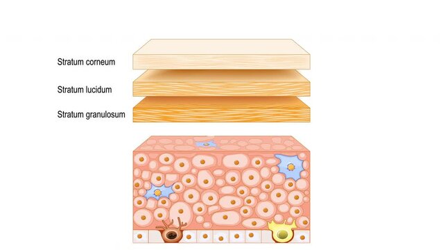
Skin epidermis structure. Skin anatomy. Cross section of the epidermis with text 2d animation
00:16
Bladder of human being in section filmed under microscope 200x for medical analysis on bright field. Sample of organ in people that stores urine from the kidneys. Study different parts of body in lab.
00:17
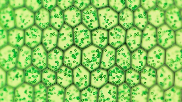
Generic green plant cells under a microscope. Seamless looping animation.
00:10
Microscopic macro close-up shot scientific research epithelial tissue biological anatomical capture
00:10
Detailed cross section of a mitochondrion revealing internal structures and interactions with surrounding molecules, emphasizing energy production and metabolic processes
00:06
Backgrounds of human cells tissue of lung human under the microscope in pathology lab.View in microscopic of ductal cell carcinoma, adenonocarcinoma from human breast cancer, tissue section by H and E
00:30
Blood vessel cross section microscopic image anatomy
00:05
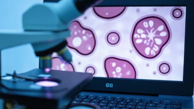
Modern microscope and human tissue section slide with computer monitor show glandular image.Medical patholology and cytologytechnology concept.Selective focus
00:05
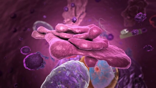
Specialized subunit called organelle inside Eukaryote, focus on golgi apparatus
00:15
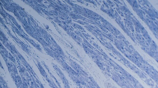
Cardiac muscle section, stained, view in microscope bright field
00:18
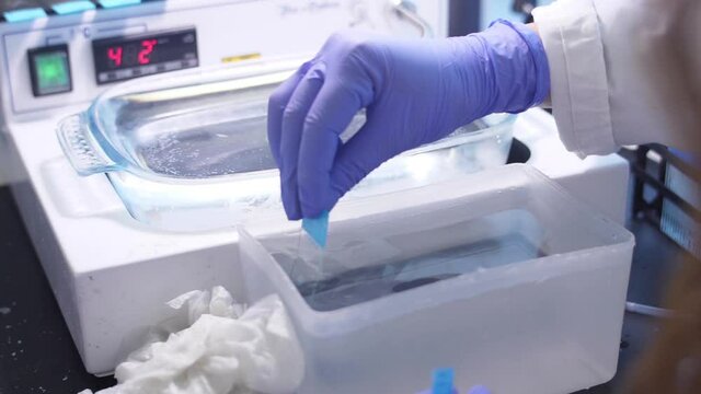
paraffin section being mounted on a microscope slide laboratory slow motion
00:20
Cross section of corn root under microscope in bright field.  100x times magnification. Main cereals in cattle nutrition. Micro botany. Detailed structure of plant roots
00:19
View in microscopic of glandular epithelial cell, adenonocarcinoma from human lung cancer, tissue section by H and E stain.Pathology diagnosis.Medical concept. Under microscope, magnification 600X
00:17
cotton stem - cross section cut under the microscope – microscopic view of plant cells for botanic education
00:30
Animated Plant Cohesion Transpiration Process
00:48
Cross section of chloroplast.
00:22
A microscopic look at the distal tubules the final section of the nephron responsible for finetuning the bodys water and electrolyte balance before urine is excreted.
00:08
Microscopic View of Plant Stem Cross Section, Cellular Structure, Tissue Detail
00:26
3D rendered Animation of moving Mitochondria Cells.
00:16
Synthetic Biology concept, microscopic plant pollen grain and spore cross section with glowing pollen sac cells and textured
00:05
Backgrounds of Characteristics Tissue of Human under microscope in biology lab.
00:19
Dense connective tissue of human whole mount filmed under microscope 100x on bright field. Macro isolated footage of collagen fibers arranged in organized manner. Study its functions and role in body.
00:24
Creative ebru background with abstract acrylic color painted waves. Beautiful marbling texture. Handmade marble surface.
00:17
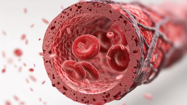
Blood vessel artery cross section showing red blood cells flowing with detailed medical illustration of circulation and vascular structure highlighting blood flow and cellular components dynamic 3D
00:05
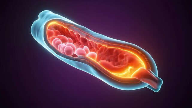
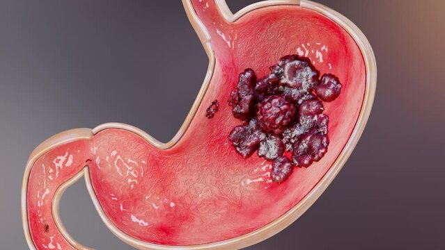
Stomach cancer. stages tumor growth in digestive system, Peptic Ulcer, Cancer attacking cell. gastric disease concept. symptoms, malignant cancerous, viruses and bacteria, Cross section, 3d render
00:10
Part of the lens is blurred on the sides. Blood cells are moving in liquid. Close up view under the microscope.
00:16
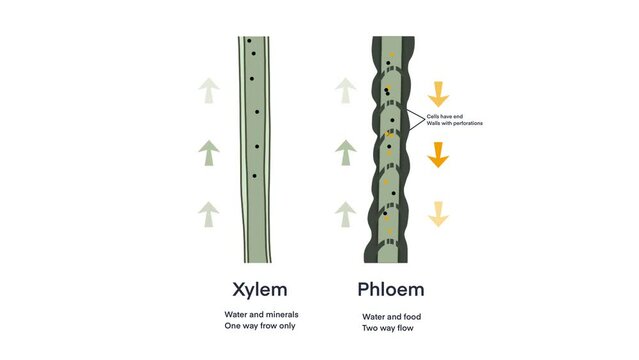
Xylem and phloem water and minerals transportation system outline diagram, Scientific Designing of Xylem And Phloem Scheme, Nutrient And Mineral Transportation, plants transport nutrient and water
00:14
Medical scientist specialist cutting cucumber with scalpel and looking with magnifying glass in food laboratory. Genetic Modifications of Product. Microbiologist analyzing half-cut lab-grown cucumber.
00:22
Close up of a structure being analyzed and watched under a microscope. Locked down real time close up shot
00:17
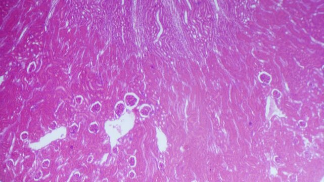
Kidney longitudinal section under microscope 4x objective bright field
00:19
Flight through an Eukaryote, showing organelles - 3d illustration
00:30
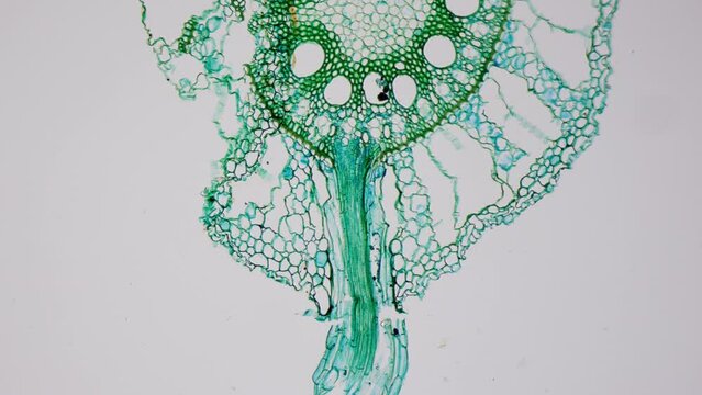
Microbotany. Cross section of green leaf under microscope, 100x magnification. Vegetative organ of plant whose main functions are photosynthesis, gas exchange and transpiration
00:20