Ein Link zum Festlegen deines Kennworts wurde gesendet an: Um in Zukunft auf deine Käufe zugreifen zu können, benötigst du ein Kennwort.
Erstellen ohne Grenzen – unbegrenzte Downloads plus leistungsstarke KI-Bearbeitung.
Neue Filter hinzugefügt
Sortieren nach

33.041 Ergebnisse für microscopy in „Videos“

Dynamic blood cells with glucose molecules — vivid biomedical visualization of sugar–blood interaction
00:05
Female Doctor Engaged In Precise Clinical Research Within Hightech Automated Laboratory Environment
00:10
Virus particle near DNA strands, medical research and virology concept, molecular biology, infection study, biotechnology science, dark realistic macro laboratory visualization
00:10
Microscopic animation of glowing particles within cell-like structures
00:10
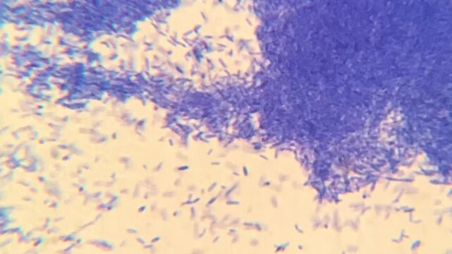
High resolution microscopy of bacteria colonies
00:44
Highdetail depiction of mitochondria in cellular energy production
00:10
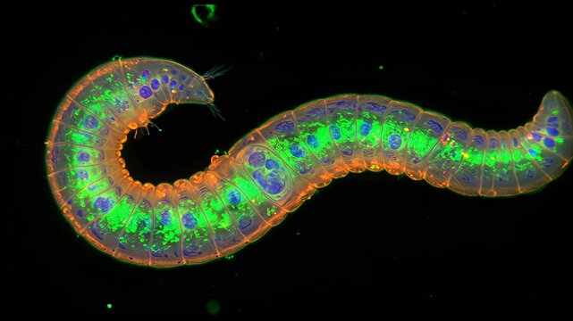
Fluorescent microscopy of Caenorhabditis elegans, with glowing neurons and transparent body structure under lab lighting
00:10
Electron Microscopy Enables Precise Scientific Examination of Glowing Biological Cellular Material
00:10
Annelids moving with rotifer colony in background under 40x magnification darkfield microscopy. 4K scientific microscopy footage.
00:27
Resistance to antibacterial agents, growth of bacteria in the intestines, microscopy of bacteria
00:10
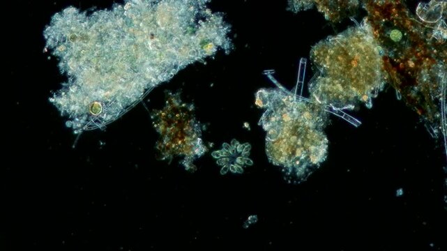
Microscopic view of paramecium and amoeba actively swimming in a drop of pond water. Dark-field microscopy lighting makes the microorganisms glow vividly against a deep black background.
00:05
Microscope cell tissue fiber biology medical closeup with blood red cells, bacteria biofilm, cellular surface network, floating particles, inflammation pathology research, abstract biomedical texture
00:08
Closeup of microscope with lens and laboratory glass setup
00:14
Biomedical animation of clostridium botulinum.
00:15
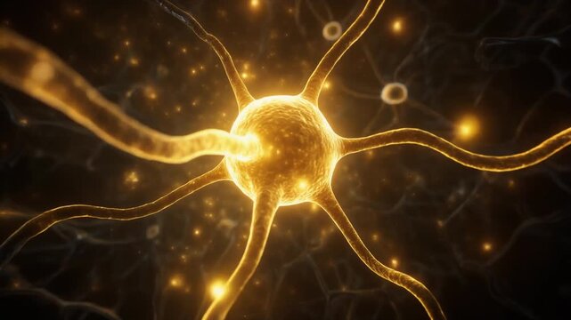
Cinematic close-up visualization of a glowing neuron cell body with branching dendrites and axon transmitting electrical impulses and synaptic light across a dark neural network
00:09
Two scientists in protective gear using microscope and advanced holographic interface to analyze DNA data in a high-tech laboratory.
00:16
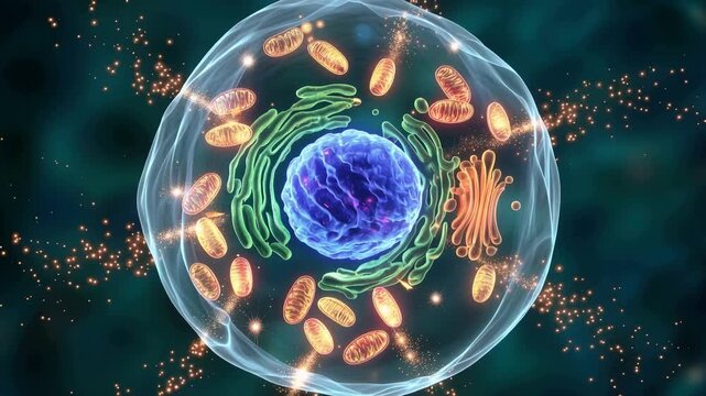
Detailed illustration of a cell with various organelles and structures highlighted.
00:07
Fibroblast cell neuron network microscopic biology glowing membrane organic structure molecular tissue science research connective extracellular matrix scientific visualization cellular anatomy
00:05
Atomic Surface Patterns via Quantum Tunneling Microscopy Generative AI
00:10
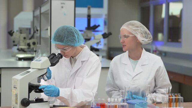
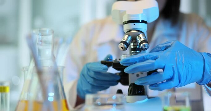
Female scientist microscope laboratory focusing on moving sample through eyepiece; gloved hands adjust controls in blue lit clinical research closeup.
00:21
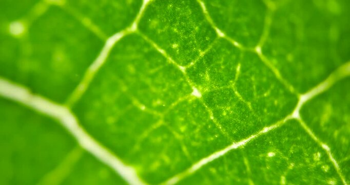
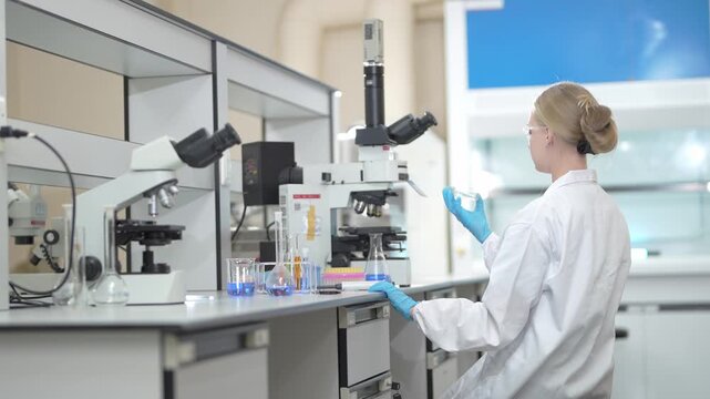
Cell Structure View of Leaf Surface Showing Plant Cells For Education. Leaf in Macro Shot Background. Bright Green Leaves of Plant or Tree With Texture and Pattern Close Up
00:05
A brightfield microscopy image of hepatocytes revealing their dual function in detoxification and the production of essential proteins such as albumin and clotting factors.
00:07
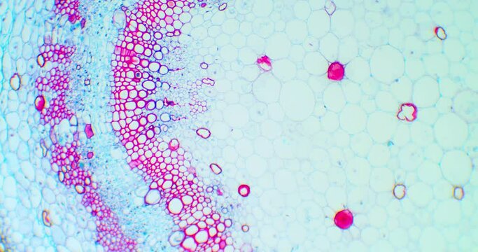
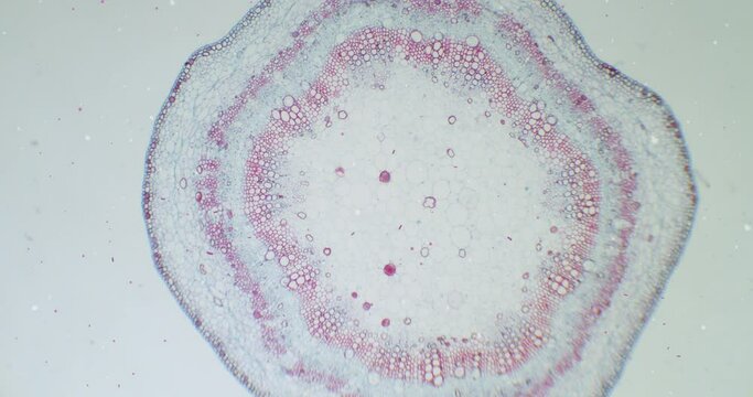
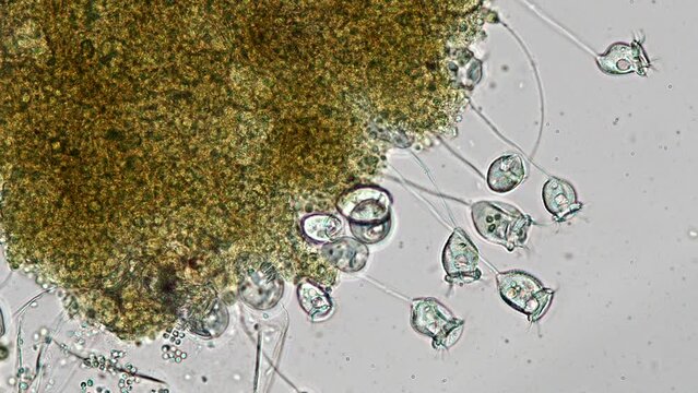
Cotton stem under a microscope with moving germs, microbes and bacteria. Infected view. Microscopic view of plant cells for botanic education. Cotton under a scope. A plant stem microscopic wideshot.
00:26
An intricate view of neurons showcasing synaptic connections and electrical activity, highlighting the beauty and complexity of the nervous system in amazing detail.
00:05
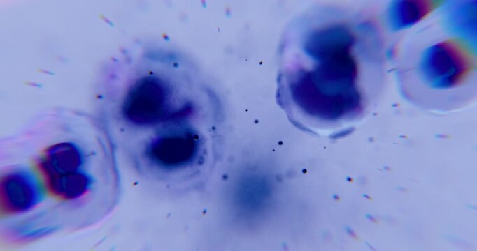
White blood cells under microscopy view, WBC under microscope
00:11
Hyper-realistic cinematic visualization of interleukin molecules activating T-cells in molecular dynamics
00:05
3D animation: Green glowing plankton, algae, and diatoms float in dark water. Microscopic aquatic life, darkfield microscopy style. Science, biology visualization.
00:10
Microscopic view of sperm cells swimming in blue fluid, fertility science and reproductive medicine concept sperm cells, spermatozoa, fertility, reproductive medicine, conception
00:05
Ciliated protozoa inside cysts surrounded by large amounts of bacteria under 400x brightfield microscopy. Detailed 4k microscopic view of microbial interaction
00:20
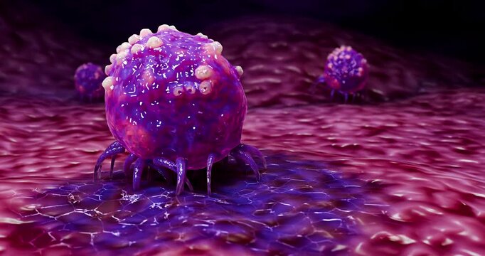
3D visualization of a virus or cancer cell inside the vascular system, medical microscopy.
00:10
Swarm of navicula diatoms swimming dynamically sampled in Japan. Microscopy footage 400x magnification time lapse.
00:57
Regeneration process.Tissue repair. Growth mechanisms. Natural regeneration. Biological adaptation. Cellular growth. A detailed visualization of cells with glowing effects and particles.
00:05
Laboratory research activities scientific lab image series professional setting close-up view microscopy techniques for education
00:09
Close-up view of red blood cells moving through a fluid medium, highlighting their shape and structure intricacies.
00:10
Female lab tech picking up pipette, aspirating sample for microscopy with gold stars overlaying
00:10
Molecule chemistry science cell biotechnology molecular nad longevity research laboratory featuring glowing molecular structure near cell surface and membrane DNA microscopy genetics medical biology
00:08
Deep Dive Into the Structure and Function of Neurons in the Human Nervous System
00:05
Healthy human liver organ anatomy with translucent glowing bile duct and branching vascular vessel tree transparent liver cross section illustrating illuminated bile duct network, microscopic
00:08
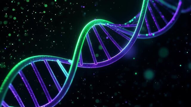
Blue microscopic DNA double helix strand molecular structure glowing genetic science genome
00:05
Close-Up of Solid Lipid Nanoparticle Aggregates Under Microscope for Scientific Research
00:05
Close-up view of vibrant green microalgae cells in detail.
00:10
Detailed analysis of antimony grains in a quality control setting focusing on texture and purity for industrial standards
00:06
Strömende rote Blutkörperchen
00:05
Cell metabolism, A vibrant, scientific illustration of a mitochondrion, featuring intricate details and glowing energy, representing cellular power and function.
00:05
Protozoa under microscope, Ciliate microorganism
00:29
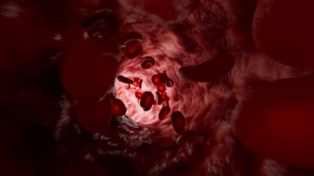
Red Blood Cell in an artery, Blood Flow, medical human health-care Concept Background, 3d animation.
00:12
Microscopic view of a neuron synapse with neurotransmitter release and signal transmission.
00:10
Red web under a microscope. Moving spider web. Vibrating thriller web under a scope. Abstract background under microscope. A lot of stranger things strings under extreme close-up. Creepy microworld.
00:30
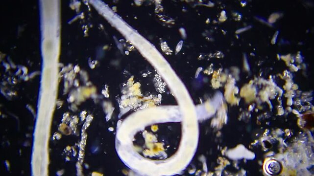
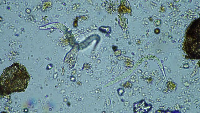
Nematodes actively moving in blue water droplet under 100x magnification brightfield microscopy. 4K scientific microscopy footage.
00:27
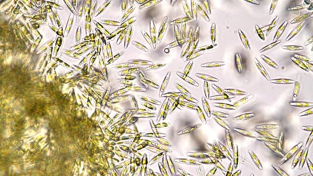
Diatom under the optical microscope
00:35
Glowing Cells Interacting in Dark Space with Dynamic Energy and Detailed Structure
00:08
Zinc Drop under Microscope Smooth Focus Magnification 100x, Visible Surface Texture Metallic Shine, Chemical Laboratory Research Science, Scientific Metallurgy Material Analysis Detailed View
00:13
3D Illustration of Ebola Virus Disease
00:05
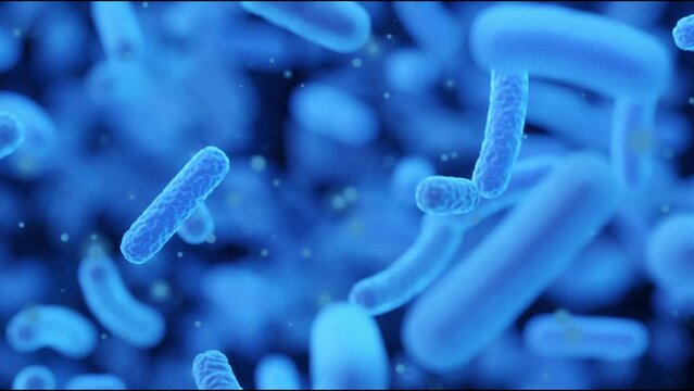
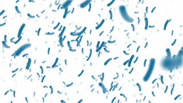
A close up of a bunch of blue bacteria. Resistance to antibacterial agents, growth of bacteria in the intestines, microscopy of bacteria.
00:10
Colorful 3D DNA Helix Model Representing Genetic Science, Biology and Molecular Research
00:05
Abstract representation of neuron cells showcasing synapses and connections within the human brain, depicting intelligence and neural networks in vivid detail
00:10
Dynamic red blood cells flow through a vibrant, microscopic environment as the camera steadily tracks their motion, creating a cinematic, educational visual experience.
00:10
Microscopic View of Giardia Lamblia Parasitic Protozoan in Blue Background. Concept of Microbiology, Parasite Study, Cellular Biology, Protozoan Research, microbe, pathogen, bacteria
00:10
Scientific analysis of food samples in a laboratory with detailed observations and microscopy
00:13
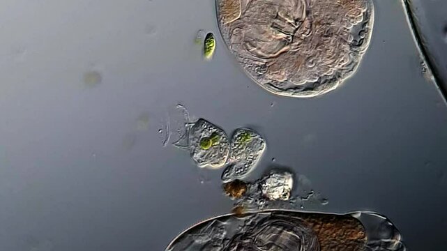
fresh water ciliate, Paramecium bursaria, with ingested green algae circulating in its cytosol. 450x magnification, phase contrast microscopy
00:57
isolated neuron with red nucleus and blue mesh envelope, slow approach revealing membrane tension and surrounding microfilaments, scientific visualization for microscopy and virology
00:10
Microscope view of chromosomes with a glowing third copy of chromosome 21 representing genetic mutation behind Down syndrome.
00:05
Microscopic analysis of a chemical substance in a petri dish in a laboratory
00:14
Hormone molecule binds to cell surface receptor on membrane, showing ligand docking and biochemical signaling action
00:13
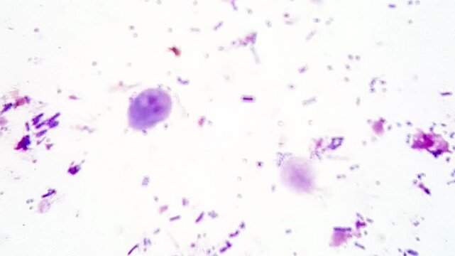
Giardia Wright's stain under microscopy
00:18
Abstract microscopic view of cells with glowing nucleus and soft texture. Concept for biology, life, medicine, science, health, and innovation
00:06
Stunning Digital Representation of DNA Helix in Futuristic Blue Tones
00:05
Bright laboratory-style abstract 3D microscopy visualization, multiple types of viruses and bacteria in vivid contrasting colors, fine structural details on surfaces, floating dyna
00:10
Large nematodes aggregation actively moving in blue water droplet under 40x magnification brightfield microscopy. 4K scientific microscopy footage.
00:26
Paramecium and other microorganisms under the microscope at 100x Magnification
00:19
In a high-tech laboratory, a scientist of Asian descent examines a holographic model of a virus, highlighting its intricate details and structure. The atmosphere is innovative and focused
00:10
Cotton stem under a microscope with a focus on germs, microbes and bacteria. Infected view. Microscopic view of plant cells for botanic education. Cotton under a scope. Microscopic plant stem
00:25
Clusters of cells cling together under microscopy.
00:40
The nematodes (Nematoda), roundworms or eelworms constitute the phylum Nematoda. Species in the phylum inhabit a broad range of environments. Found in freshwater crabs.
00:16
Microbial green chemistry microscopy of algae and microorganism culture water. glass vial and petri showing diverse single celled life soft warm
00:05
Red blood cells move in human blood stream during a close-up view under a microscope showing various shapes and sizes of the cells in a dark background
00:10
Visual representation of mitochondria highlighting structure and function in cellular energy production during biological processes
00:10
examination of a slide with a tissue sample for pathology microscopy
01:17
Firefly flying through bloodstream with red blood cells and sugar cubes vibrant medical illustration
00:05
Beautiful and very active single-cell marine microalgae found in the bucket of a fisherman. Microscope footage, time lapse, 200x magnification, bright field microscopy
00:43
Research lab scene, Advanced molecular testing environment with various diagnostic tools present, Innovative biomedical research setting featuring colorful reagents and precise imaging devices
00:05
Microscopic depiction of nerve signal transmission at cellular level
00:05
Advanced Scientific Analysis: A futuristic interface displays detailed scientific data, highlighting intricate biological structures with precision, symbolizing cutting-edge research and innovation.
00:10
A high-resolution depicting red blood cells filled with hemoglobin against a microscopic background.
00:10
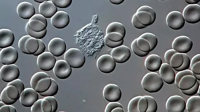
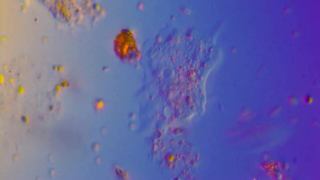
Movement of freshwater Amoebae under microscopy
00:33
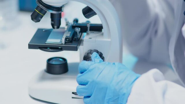
Scientist adjusts microscope objectives and focuses under blue laboratory lighting, gloved hands rotating lenses and peering closely at samples.
00:33
Animation Microscopic blue nipah virus Particles in Medical Science Concept.
00:06
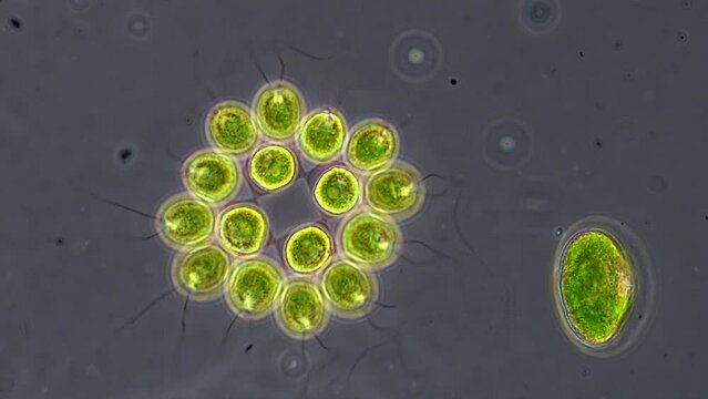
Microscopy: Plankton Translucent body, eye, antennae, internal details. Green/yellow spiral algae, blue background.
00:10
Oligochaete worm moving among detritus with visible digestive tract under 40x magnification brightfield microscopy. 4K scientific microscopy footage.
00:15
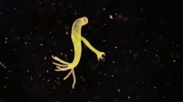
Hydra under the microscope at 40x Magnification
00:35
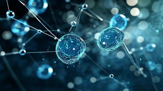
Molecule network technology for skincare science, nanotechnology chemistry, glowing blue molecular structure with connected nodes, serum delivery innovation, cellular repair research
00:10
Leukocytes moving in fluid environment under a microscope
00:10
Close-up shot of sample slide and microscope with metal lens at laboratory.
00:18
The nematodes (Nematoda), roundworms or eelworms constitute the phylum Nematoda. Species in the phylum inhabit a broad range of environments. Found in freshwater crabs.
00:17
Microscopy of parasite worm nematode (Strongyloides stercoralis). Movement of active parasite infection form in magnification 150x.
00:11
Bacterial infection Yersinia pestis microscopy fluorescent microscopic animation
00:10
Macro image reveals oval-shaped Saccharomyces cerevisiae yeast cell with brownish-gold hue, Ai Generated.
00:10
Pan across human sperm cells seen under high magnification.
00:28
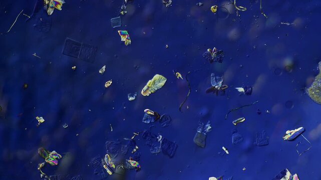
Melting ice under polarized light microscope
00:21
fresh water micro organism, Heliozoa, with its beautiful filaments spreading out like sun rays. 400x magnification, dark field microscopy
00:49