Ein Link zum Festlegen deines Kennworts wurde gesendet an: Um in Zukunft auf deine Käufe zugreifen zu können, benötigst du ein Kennwort.
Erstellen ohne Grenzen – unbegrenzte Downloads plus leistungsstarke KI-Bearbeitung.
Neue Filter hinzugefügt
Sortieren nach

111 Ergebnisse für microconidia in „Videos“

Spread of new life and evolution in universe. Abstract global warming and climate change 3d animation loop background concept for growth, blue renewable energy, microbial biofuel or hydrogen power.
00:20
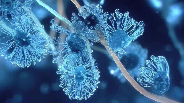
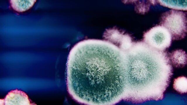
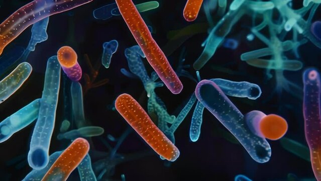
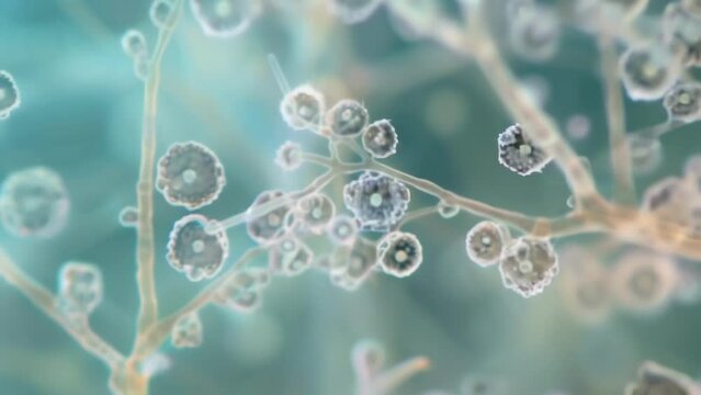
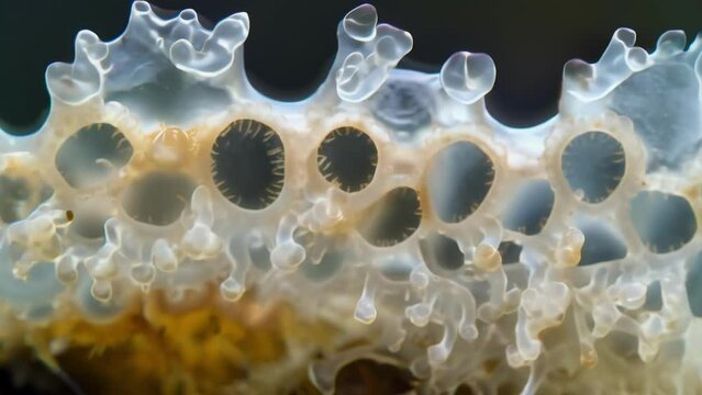
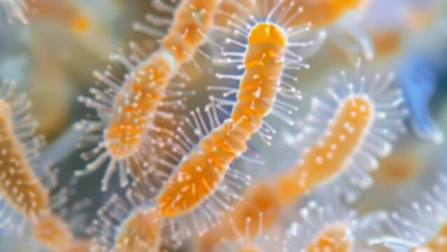
macro microscope video of a microscopic conidia the organisms detailed structure. The conidia is composed of a rigid cell wall that appears light blue in color and
00:06
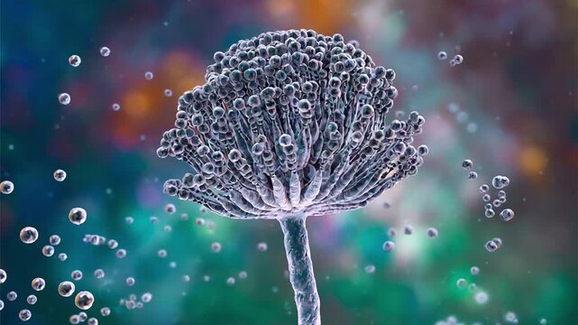
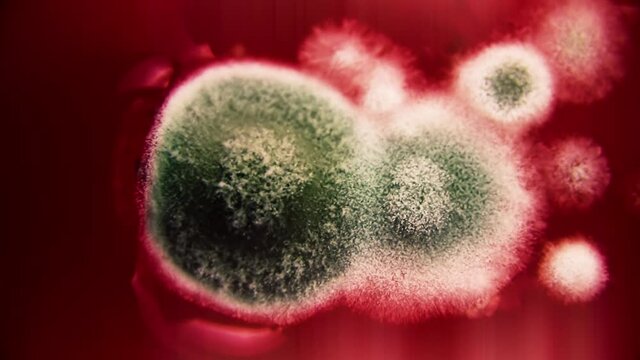
Aspergillus, Mold fungi can cause lung infection
00:08
Penicillium chrysogenum species of fungus in a mold, rotating close up shot
00:19
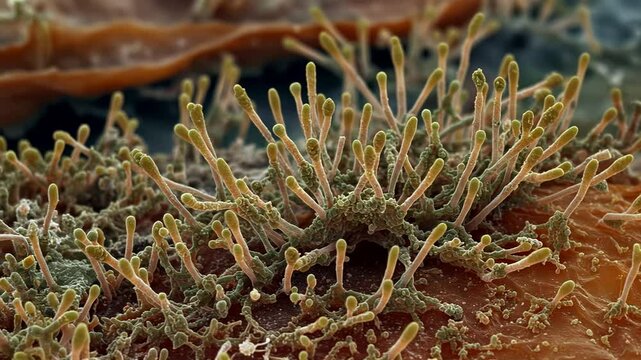
High-magnification image of Aspergillus fungi colonies on spoiled fruit, with radiating conidiophores and spore chains under soft lighting
00:10
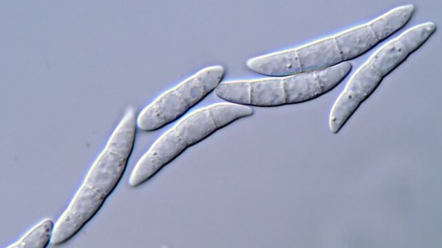
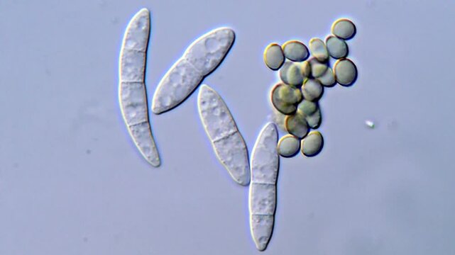
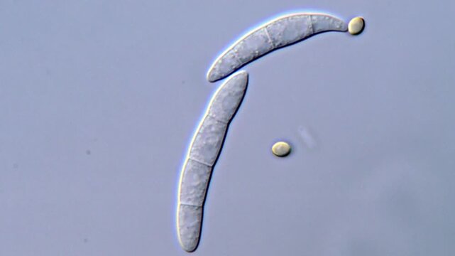
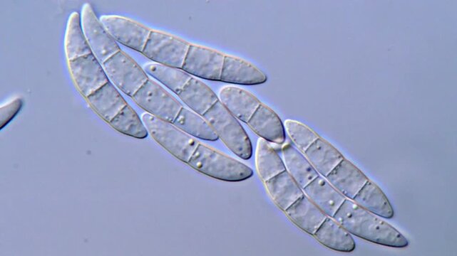
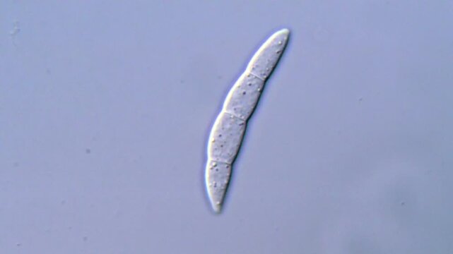
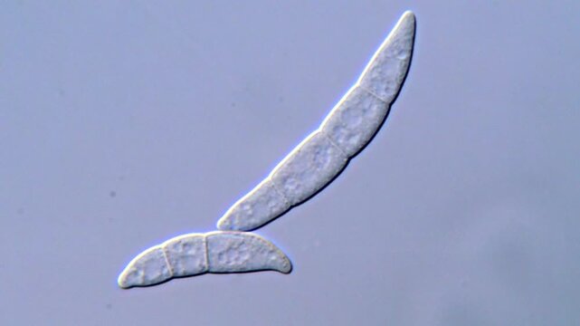
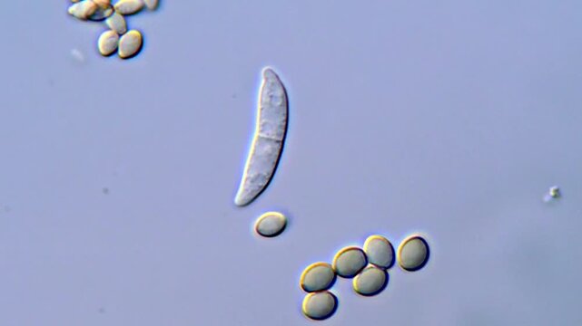
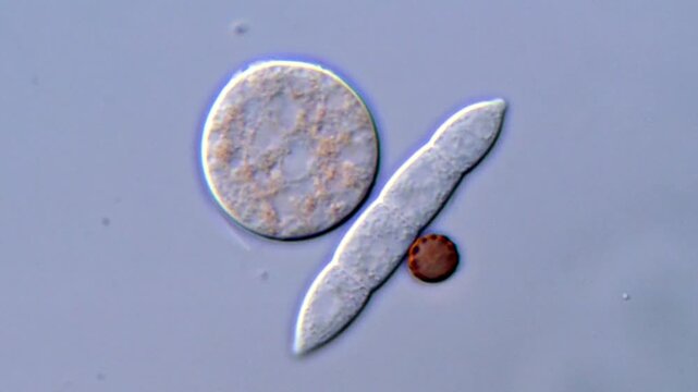
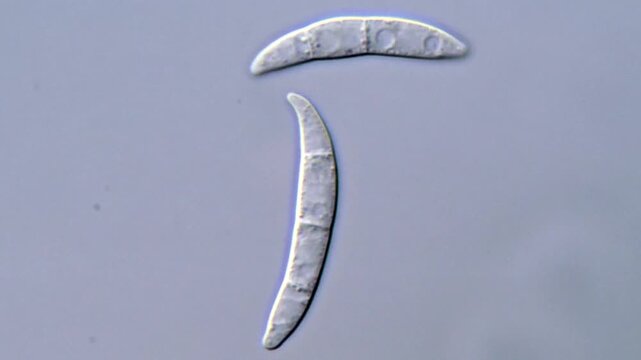
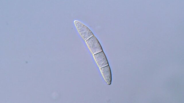
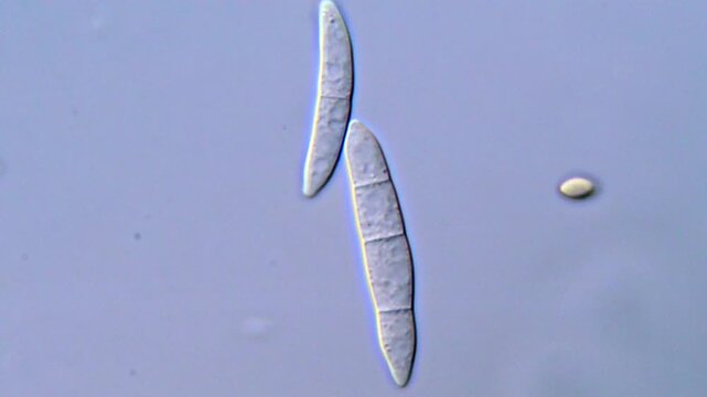
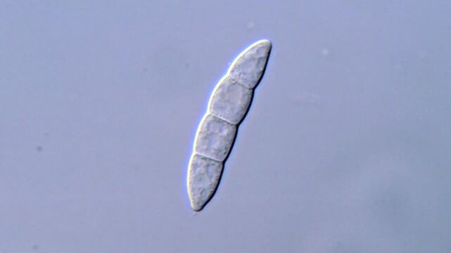
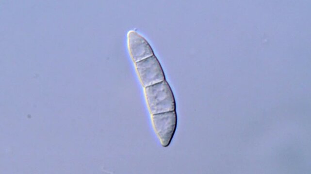
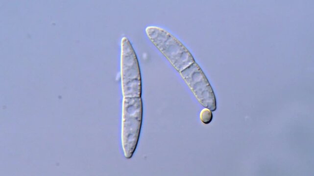
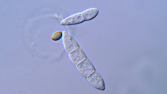
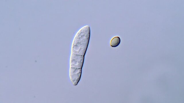
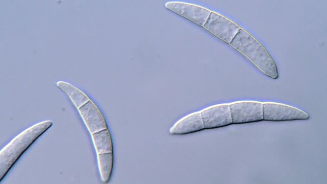
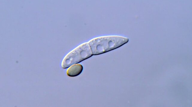
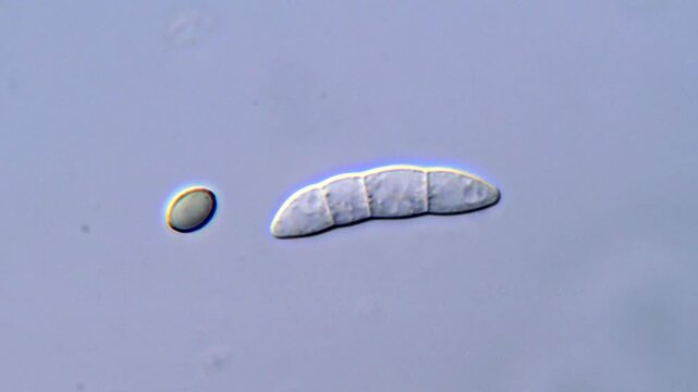
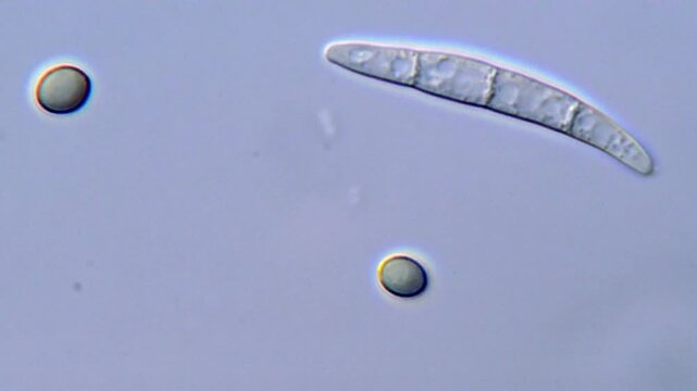
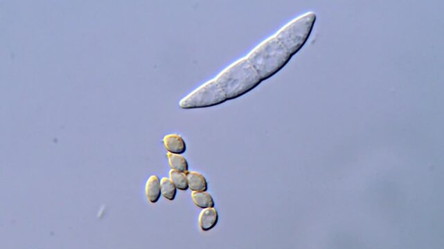
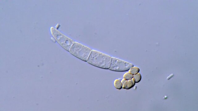
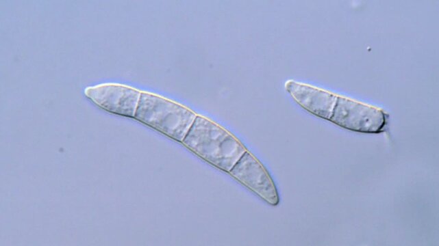
High Resolution video of fusarium fungi spore under microscopic view
00:13
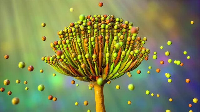
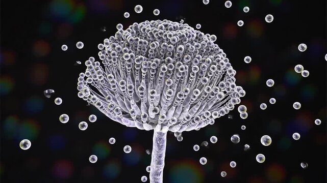
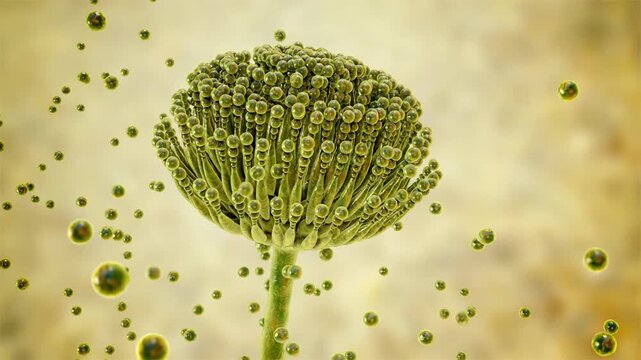
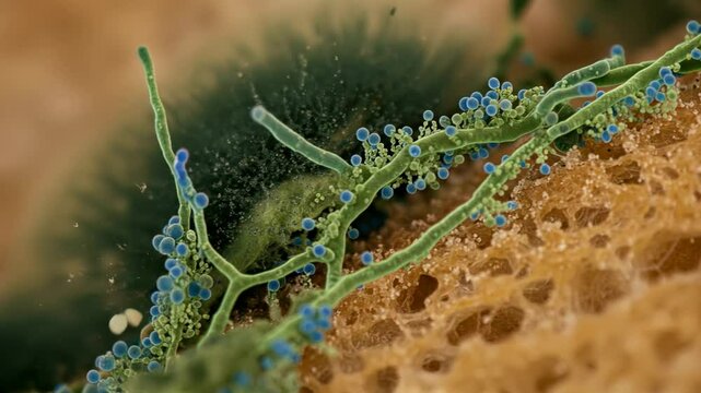
Aspergillus fungal structures, 3D animation showing slowly rotating hyphae and conidia, illustrating opportunistic mould associated with respiratory infections.
00:10
Fungal spores in the liquid. Viral bacterium in the blood.Fungal infection. Distribution and multiplication of fungi and bacteria. Virus attack.

00:18
A group of mushrooms on a dark, wet surface with reflections and bokeh in the background
00:05
3D rendered medical animation of the Cladosporium mold
00:13
macro microscope video of a microscopic conidia the organisms detailed structure. The conidia is composed of a rigid cell wall that appears light blue in color
00:05
Animation of Cladosporium mould
00:13
Delicate red coral branch with blue marine underwater mood pink microscopic filament branching
00:05
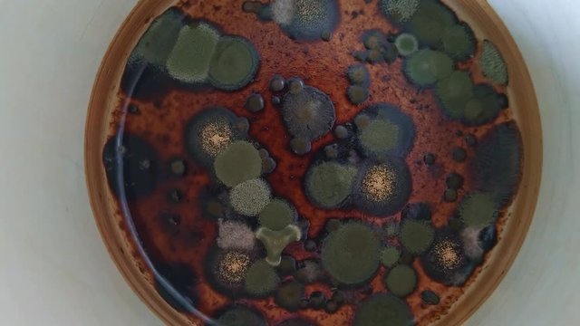
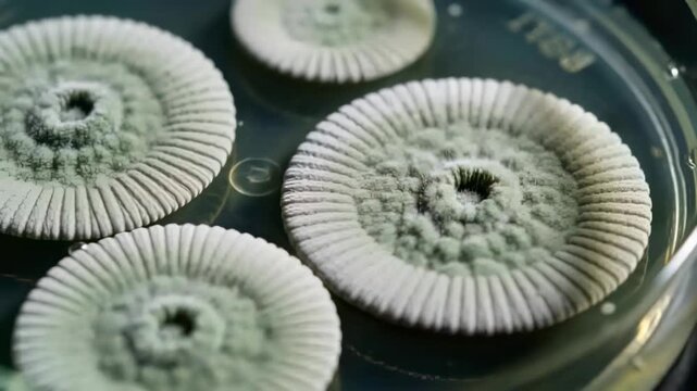
Petri dish containing fungal culture with orange-brown hyphae and spores growing on agar.
00:05
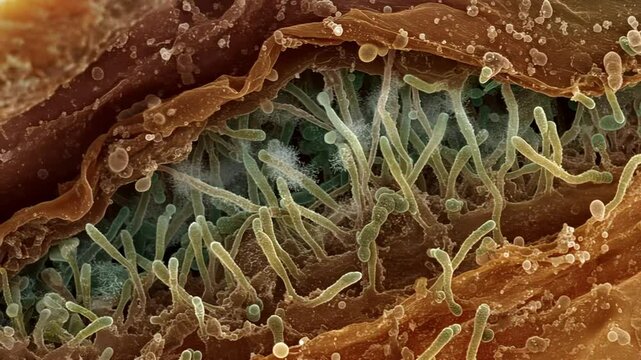
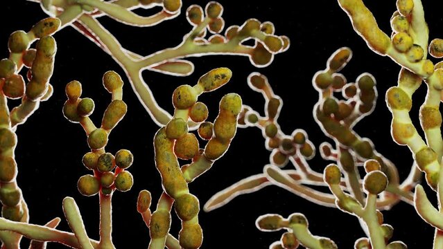
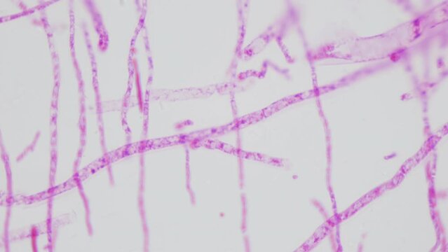
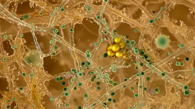
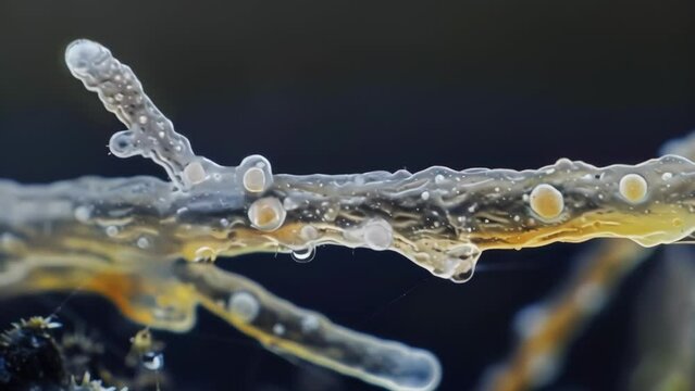
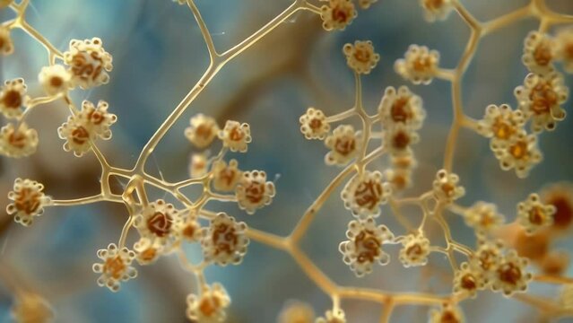
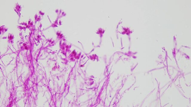
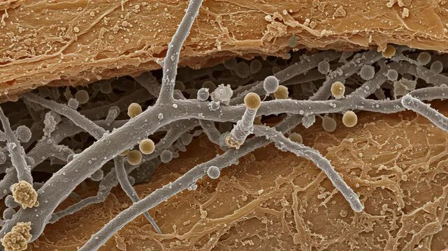
Microscopic view of branching fungal hyphae with spores and conidia
00:08
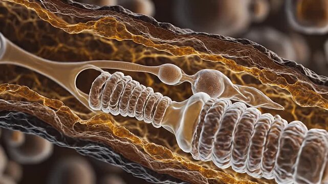
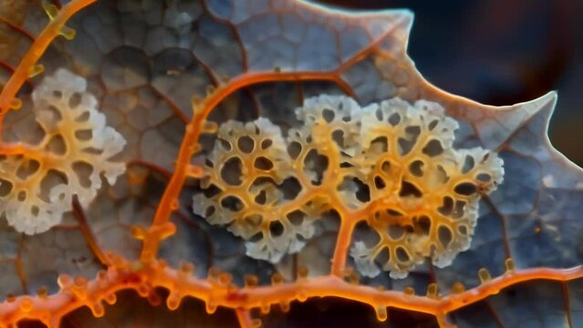
Microscopic View of Fungal Hyphae with Conidiophores and Conidia in Warm Tones
00:08
Spoiled tomato close up on white background
00:24
Scientific Animation of Floating Fungal Spores and Mycelium.
00:08
Coccidioidomycosis with medical digital technology concept
00:18
Producers of antibiotic penicillin. Mold under microscope. Detailed anatomy of fungi of genus Penicillium. Multiple magnification. Toxic metabolites.Three-tiered tassels with conidia, complete mushrom
00:26
Spread of new life and evolution in universe. Abstract global warming and climate change 3d animation loop background concept for growth, green renewable energy, microbial biofuel or hydrogen power.
00:20
A series of sequential images captured under a light microscope showing the movement of conidia spores as they are released from a . AI generation.
00:05
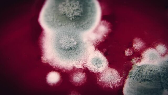
Time-lapse recording capturing fungal colonies expanding and darkening on petri dish agar
00:05
Microscopic visualization of Stachybotrys, a genus of mold.
00:10
 Microscopic view of Penicillium mold growing on a slice of bread, showing branching hyphae and spore-producing structures in vivid detail
00:10
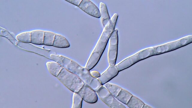
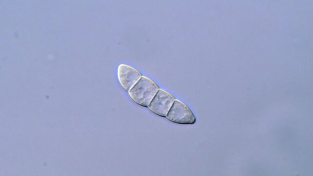
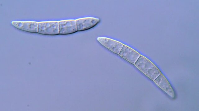
Fusarium Spores and Hyphae Under Compound Microscope
00:13
Aspergillus fungal structures, 3D animation showing slowly rotating hyphae and conidia, illustrating opportunistic mould associated with respiratory infections.
00:10
Fungal spores in the liquid. Viral bacterium in the blood.Fungal infection. Distribution and multiplication of fungi and bacteria. Virus attack.
00:18
Mushrooms close-up in nature with bokeh background
00:05
ゆったりとした時が流れる湖 | 秦野 | 震生湖
00:55
Visualization on different focal planes of a sclerotium of botrytis with mycelial germination
00:04
A network of branching hyphae each ending in a of conidia forming a sprawling and complex web of fungal reproduction. . AI generation.
00:06
A colorful image revealing the diverse shapes and sizes of conidia spores from different fungal species. Some spores are ovoid while . AI generation.
00:06
A crosssection of a spore print displaying a variety of conidium shapes and sizes ranging from elongated and tapered to short and . AI generation.
00:06
Fusarium Fungus Growth Seen Through Microscope Lens
00:17
Detailed Microscopy of Fusarium Fungus Hyphae
00:21
Fungus spore under DIC microscope, Mold fungi
00:14
An electron microscope image revealing the intricate weblike structure of a conidiophore the specialized structure responsible for . AI generation.
00:06
A closeup of a single conidium revealing its distinctive shape and detailed structure under high magnification. . AI generation.
00:05
A highly detailed image of a conidium releasing spores showcasing its complex structure and function. . AI generation.
00:06
A highly magnified image of a single conidium showing its spindlelike shape and numerous spiky projections that provide a means of . AI generation.
00:06
Fusarium Spore Microscopy for Scientific Observation
00:15
A single elongated conidium with a smooth glossy surface branching off from a larger fungal structure. . AI generation.
00:06
Mold fungi spore under DIC microscope view
00:18
A collection of brightly colored conidia ranging from vibrant blues and greens to deep oranges and purples showcasing the diversity . AI generation.
00:06
A vast field of conidia each one a different color forming a beautiful mosaic of shapes and patterns as they spread and multiply. . AI generation.
00:06
Aspergillus fungal structures, 3D animation showing slowly rotating hyphae and conidia, illustrating opportunistic mould associated with respiratory infections.
00:10
A timelapse image capturing the rapid growth of a fungal colony with small conidia emerging and quickly multiplying to cover the surface . AI generation.
00:06
Fungi spore under DIC microscopy view 1000x magnification
00:14
A closeup view of a single conidium showing its spherical shape and the intricate network of branching structures that allow it to . AI generation.
00:05
Aspergillus fungal structures, 3D animation showing slowly rotating hyphae and conidia, illustrating opportunistic mould associated with respiratory infections.
00:10
A crosssection of a conidium revealing the intricate network of cell walls and filaments that make up its structure. . AI generation.
00:06
Plant pathogenic fungi spore under microscopy view 1000x magnification
00:17
Microscopic Observation of Fusarium Pathogenic Spores
00:14
Fungal spore in mold rice under microscope
00:32
A side view of a conidium showing the complex patterns of the outer layer and the visible pores where spores will be released. . AI generation.
00:06
 Microscopic view of Penicillium mold growing on a slice of bread, showing branching hyphae and spore-producing structures in vivid detail
00:10
High Resolution Microscopy of Fusarium Spores, 1000x magnification
00:13
Fungal spores in the liquid. Viral bacterium in the blood.Fungal infection. Distribution and multiplication of fungi and bacteria
00:41
Highly magnified image showing numerous conidia clumped together their elongated shapes and spikes resembling a of ly caterpillars. . AI generation.
00:06
Penicillium. Producers of the antibiotic penicillin under microscope. Anatomy of fungi of the genus Penicillium. 100x times magnification. Enzyme production, medicine usage. Three-tiered tassels
00:19
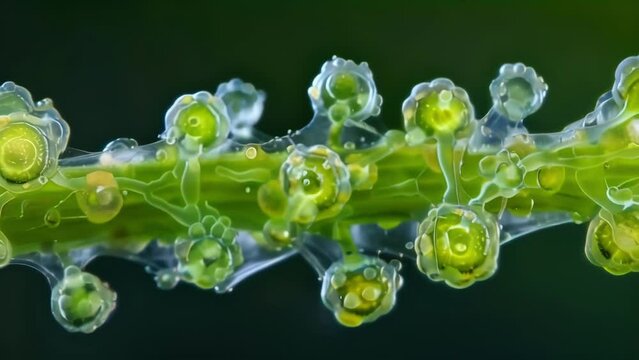
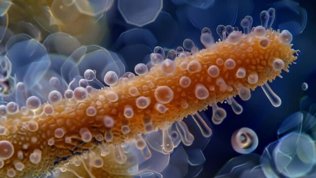
A of conidia clinging to a plant stem with their unique structures and textures helping them to cling and thrive in their natural . AI generation.
00:06
Fusarium spore Observed with Microscopic Detail
00:13
Mold Hyphal Structures and Spores Under Microscope
00:16
Closeup view of an individual conidium with multiple spores forming a circular pattern resembling a flower. . AI generation.
00:05
Fungi spores under microscopy view 1000x magnification
00:18
Fungal spores in the liquid. Viral bacterium in the blood.Fungal infection. Distribution and multiplication of fungi and bacteria
00:08
A highresolution image of a conidium spore settled on the surface of a leaf. The spore is surrounded by a halo of tiny branching filaments . AI generation.
00:06
A highly magnified image of a conidium releasing spores through its specialized opening a vital tactic for survival and propagation . AI generation.
00:05
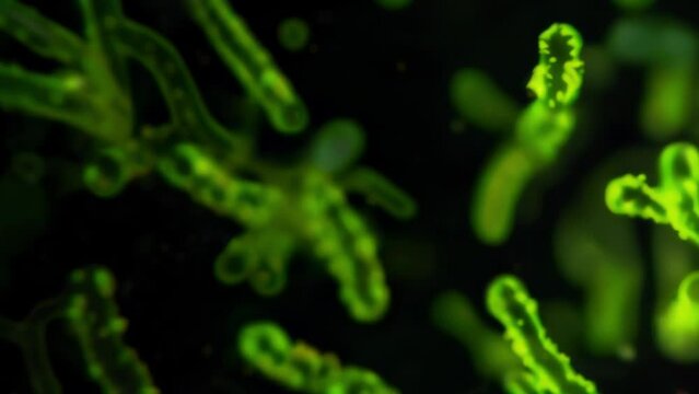
A fluorescent microscopy image showing the glowing green conidia against a black background emphasizing their microscopic but powerful . AI generation.
00:06
A magnified view of a single conidium spore revealing its rugged outer surface and elongated shape. The spore is surrounded by smaller . AI generation.
00:06
Fungi spores under microscope, microorganism under microscopy
00:13
Spoiled tomato close up on white background
00:28
Fusarium Fungal Sporangium Captured in Detail
00:12
Fungi under microscope
00:14
High-magnification image of Aspergillus fungi colonies on spoiled fruit, with radiating conidiophores and spore chains under soft lighting
00:10
Fungi spores in high Resolution under microscopy
00:15
Fusarium Spore Morphology Under High Resolution
00:14
Fungal spores in the liquid. Viral bacterium in the blood.Fungal infection. Distribution and multiplication of fungi and bacteria. Virus attack.
00:07
 filamentous fungi spore under microscopy view
00:13
Fungus in mold rice under microscopy view
00:13
An aerial view of a petri dish filled with conidia each one growing and spreading in a controlled environment displaying their resilience . AI generation.
00:06
 Microscopic view of Penicillium mold growing on a slice of bread, showing branching hyphae and spore-producing structures in vivid detail
00:10
Fungi spores under microscope
00:13
Fusarium Fungus Spore Microscopy in ultra HD Video
00:13
ゆったりとした時が流れる湖 | 秦野 | 震生湖
00:55
ゆったりとした時が流れる湖 | 秦野 | 震生湖
00:55
Plant pathogenic fungi spore, 4K Microscope video
00:11
ゆったりとした時が流れる湖 | 秦野 | 震生湖
00:55
Microscopic Observation of Fusarium Pathogenic Spores, 1000x
00:13
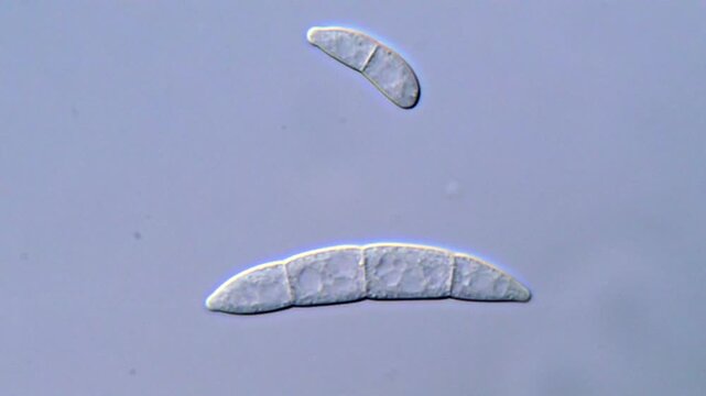
Fusarium Macroconidia Observed Under Microscope, 1000x magnification
00:12
Phytopathogenic fungi spore under microscopy view
00:17
Pathogenic Fusarium Spores Viewed Under Microscope
00:14
Spore in rice under microscopy view, fungi spore
00:12
ゆったりとした時が流れる湖 | 秦野 | 震生湖
00:55
Fungal spore under DIC microscope
00:12
Spore of fungi under microscope
00:13
High Definition Microscopic Footage of Fusarium Spores
00:11
ゆったりとした時が流れる湖 | 秦野 | 震生湖
00:55
Fungi spore under microscope, High Resolution video
00:13