Ein Link zum Festlegen deines Kennworts wurde gesendet an: Um in Zukunft auf deine Käufe zugreifen zu können, benötigst du ein Kennwort.
Kreativität ohne Ende – unbegrenzte Downloads plus leistungsstarke KI-Bearbeitung.
Neue Filter hinzugefügt
Sortieren nach

2.526 Ergebnisse für fibula in „Videos“

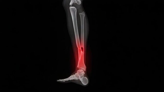
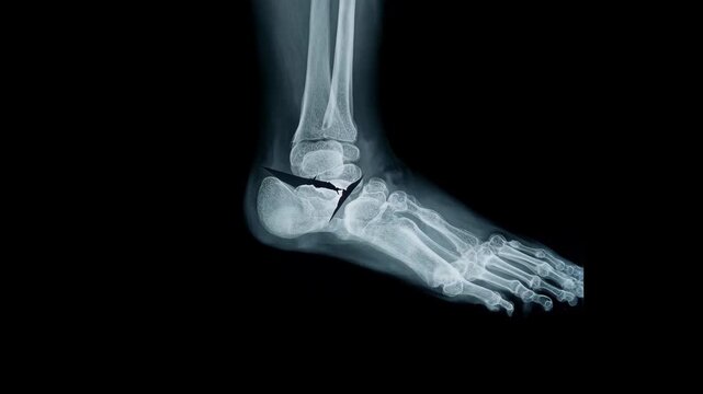
Medical x ray of a broken tibia
00:05
HD
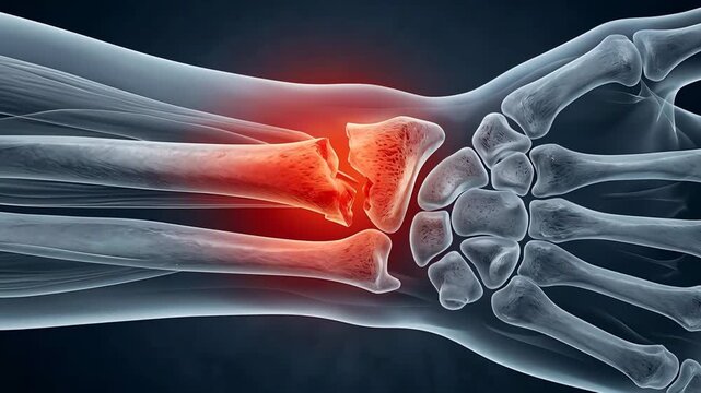
Close Up Medical Illustration Of A Broken Wrist Bone Showing Severe Pain And Inflammation On A Dark Blue Background
00:05
HD
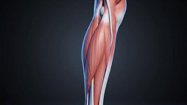
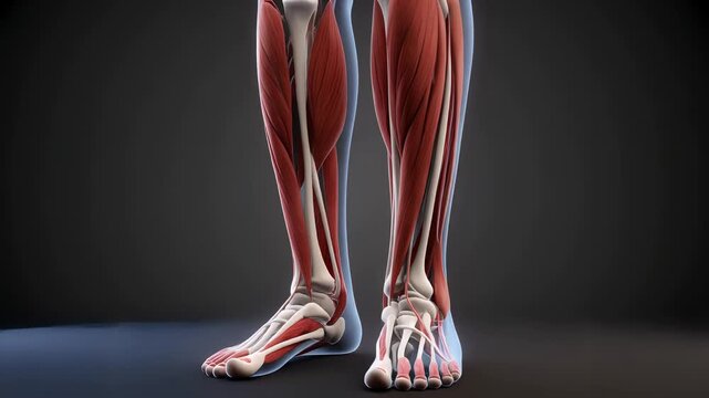
Detailed 3D anatomical rendering of the human calf muscles, showcasing the complex muscular and skeletal structures against a dark background, for human muscular system education.

00:05
HD
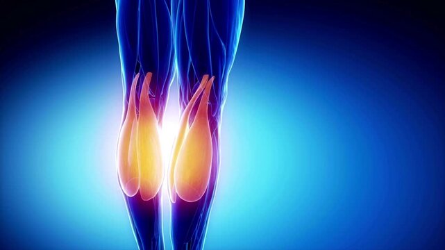
Leg Muscles X-Ray View 3D Animation | Human Leg Anatomy Medical Scan Visualization 4K
00:15
HD
Bubbles and metallic objects submerged in green liquid, concept of innovation
00:08
HD
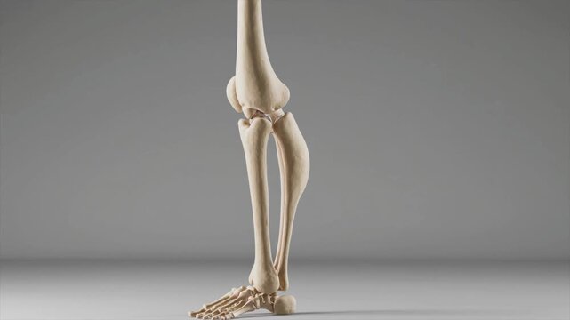
Anatomy of human lower leg skeletal structure with tibia and fibula bones in medical visualization skeleton bone orthopedic healthcare scientific
00:10
HD
Broken Fibula Bone Pain Anatomy Medical Concept
00:12
HD
Knee Anatomy: Detailed medical illustration highlights the knee joint, emphasizing bone structure with a focus on inflammation.
00:10
HD
3D Medical Illustration of Quadriceps Muscle and Patellar Tendon in Knee Joint Anatomy
00:08
HD
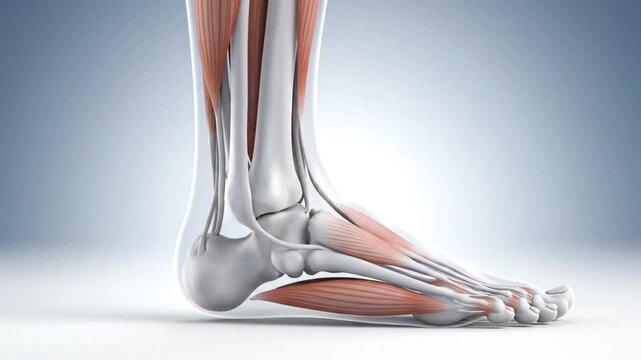
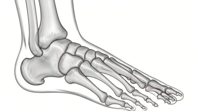
Detailed 3D Anatomy of the Human Foot and Ankle with Bones and Muscles.
00:08
HD
Lumbar Disc Herniation and Sciatic Nerve Pain Visualization - This 3D medical illustration depicts a herniated lumbar disc compressing a nerve root, causing inflammation and radiating pain.
00:08
HD
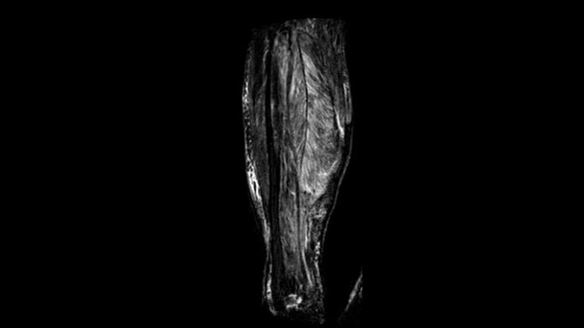
Coronal MRI Scan of Human Lower Legs Showing Tibia Fibula Calf Muscles and Soft Tissue Structure
00:16
HD
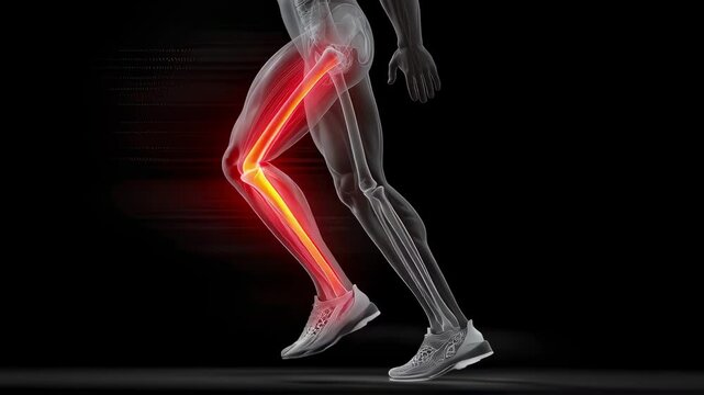
Semi transparent legs of an athlete running showing glowing tibia and fibula bones, highlighting a stress fracture, against a black background
00:05
HD
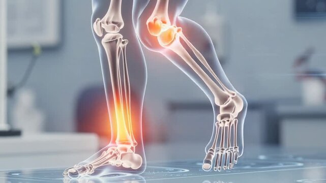
Detailed digital illustration of lower leg bones highlighting pain area in medical setting
00:05
HD
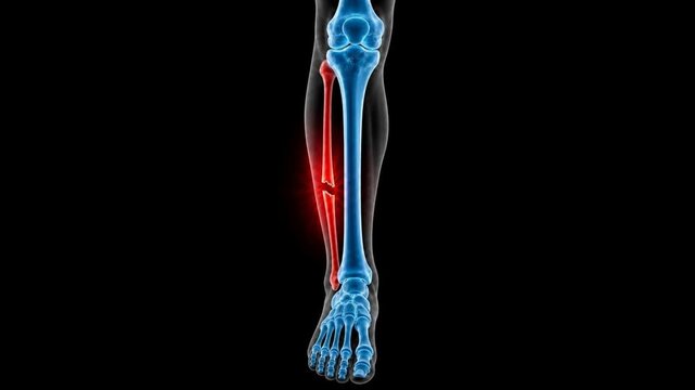
Medical 3D animation showing a fibula bone fracture in the human lower leg
00:06
HD
Pediatric Normal Both Knee X-ray Showing Healthy Bone Structure
00:15
HD
Detailed anatomical model of a human knee joint showing progressive angles and components, including femur, tibia, fibula, and metallic prosthetic elements in a dark background
00:08
HD
Hands of doctor in latex gloves holding a knee prosthesis. Materials for knee joint replacement. Close up.
00:12
HD
A vivid representation of human skeleton anatomy focuses on the knee joint, showcasing intricate details of the bones and surrounding structures in a striking green hue.
00:06
HD
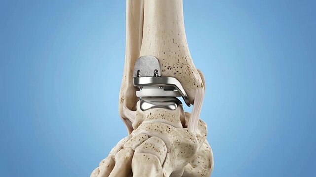
Close up 3D medical animation showcasing a metallic ankle joint prosthesis surgically implanted into a human tibia fibula and tarsal bones with
00:05
HD
Detailed anatomical rendering of human leg muscles in a blue-toned scientific illustration on a dark background.
00:05
HD
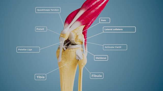
3D rendering of human raising knee, zooming into joint anatomy with labeled tendons, bones, ligaments, and cartilage. Ideal for medical, scientific, and educational use
00:08
HD
human ankle bone Isolated 3D hologram. Medical concept. new technology
00:08
HD
Anatomy of running legs showing bones while exercising on a treadmill in a gym.
00:08
HD
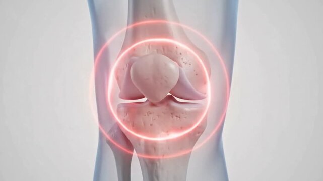
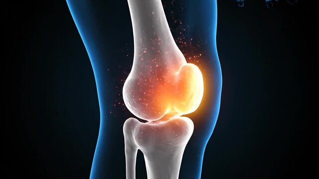
Detailed anatomical illustration of a human knee joint with a red glow indicating pain or inflammation.
00:06
HD
The Human Knee Joint in Pain: An abstract, artistic representation of a human knee joint glowing with intense red, highlighting areas of pain and inflammation.
00:10
HD
3D Medical Illustration of Human Calf Muscle Contraction and Foot Plantarflexion
00:10
HD
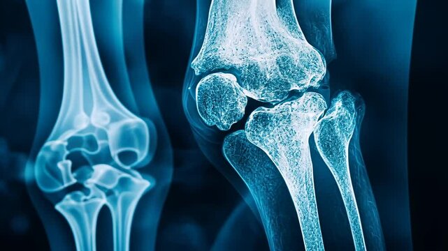
Anatomical knee joint x-ray examination for diagnostic analysis and bone structure, side view
00:10
HD
3D model of human tibia and fibula leg bones, lower leg anatomy
00:10
HD
Isolated Human Knee Joint Skeleton CT Scan
00:16
HD
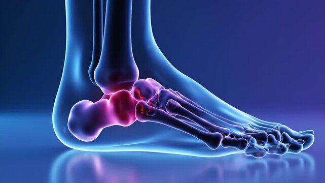
Animation displaying foot bones with inflamed calcaneus, showcasing anatomy and potential pain points related to bone injury and musculoskeletal disorders requiring medical attention.
00:10
HD
Human Leg Anatomy with Pain Highlight 3D Illustration | Muscle Bone Nerve and Joint Medical Concept
00:12
HD
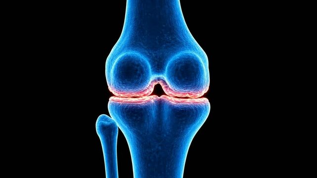
3D medical animation of human knee joint anatomy with blue x-ray style bones and highlighted meniscus cartilage on a black background.
00:06
HD
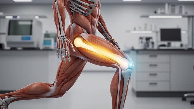
Anatomy of the Human Leg Performing a Lunge Exercise Showing Muscle Engagement and Bone Structure in a Medical Lab
00:06
HD
X-ray of a broken ankle showing a fracture in the fibula and surrounding bones taken in a medical setting
00:09
HD
Glowing Bone Held by Gloved Hands.
00:08
HD
Digital X-ray visualization of human knee joint. Orthopedic anatomy, injury diagnosis and joint health, copy space for text
00:05
HD
A detailed illustration showcasing the intricate mechanics of a knee joint, highlighting the bones, ligaments, and tendons essential for movement.
00:05
HD
Medical illustration highlighting pain and inflammation in the human lower leg bones with a glowing effect
00:08
HD
Knee Anatomy: A close-up view highlights the intricate structure of a knee joint. The image, illuminated with a cool blue glow.
00:10
HD
Ankle Joint Pain Injury 3D Medical Animation Foot Anatomy Achilles Tendon Bone X-Ray Glowing Dark Background
00:06
HD
Human leg bones and joints anatomy with inflammation areas in medical hospital environment
00:05
HD
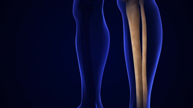
Detailed medical illustration of human leg bones showing tibia and fibula anatomy in high resolution
00:05
HD
This visualization presents the human skeleton, showcasing the intricate details of the skeletal system. Joint structures are illuminated for better understanding and study of human anatomy.
00:06
HD
Professional medical leg scan showcasing a cross-sectional analysis of the tibia and fibula for clinical evaluation.
00:56
HD
3d rendering of human leg showing hamstring and calf muscle anatomy with knee joint bone structure highlighted against a blue background
00:05
HD
Human Leg Anatomy with Pain Highlight 3D Illustration | Muscle Bone Nerve and Joint Medical Concept
00:10
HD
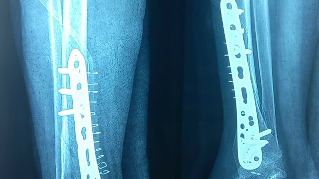
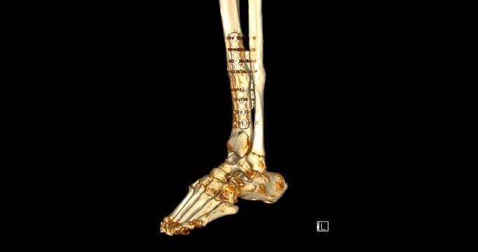
X-ray of the human leg with knee and ankle. Fractures are noted at lower shaft of tibia and head of fibula. Metallic fixator is seen in situ. Soft tissue swelling noted around the fracture site.
00:22
HD
Human Leg Anatomy Knee Joint Medical Visualization
00:05
HD
Human leg anatomy illustration showing glowing calf muscles and bones
00:10
HD
X-ray image of an ankle joint, showcasing bones and structure for medical analysis.
00:04
HD
Anatomical Study of Human Leg Muscles and Bones in Detailed 3D Render Against
00:05
HD
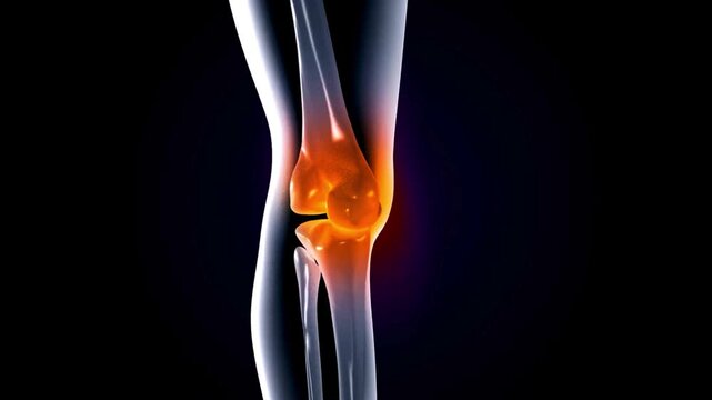
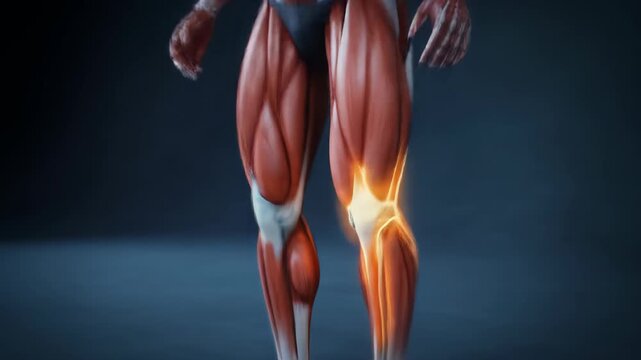
Detailed 3D animation of a male human anatomy model highlighting the knee joint with glowing orange pain or inflammation, showing anatomical structures and muscle tendons
00:15
HD
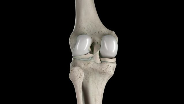
Animation of the knee bones
00:13
HD
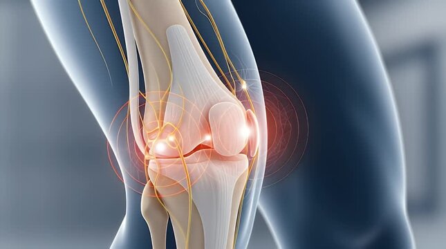
Detailed 3D anatomical illustration showcasing the human knee joint experiencing significant pain and inflammation, emphasizing nerve pathways and bone structure for medical and educational purposes
00:05
HD
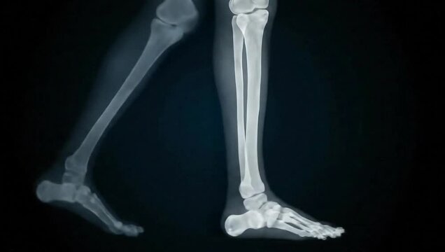
Lateral X-ray view of human legs and feet in a walking motion, showing skeletal bone structure including the tibia, fibula, ankle joint, and metatarsals against a black background.
00:08
HD
Human Skeleton Model Rotating on Black Background.
00:10
HD
Anatomical 3D Rendering of Human Knee Joint Showing Patella Cartilage Inflammation and Pain With Red Highlighted Area Over Black Background Corporate Presentation Medical Education
00:08
HD
Human Skeletal System and Nerves: Anatomical Illustration
00:07
HD
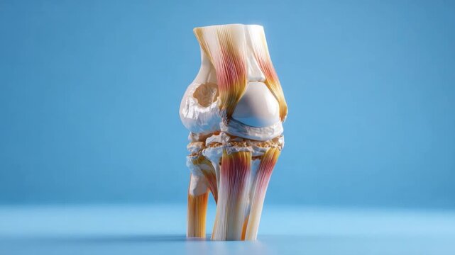
Anatomical Marvel: A detailed anatomical model of a knee joint sits against a soft blue backdrop, revealing the intricate network of bones, cartilage, and ligaments.
00:10
HD
3D Medical Illustration of Human Knee Joint and Ligaments Movement
00:10
HD
Skeleton dancing with arms raised on a green background in a playful and lively animation halloween skelton green screen video
00:10
HD
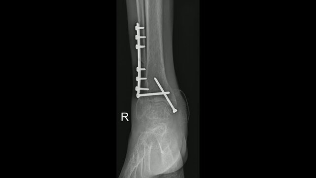
Ankle Joint Fracture Fixation X-Ray Post-Surgical Assessment,
00:15
HD
3D Medical Animation of Knee Anatomy, Patella Structure, and Lower Body Skeleton
00:08
HD
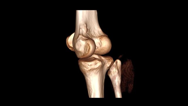
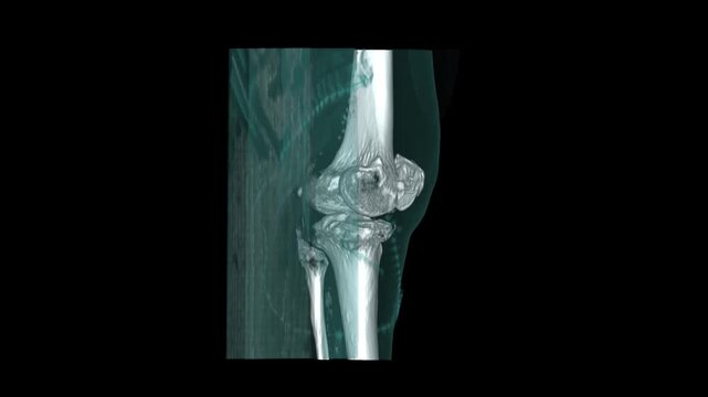
3D volume rendered CT scan reconstruction of a left knee joint showing femur patella tibia and fibula bones
00:16
HD
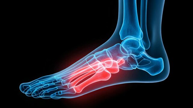
Anatomical illustration of a human foot and ankle showcasing inflamed bones and joints, highlighting pain and discomfort associated with foot conditions and injuries
00:10
HD
Human Leg Anatomy with Pain Highlight 3D Illustration | Muscle Bone Nerve and Joint Medical Concept
00:19
HD
Medical illustration of a knee joint highlighting cartilage, bones, and inflammation.
00:08
HD
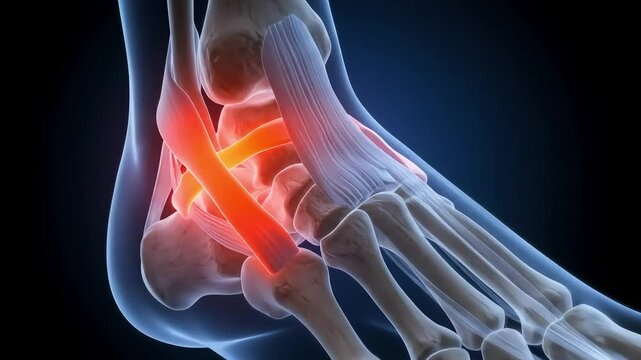
Anatomical Visualisation of Foot Sprain with Highlighted Ligament Injury
00:16
HD
CT scan of ankle joint showing a fracture in the tibia and fibula bones. Detailed medical imaging perfect for educational, diagnostic, and healthcare content.
00:22
HD
A close-up view of a human knee joint, showing the bones, muscles, and ligaments.
00:08
HD
Professional Workspace Modern Office Setup for Virtual InPerson Meetings
00:05
HD
Runner with leg pain demonstrating muscle inflammation and knee joint discomfort during exercise showcasing potential sports injury
00:10
HD
Animated Foot Anatomy Detailed 3D Visualization of Bones and Structure on Blue Background
00:08
HD
Wireframe runner legs highlighting a stress fracture injury during running, providing a visualization of the impact and stress on bones during physical activity
00:05
HD
Rotating 3D Human Skeleton Animation with Detailed Bone Structure and Transparent Body Outline on Dark Blue Background, symbolizing medical education and anatomical study
00:05
HD
Human Skeleton 3D Rendering Rotation Animation.
00:10
HD
A detailed side view of a full human skeleton model sitting upright on a black stool against a plain grey background.
00:06
HD
Detailed Human Skeleton Leg Anatomy Articulated Bones and Joints Displayed in a Studio Setting with Dramatic Lighting and Dark Background for Medical Educational Purposes
00:06
HD
Knee Anatomy and Pain: A detailed, illuminated illustration of human knee anatomy, showcasing pain and inflammation, ideal for medical or health-related projects.
00:10
HD
Man Sitting Medical Exam Table Holding Ankle Fracture Pain Glowing X-Ray Bone Anatomy Overlay Crack Visible Clinic Hospital Background
00:07
HD
X ray of a human leg and lower torso with highlighted knee joint
00:08
HD
Sciatica sciatic nerve pain radiating down leg from spine through pelvis to foot muscle anatomy, inflamed nerve pathway and lower back hip spine anatomy illustration for medical education
00:08
HD
Academic 3D MRI Visualization of Lower Leg Cross-Section and Tibia Fibula Skeletal Architecture
00:11
HD
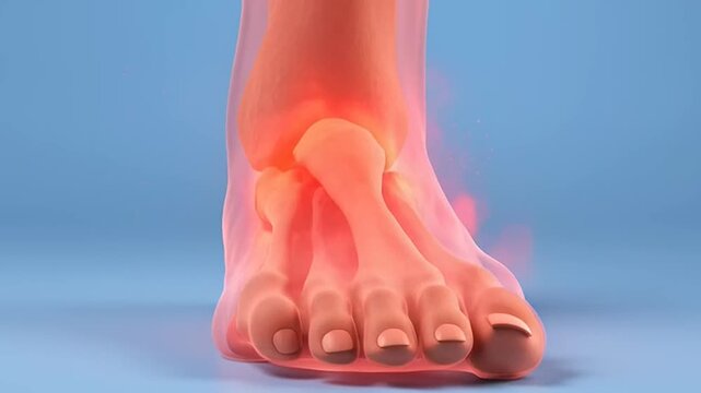
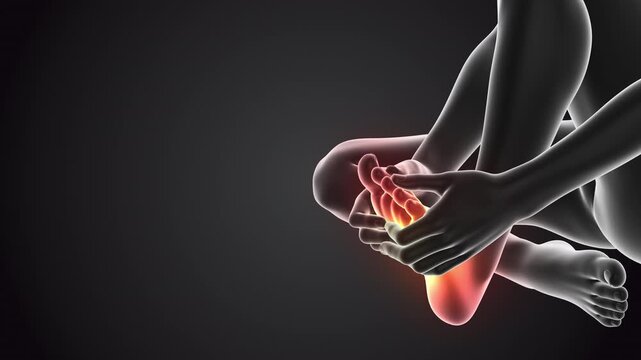
Human having pain in feet
00:08
HD
3d rendering of human foot and ankle bones, highlighting the skeletal structure and anatomy
00:08
HD
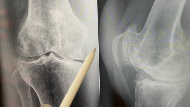
Plain X ray of the knee shows apparent joint osteoarthritis according to Kellgren and Lawrence system, subchondral sclerosis, knee joint inflammation
00:21
HD
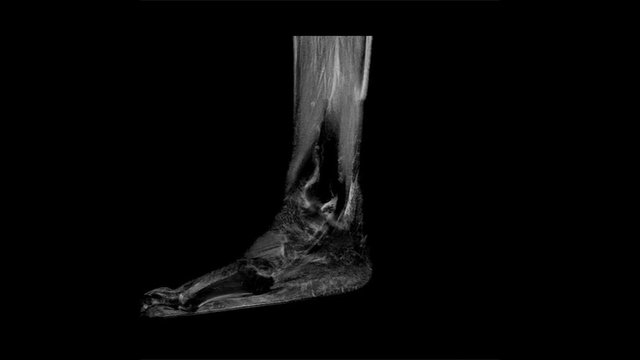
Time Lapse video animation of R.M.N. Nuclear magnetic resonance, of human foot, with cross-section and sagittal section.
00:06
HD
MRI foot scan for a diagnostic tool used to image the structures of the foot and ankle following symptoms: pain  both feet, swelling, redness or warmth, numbness or tingling and stiffness.
00:12
HD
HIP joint skeleton x-ray scan in blue
00:14
HD