Посилання, за допомогою якого можна встановити пароль, надіслано на цю адресу: Щоб отримати доступ до своїх покупок у майбутньому, вам буде потрібен пароль.
Отримайте 3 відео або 25 зображень у безкоштовній пробній версії
Додано нові фільтри
Сортувати за

206 результатів за запитом pathogenicity у категорії відео
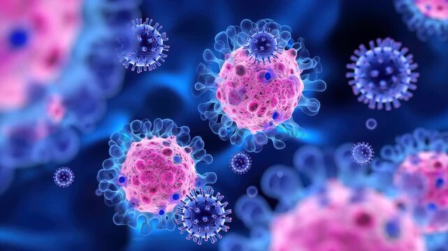
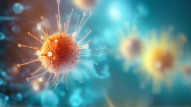
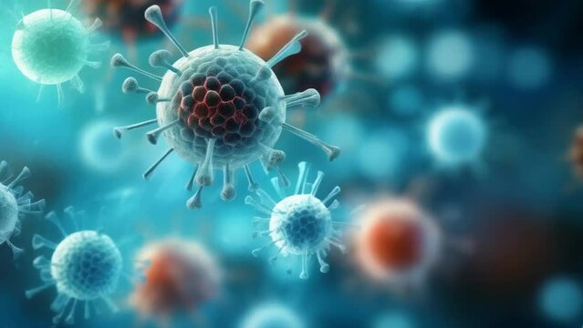
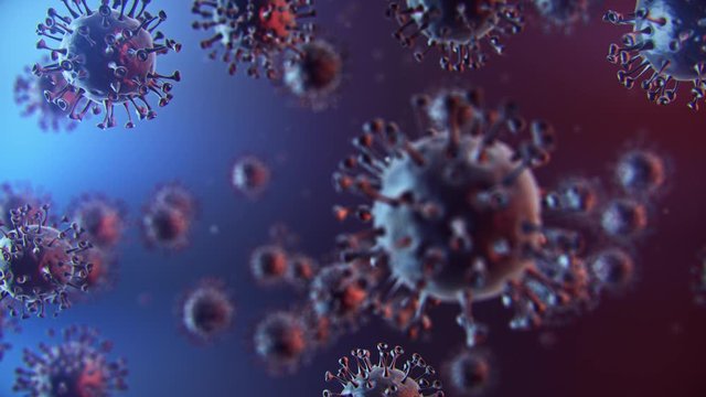
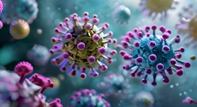
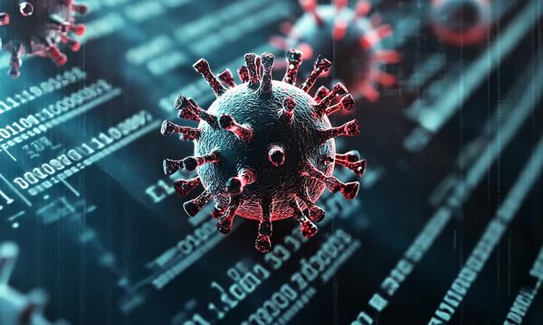
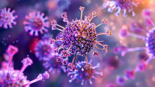
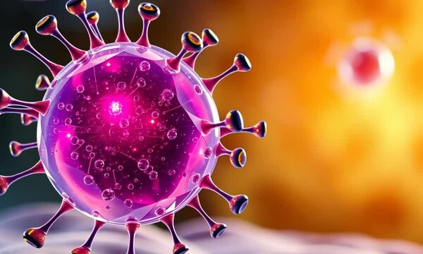
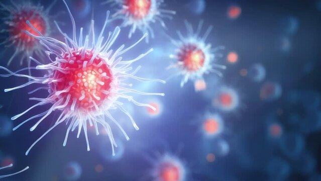
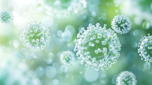
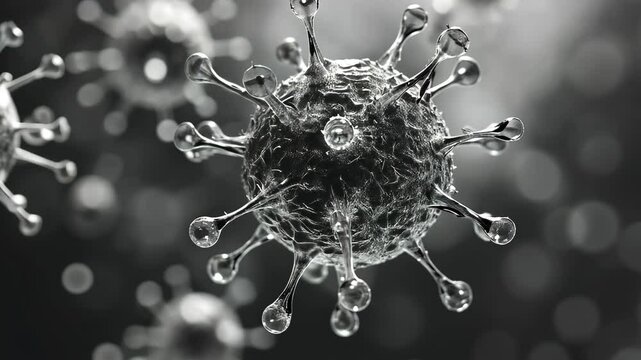
Microscopic view of virus particles with water droplets, representing pathogenicity and transmission.
00:10
HD
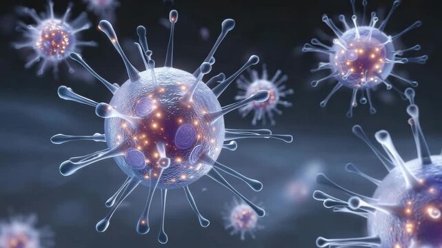
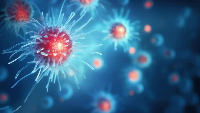
This clip explores the intricate design of virus particles, showcasing their structure and the role they play in disease transmission. Learn about their interaction with host cells
00:10
HD
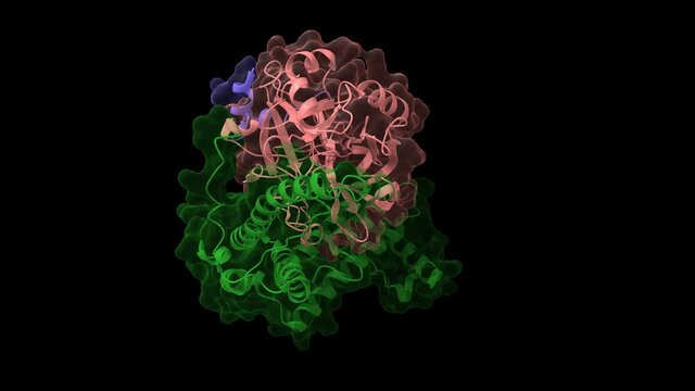
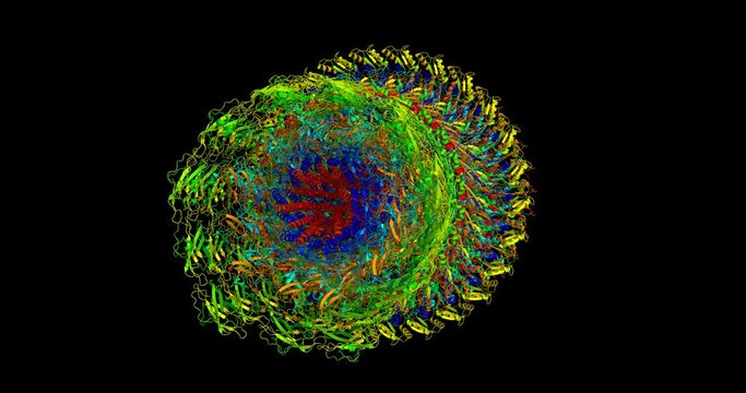
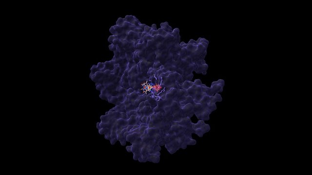
Staphylocoagulase (green) complexed with human thrombin heterodimer. Animated 3D cartoon and Gaussian surface models in two purpendicular projections, PDB 1nu7, black background
00:23
HD
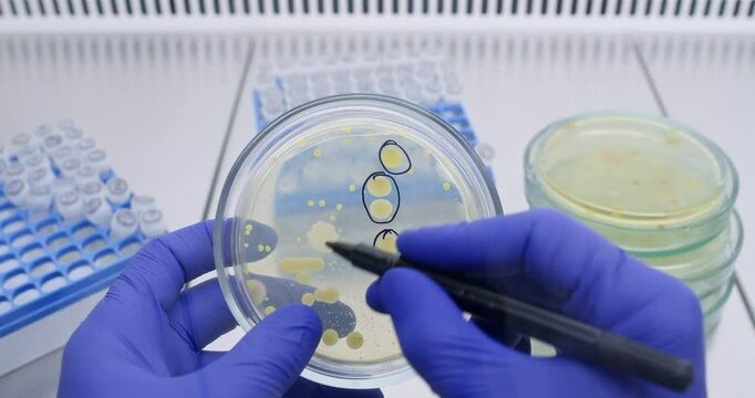
A doctor examines a petri dish with bacterial colonies and signs the dish with a black marker. Bacteria diagnostics by agar plating.
00:16
HD
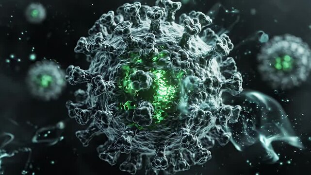
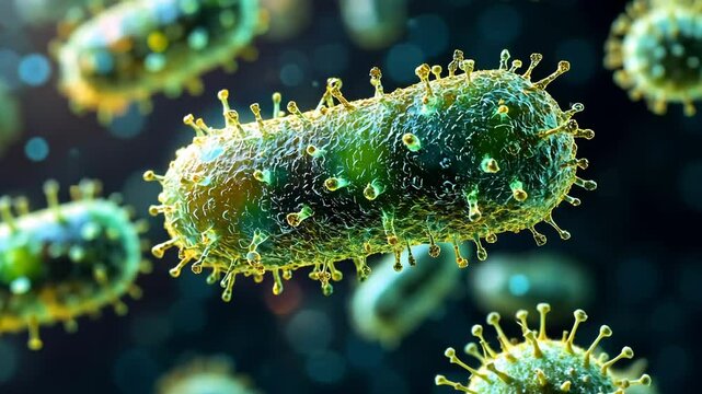
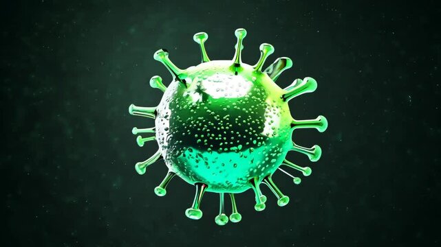
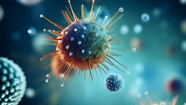
Microscopic Virus Structure in Dark Environment - A detailed 3D video shows a microscopic virus with a glowing green core, floating in a dark, atmospheric environment filled with tiny particles.
00:08
HD
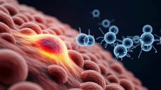
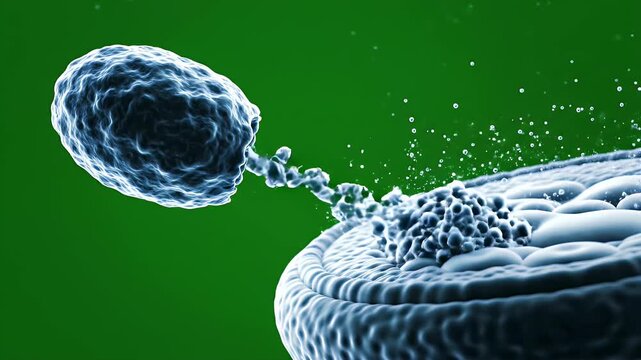
Virulent microorganisms penetrating cellular membrane, triggering inflammatory response and systematically compromising biological integrity through damaging viral invasion
00:06
HD
Flagellar motor-hook complex, natural nanomachine, 3D molecule
00:24
HD
Virus moving close-up. Microbiology and virology concept. High quality 4k footage
00:05
HD
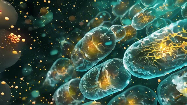
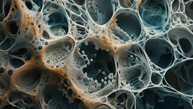
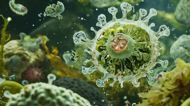
Delicate branching cells embedded in a tough gelatinous material.
00:08
HD
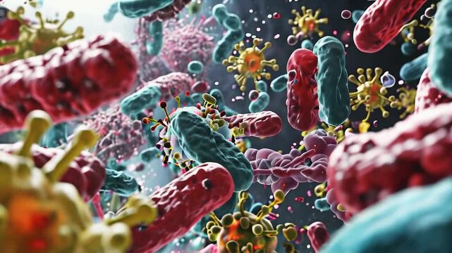
Exploring the intricate details of viruses and bacteria in high definition digital imagery
00:10
HD
animation of bacteria 01
00:19
HD
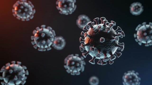
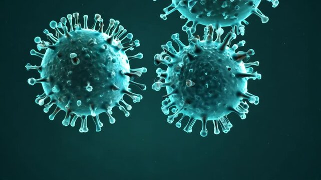
Multiple virus particles floating in a dark environment, 3D rendering of virus-43, microscopic view
00:05
HD
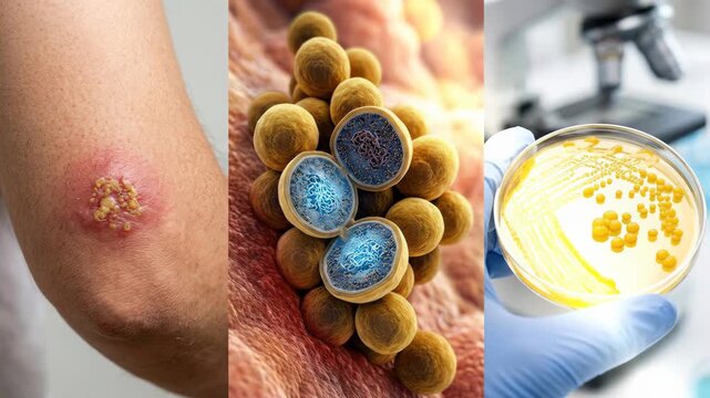
Pathogen investigation reveal: Staphylococcus cell examine, human skin lesion develop, laboratory sample grow, infection cause study, diagnosis method find.
00:08
HD
Corona Virus COVID-19 medical animation with realistic virus model. Microscope close up 3D rendering of SARS-CoV-2, responsible for pandemic
00:30
HD
Exploring various viruses under a microscope with intricate details and vibrant colors
00:05
HD
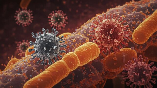
Microscopic view of virus showing pathogens attacking cells
00:05
HD
Museum Quality Anatomical CGI Visualization of Microsporidia Interacting with Host Cell Membrane
00:08
HD
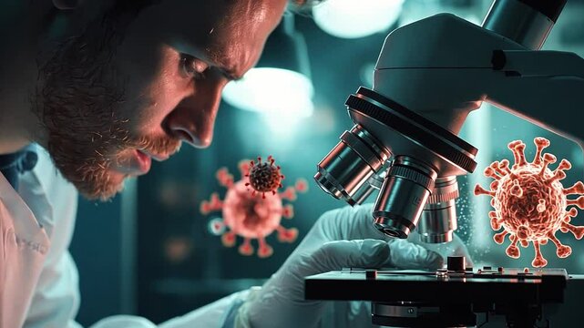
Close-up of a scientist examining a glowing zombie virus under a microscope, highlighting the ominous threat of infection.
00:10
HD
Visualization of virus particles showcasing their structure and characteristics in a scientific context
00:05
HD
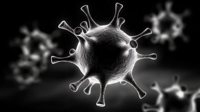
Microscopic View of a Virus: A Close-Up on Viral Structure and Pathogenicity
00:05
HD
Understanding virus structure and function in biological contexts
00:10
HD
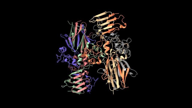
Crystal structure of Haemophilus influenzae adhesive protein HiaBD. Animated 3D cartoon and molecular surface models, chain id color scheme, PDB 1s7m, black background.
00:29
HD
microbiology, cell
00:07
HD
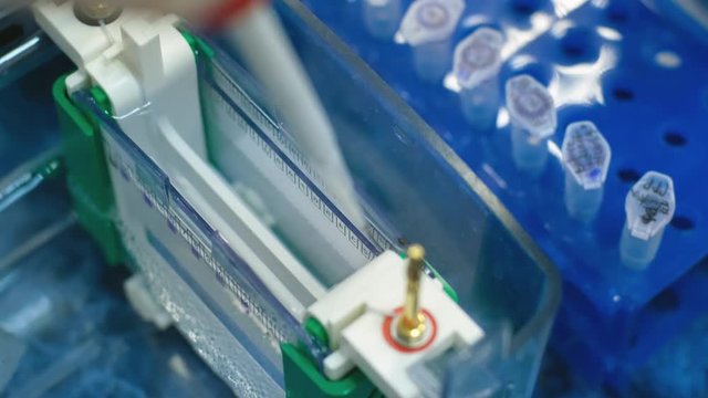
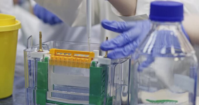
Scientific experiment with human DNA. A scientist uses a thin pipette to place samples with DNA fragments on an agarose gel to separate DNA fragments.
00:12
HD
Atemwegsinfektionen durch Viren
00:24
HD
Understanding the role of viruses in cellular processes and disease transmission
00:08
HD
Virus moving close-up. Microbiology and virology concept. High quality 4k footage
00:07
HD
A microscopic view of the intermembrane space of a mitochondrion showing the accumulation of protons during the electron transport chain for ATP synthesis.
00:07
HD
Detailed Rotation of a Coronavirus Cell Under Microscopic View Generative AI
00:10
HD
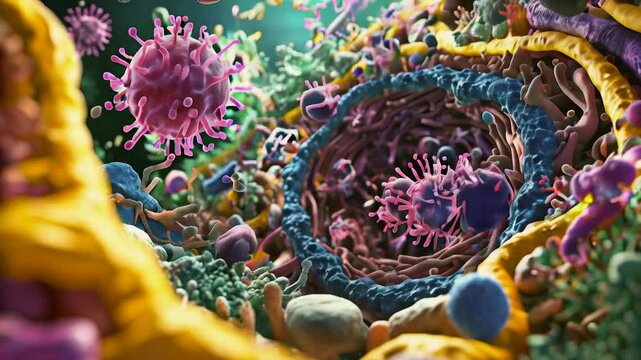
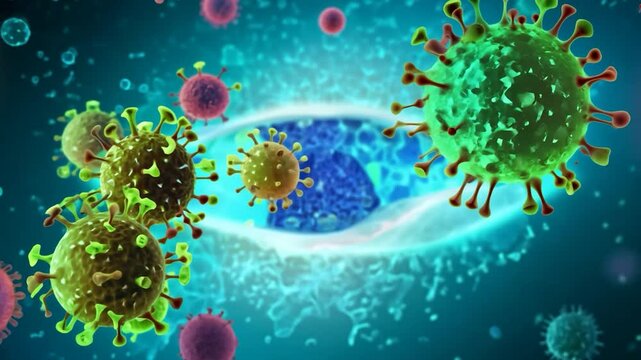
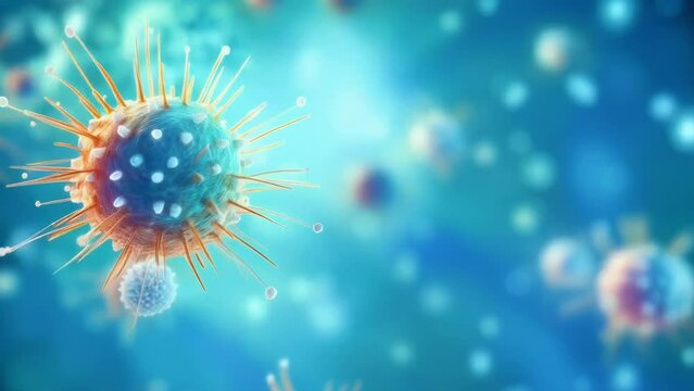
This illustration depicts various viruses in vibrant colors engaging with a human cell, highlighting the complexity of cellular interactions at a microscopic level
00:10
HD
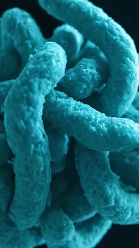
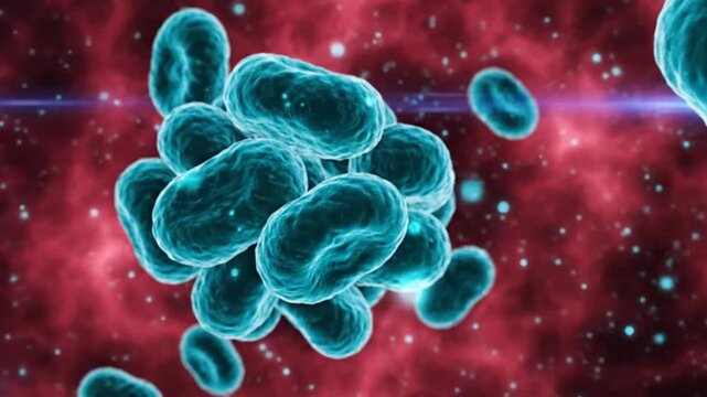
Detailed 3D Rendering of Vibrio Cholerae Bacteria Microscopic View in Cyan Lighting Showing Rod Shape Structure and Virulence Factors for Medical
00:08
HD
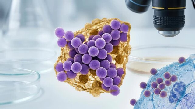
Bacterial cell display: Staphylococcus microbe, grape cluster, infection pathogen, cellular growth, microscope observe, medical science, disease cause, organism replicate.
00:08
HD
Corona Virus COVID-19 medical animation with realistic virus model. Microscope close up 3D rendering of SARS-CoV-2, responsible for pandemic
00:30
HD
Exploring the intricate structure of bacteria and their role in the environment
00:10
HD
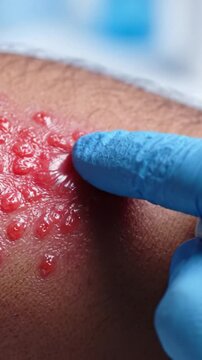
Close-Up Examination of an Inflamed Skin Condition with Fluid-Filled Bumps and Redness on the Surface Highlighting Potential Dermatitis or Infection Symptoms
00:08
HD
D Closeup Medical Images of Various Disease-Causing Viruses. Concept Medical Photography, Closeup Shots, Diseases, Viruses, Microscopic Details
00:04
HD
Dynamic Demonstration of Microscopic Life Forms Cells Viruses
00:05
HD
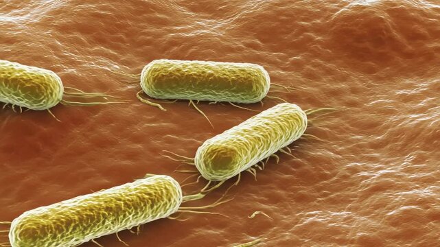
High Resolution 3D Animation of Bacteria Crawling Over Human Skin Surface
00:10
HD
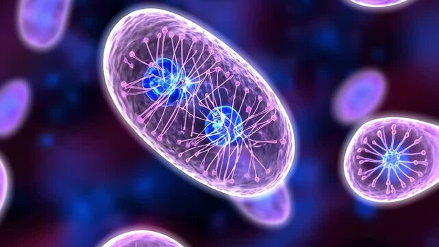
Illustration of bacteria with a focus on cellular processes like division and internal structures.
00:05
HD
Microscopic view of bacteria and microorganisms floating within a vibrant red environment highlighting their various shapes and
00:08
HD
animation of bacterias 03
00:20
HD
microbiology, cell
00:07
HD
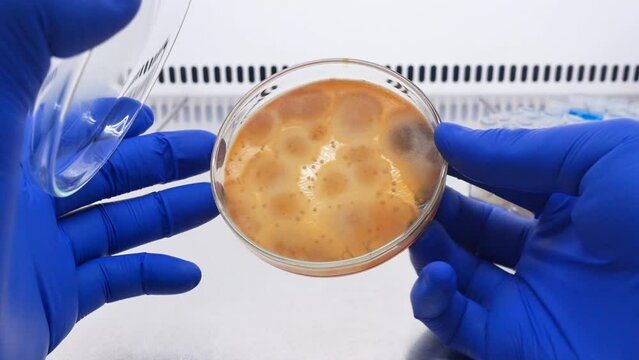
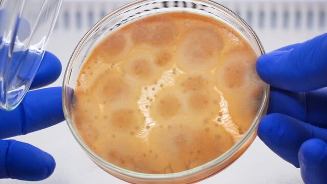
Isolation of pathogenic mold on a Petri dish in microbiology laboratory, the scientist opens the petri dish and turns it.
00:18
HD
HIV 1 gag polyprotein hexamer complexed with antiviral drug, AZT. Animated 3D cartoon and Gaussian surface models in perpendicular projections, PDB 6erm, black background
00:32
HD
Microscopic view of antibodies attacking pathogens in human body
00:05
HD
Visual exploration of virus structure showcasing intricate details of cellular mechanics
00:08
HD
Virus moving close-up. Microbiology and virology concept. High quality 4k footage
00:08
HD
Delicate branching cells embedded in a tough gelatinous material.
00:08
HD
Microscopic View of Coronavirus
00:10
HD
Virus particles are depicted interacting with host cell structures at the microscopic level, showcasing their intricate features and potential impact on health
00:10
HD
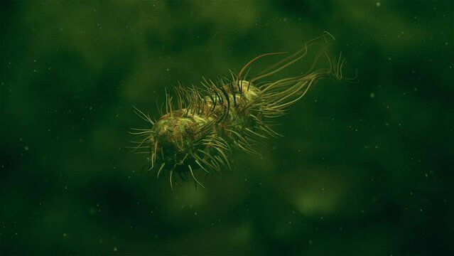
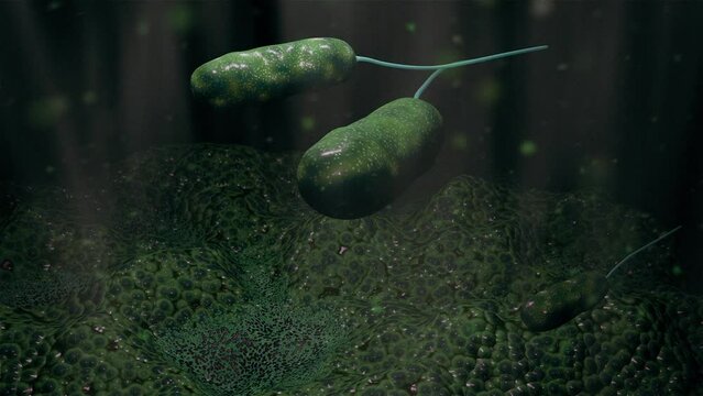
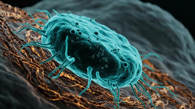
Bacterium moves across tissue surface. Microscopic microbe shows flagellum and cell details. Pathogen clings to fiber and forms biofilm. Infection process visualized in close up. Aid for research.
00:07
HD
Internal Anatomy of a Virus
00:24
HD
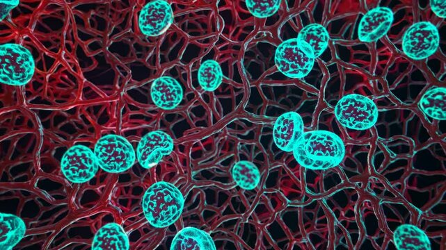
Microscopic view revealing viral and bacterial pathogens traversing intricate blood vessel networks, depicting infectious disease spreading through human circulatory system
00:19
HD
bunch of viruses are floating in the air. The viruses are brown and have a fuzzy appearance, Group of virus cells. 3D illustration, Coronavirus cells
00:10
HD
Understanding Virus Structure and Function in Cellular Environments Over Time
00:05
HD
Dynamic Structure Presentation of Virus from Micro Perspective
00:05
HD
Microscopic Pathogen Cell Structure Scientific Research Application
00:05
HD
Symbolische Darstellung eines Virus in Laborumgebung
00:05
HD
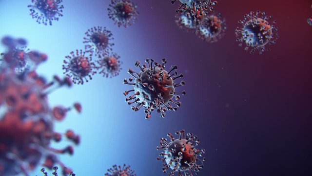
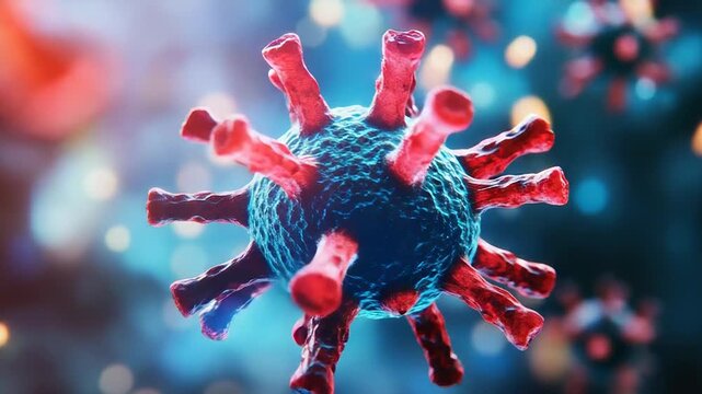
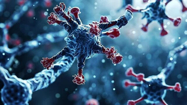
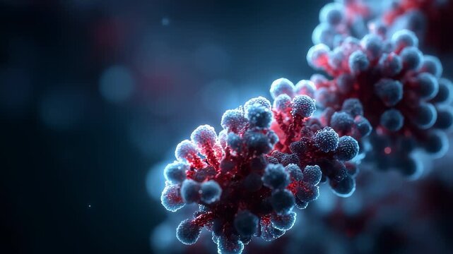
Detailed Close-Up of Virus Structure with Red and Blue Elements
00:10
HD
Exploring the structure and features of a virus under microscopic analysis in a lab setting
00:10
HD
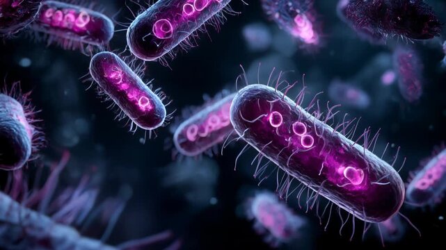
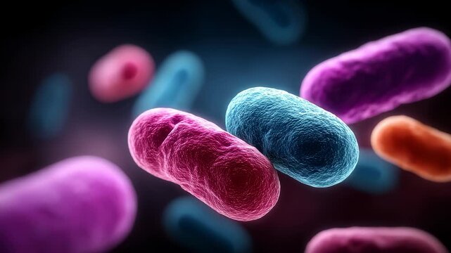
Colorful World of Bacteria Under Microscopic View
00:05
HD
microbiology, cell
00:07
HD
A scientist marks the growth zone of bacterial colonies on a petri dish in a laboratory, close-up.
00:23
HD
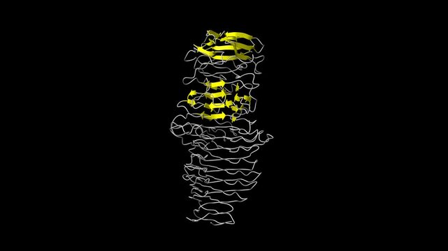
Structure of a novel Hyaluronidase (Hyal_Sk) from Streptomyces koganeiensis. Animated 3D cartoon and molecular surface models, secondary structure color scheme, PDB 4ufq, black background.
00:29
HD
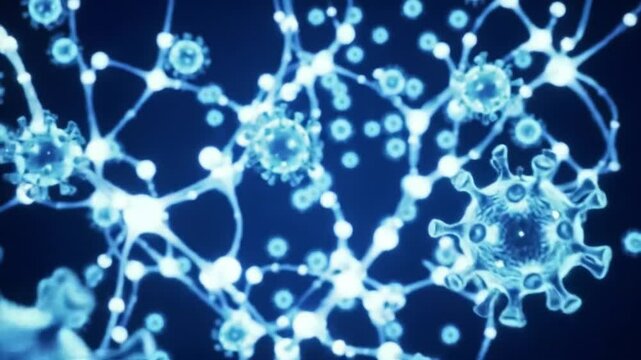
Transparente, schwebende Zellen – runde Mikrostrukturen für Wissenschaft, Biologie, Forschung, Kommunikation & virale Prozesse in Bewegung, blau
00:12
HD
bacteria space
00:07
HD
Virus moving close-up. Microbiology and virology concept. High quality 4k footage
00:08
HD
A microscopic view of the intermembrane space of a mitochondrion showing the accumulation of protons during the electron transport chain for ATP synthesis.
00:08
HD
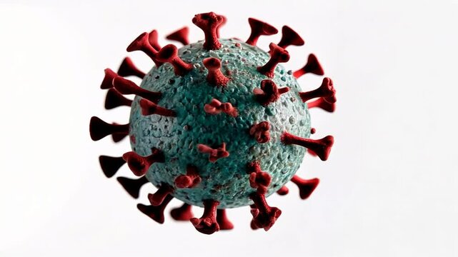
Microscopic View of a Virus Particle Detailed 3D Render
00:10
HD
Bacterial flagellar motor ring, MOTOR PROTEIN, 3D molecule, spinning 4K
00:24
HD
Video animation Virus bacterial infection in zoom view. Microorganism in laboratory diagnosis
00:11
HD
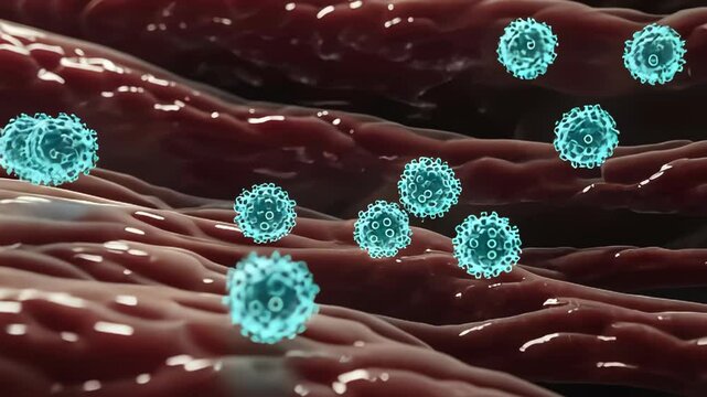
Dynamic interaction of virus particles with organic tissues highlighting cellular processes and viral mechanisms
00:05
HD
bunch of viruses are floating in the air. The viruses are brown and have a fuzzy appearance, Group of virus cells. 3D illustration, Coronavirus cells
00:10
HD
Microscopic view of virus particles showcasing their unique structure and biology in detail
00:09
HD
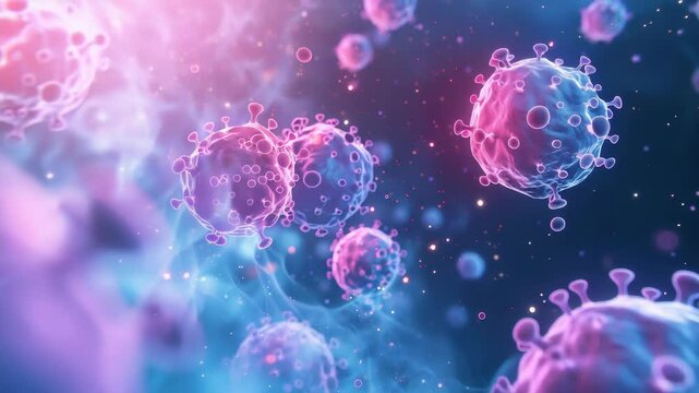
Microscopic World of Virus Cells on Blue Background
00:05
HD
microscopic image of viruses attacking a human cell
00:05
HD
日本のCOVID-19ワクチン接種券
00:08
HD
Immune system cells attacking pathogens, a microscopic battle
00:10
HD
Close-up scientist hands in gloves sorts small test tubes in a laboratory tripod. Scientific experiment with human DNA.
00:08
HD
A scientist uses his hand to move and sort small test tubes with samples in a cryogenic container, close-up.
00:16
HD
A scientist prepares DNA electrophoresis in a special device, selects a test tube with a sample. Preparation of protein electrophoresis.
00:11
HD
Orange mold spores on a solid medium in a petri dish, close-up.
00:18
HD
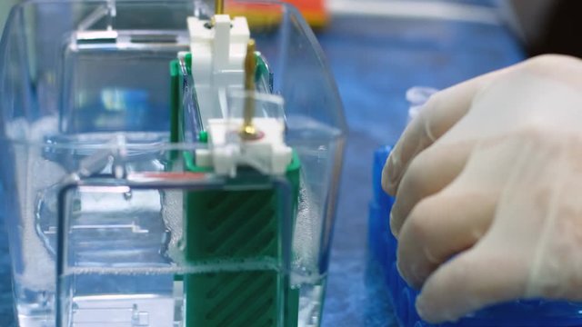
Study of protein and nucleic acids using vertical electrophoresis.
00:10
HD
Virus moving close-up. Microbiology and virology concept. High quality 4k footage
00:05
HD
Virus moving close-up. Microbiology and virology concept. High quality 4k footage
00:08
HD
Virus moving close-up. Microbiology and virology concept. High quality 4k footage
00:05
HD
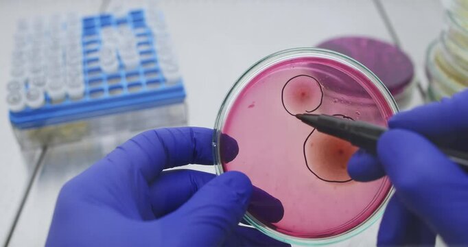
A scientist circles bacterial colonies in a petri dish with a black marker while conducting research in a lab.
00:24
HD
Virus moving close-up. Microbiology and virology concept. High quality 4k footage
00:05
HD
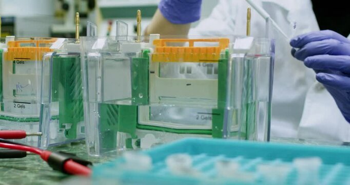
A group of scientists are working on adding protein samples to polyacrylamide gel electrophoresis.
00:13
HD
Virus moving close-up. Microbiology and virology concept. High quality 4k footage
00:05
HD
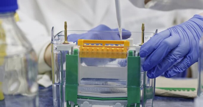
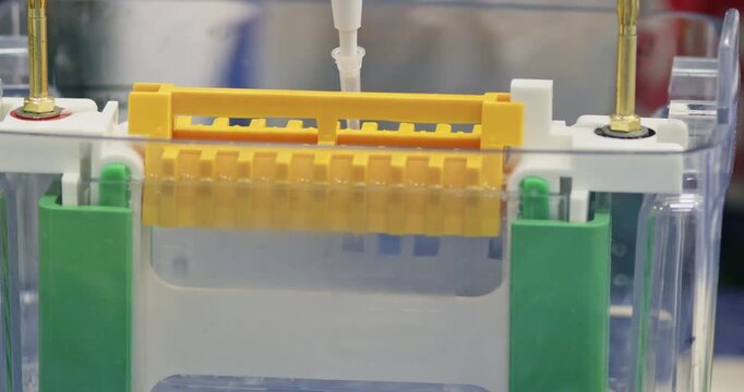
A scientist inserts a DNA sample into a thin polyacrylamide gel using a thin tip of a dispenser to perform nucleic acid electrophoresis. Inserting samples for separation.
00:09
HD
Dynamic Presentation of Viral Cells from a Microscopic Perspective
00:05
HD
Using a thin nozzle, DNA is introduced into the cells of an acrylamide gel, and DNA molecules are separated using electrophoresis.
00:12
HD
Vertical electrophoresis, filling the space to form a gel for electrophoresis, using a dispenser with a tip.
00:16
HD
Medical Visualization of Microscopic Virus Pathogen Structure
00:05
HD
Microscopic World Virus Cell Medical Health Science Background
00:05
HD