Посилання, за допомогою якого можна встановити пароль, надіслано на цю адресу: Щоб отримати доступ до своїх покупок у майбутньому, вам буде потрібен пароль.

Dr_Microbe

Trypanosoma brucei parasites, 3D animation showing slowly rotating flagellated protozoa with undulating membrane, illustrating African trypanosomiasis.
00:10
Trichomonas vaginalis trophozoites, 3D animation showing slowly rotating flagellated protozoa, illustrating sexually transmitted parasitic infection.
00:10
Trichomonas vaginalis trophozoites, 3D animation showing slowly rotating flagellated protozoa, illustrating sexually transmitted parasitic infection.
00:10
Trichomonas vaginalis trophozoites, 3D animation showing slowly rotating flagellated protozoa, illustrating sexually transmitted parasitic infection.
00:10
Clostridium tetani bacterium, 3D animation showing rod-shaped anaerobic cell with terminal spherical endospore giving drumstick appearance.
00:13
Helicobacter pylori bacteria, 3D animation showing curved, spiral-shaped rods with terminal bundles of fine flagella, demonstrating characteristic morphology for gastric colonisation.
00:10
Clostridium tetani bacterium, 3D animation showing rod-shaped anaerobic cell with terminal spherical endospore giving drumstick appearance.
00:13
Streptococcus pyogenes bacterium, 3D animation showing spherical Gram-positive cocci arranged in chains, representing characteristic group A streptococcal morphology.
00:10
Streptococcus pyogenes bacterium, 3D animation showing spherical Gram-positive cocci arranged in chains, representing characteristic group A streptococcal morphology.
00:16
Streptococcus pyogenes bacterium, 3D animation showing spherical Gram-positive cocci arranged in chains, representing characteristic group A streptococcal morphology.
00:15
Streptococcus pyogenes bacterium, 3D animation showing spherical Gram-positive cocci arranged in chains, representing characteristic group A streptococcal morphology.
00:15
Clostridium bacteria, 3D animation showing rod-shaped obligate anaerobic bacilli with endospore formation in varying positions within the cell, demonstrating characteristic clostridial morphology.
00:13
Clostridium bacteria, 3D animation showing rod-shaped obligate anaerobic bacilli with endospore formation in varying positions within the cell, demonstrating characteristic clostridial morphology.
00:15
Clostridium botulinum bacterium, 3D animation showing rod-shaped anaerobic bacteria with subterminal endospores giving a tennis-racket appearance.
00:10
Clostridium botulinum bacterium, 3D animation showing rod-shaped anaerobic bacteria with subterminal endospores giving a tennis-racket appearance.
00:15
Clostridium botulinum bacterium, 3D animation showing rod-shaped anaerobic bacteria with subterminal endospores giving a tennis-racket appearance.
00:16
Trichomonas hominis protozoa, 3D animation showing slowly rotating flagellated intestinal microorganisms, illustrating commensal protozoan of the human digestive tract.
00:16
Streptococcus pyogenes bacterium, 3D animation showing spherical Gram-positive cocci arranged in chains, representing characteristic group A streptococcal morphology.
00:15
Streptococcus pyogenes bacterium, 3D animation showing spherical Gram-positive cocci arranged in chains, representing characteristic group A streptococcal morphology.
00:15
Clostridium botulinum bacterium, 3D animation showing rod-shaped anaerobic bacteria with subterminal endospores giving a tennis-racket appearance.
00:15
Clostridium botulinum bacterium, 3D animation showing rod-shaped anaerobic bacteria with subterminal endospores giving a tennis-racket appearance.
00:16
Clostridium botulinum bacterium, 3D animation showing rod-shaped anaerobic bacteria with subterminal endospores giving a tennis-racket appearance.
00:16
Clostridium botulinum bacterium, 3D animation showing rod-shaped anaerobic bacteria with subterminal endospores giving a tennis-racket appearance.
00:13
Parvoviruses, 3D animation showing small non-enveloped icosahedral DNA viruses, including pathogens such as parvovirus B19 and canine parvovirus.
00:14
Bacteria of different shapes within biofilm, 3D animation showing cocci and rod-shaped cells embedded in extracellular matrix, illustrating microbial community and resistance.
00:14
Staphylococcus bacteria, 3D animation showing spherical Gram-positive cocci arranged in characteristic grape-like clusters, common human pathogens causing skin and systemic infections.
00:16
Staphylococcus bacteria, 3D animation showing spherical Gram-positive cocci arranged in characteristic grape-like clusters, common human pathogens causing skin and systemic infections.
00:12
Staphylococcus bacteria, 3D animation showing spherical Gram-positive cocci arranged in characteristic grape-like clusters, common human pathogens causing skin and systemic infections.
00:08
Staphylococcus bacteria, 3D animation showing spherical Gram-positive cocci arranged in characteristic grape-like clusters, common human pathogens causing skin and systemic infections.
00:08
Parvoviruses, 3D animation showing small non-enveloped icosahedral DNA viruses, including pathogens such as parvovirus B19 and canine parvovirus.
00:14
Parvoviruses, 3D animation showing small non-enveloped icosahedral DNA viruses, including pathogens such as parvovirus B19 and canine parvovirus.
00:16
Parvoviruses, 3D animation showing small non-enveloped icosahedral DNA viruses, including pathogens such as parvovirus B19 and canine parvovirus.
00:10
Neisseria bacteria, 3D animation showing Gram-negative diplococci associated with meningococcal disease and gonorrhoea in humans.
00:13
Neisseria bacteria, 3D animation showing Gram-negative diplococci associated with meningococcal disease and gonorrhoea in humans.
00:10
Neisseria bacteria, 3D animation showing Gram-negative diplococci associated with meningococcal disease and gonorrhoea in humans.
00:10
Trichomonas vaginalis trophozoites, 3D animation showing slowly rotating flagellated protozoa, illustrating sexually transmitted parasitic infection.
00:16
Trichomonas hominis protozoa, 3D animation showing slowly rotating flagellated intestinal microorganisms, illustrating commensal protozoan of the human digestive tract.
00:13
Saccharomyces yeast cells, 3D animation showing slowly rotating budding cells, illustrating probiotic yeast and beneficial microflora used in biotechnology and gut health.
00:09
Pollen grains, 3D animation showing various airborne pollen particles of different shapes and surface textures slowly drifting in air, associated with plant reproduction and seasonal allergies.
00:12
Pollen grains, 3D animation showing various airborne pollen particles of different shapes and surface textures slowly drifting in air, associated with plant reproduction and seasonal allergies.
00:10
Pollen grains, 3D animation showing various airborne pollen particles of different shapes and surface textures slowly drifting in air, associated with plant reproduction and seasonal allergies.
00:10
Pollen grains, 3D animation showing various airborne pollen particles of different shapes and surface textures slowly drifting in air, associated with plant reproduction and seasonal allergies.
00:10
Streptococcus pneumoniae, 3D animation showing Gram-positive encapsulated diplococci, the causative agent of pneumococcal pneumonia, meningitis and septicaemia in humans.
00:12
Streptococcus pneumoniae, 3D animation showing Gram-positive encapsulated diplococci, the causative agent of pneumococcal pneumonia, meningitis and septicaemia in humans.
00:11
Streptococcus pneumoniae, 3D animation showing Gram-positive encapsulated diplococci, the causative agent of pneumococcal pneumonia, meningitis and septicaemia in humans.
00:10
Parvoviruses, 3D animation showing small non-enveloped icosahedral DNA viruses, including pathogens such as parvovirus B19 and canine parvovirus.
00:10
Neisseria bacteria, 3D animation showing Gram-negative diplococci associated with meningococcal disease and gonorrhoea in humans.
00:10
Neisseria bacteria, 3D animation showing Gram-negative diplococci associated with meningococcal disease and gonorrhoea in humans.
00:10
Neisseria bacteria, 3D animation showing Gram-negative diplococci associated with meningococcal disease and gonorrhoea in humans.
00:10
Leishmania promastigotes, 3D animation showing elongated flagellated protozoa in the promastigote stage, transmitted by sandflies and responsible for leishmaniasis in humans.
00:10
Leishmania promastigotes, 3D animation showing elongated flagellated protozoa in the promastigote stage, transmitted by sandflies and responsible for leishmaniasis in humans.
00:10
Leishmania promastigotes, 3D animation showing elongated flagellated protozoa in the promastigote stage, transmitted by sandflies and responsible for leishmaniasis in humans.
00:10
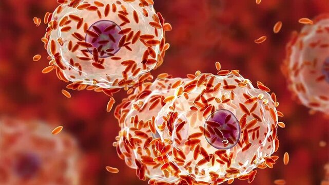
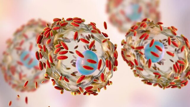
Leishmania amastigotes in macrophage, 3D animation showing intracellular round amastigote forms within human macrophages, characteristic of visceral and cutaneous leishmaniasis.
00:10
Leishmania amastigotes in macrophage, 3D animation showing intracellular round amastigote forms within human macrophages, characteristic of visceral and cutaneous leishmaniasis.
00:10
Leishmania amastigotes in macrophage, 3D animation showing intracellular round amastigote forms within human macrophages, characteristic of visceral and cutaneous leishmaniasis.
00:10
Brucella species, 3D animation showing small Gram-negative intracellular coccobacilli, causative agents of brucellosis in humans and animals.
00:10
Brucella species, 3D animation showing small Gram-negative intracellular coccobacilli, causative agents of brucellosis in humans and animals.
00:16
Brucella species, 3D animation showing small Gram-negative intracellular coccobacilli, causative agents of brucellosis in humans and animals.
00:13
Brucella species, 3D animation showing small Gram-negative intracellular coccobacilli, causative agents of brucellosis in humans and animals.
00:09
Streptococcus pneumoniae, 3D animation showing Gram-positive encapsulated diplococci, the causative agent of pneumococcal pneumonia, meningitis and septicaemia in humans.
00:16
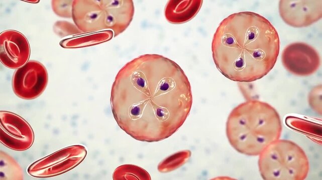
Plasmodium malariae ring stage, 3D animation showing early intraerythrocytic trophozoite within a red blood cell, associated with quartan malaria in humans.
00:10
Plasmodium malariae ring stage, 3D animation showing early intraerythrocytic trophozoite within a red blood cell, associated with quartan malaria in humans.
00:12
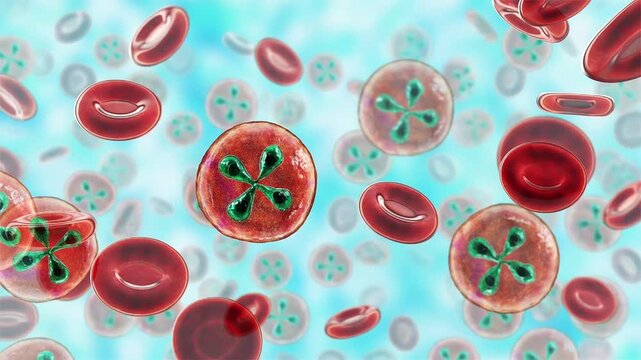
Plasmodium falciparum schizont stage, 3D animation showing mature intraerythrocytic schizont containing multiple merozoites within an infected red blood cell in falciparum malaria.
00:16
Plasmodium falciparum schizont stage, 3D animation showing mature intraerythrocytic schizont containing multiple merozoites within an infected red blood cell in falciparum malaria.
00:16
Plasmodium falciparum schizont stage, 3D animation showing mature intraerythrocytic schizont containing multiple merozoites within an infected red blood cell in falciparum malaria.
00:10
Plasmodium falciparum schizont stage, 3D animation showing mature intraerythrocytic schizont containing multiple merozoites within an infected red blood cell in falciparum malaria.
00:10
Plasmodium falciparum ring stage, 3D animation showing early intraerythrocytic trophozoites within red blood cells, characteristic of falciparum malaria in humans.
00:14
Plasmodium falciparum ring stage, 3D animation showing early intraerythrocytic trophozoites within red blood cells, characteristic of falciparum malaria in humans.
00:10
Plasmodium falciparum ring stage, 3D animation showing early intraerythrocytic trophozoites within red blood cells, characteristic of falciparum malaria in humans.
00:10
Junin virus particle, 3D animation showing enveloped arenavirus with granular ribonucleoprotein structure, causative agent of Argentine hemorrhagic fever.
00:10
Junin virus particle, 3D animation showing enveloped arenavirus with granular ribonucleoprotein structure, causative agent of Argentine hemorrhagic fever.
00:10
Junin virus particle, 3D animation showing enveloped arenavirus with granular ribonucleoprotein structure, causative agent of Argentine hemorrhagic fever.
00:10
Gallbladder with stones, 3D animation showing cholelithiasis. Gallstones can block bile flow and cause pain, inflammation, or infection requiring medical or surgical treatment.
00:12
Foot-and-mouth disease virus (FMDV), 3D animation showing viral particles, a highly contagious RNA virus causing severe disease in cloven-hoofed animals like cattle, pigs, sheep, and goats.
00:10
Foot-and-mouth disease virus (FMDV), 3D animation showing viral particles, a highly contagious RNA virus causing severe disease in cloven-hoofed animals like cattle, pigs, sheep, and goats.
00:10
Foot-and-mouth disease virus (FMDV), 3D animation showing viral particles, a highly contagious RNA virus causing severe disease in cloven-hoofed animals like cattle, pigs, sheep, and goats.
00:10
Foot-and-mouth disease virus (FMDV), 3D animation showing viral particles, a highly contagious RNA virus causing severe disease in cloven-hoofed animals like cattle, pigs, sheep, and goats.
00:10
Dengue virus bound with neutralizing human antibodies, 3D animation showing flavivirus particles coated by immunoglobulins, illustrating antiviral immune response.
00:10
Dengue virus bound with neutralizing human antibodies, 3D animation showing flavivirus particles coated by immunoglobulins, illustrating antiviral immune response.
00:10
Dengue virus bound with neutralizing human antibodies, 3D animation showing flavivirus particles coated by immunoglobulins, illustrating antiviral immune response.
00:10
Banna virus particle, 3D animation showing icosahedral double-stranded RNA arbovirus with segmented genome, associated with mosquito-borne febrile illness.
00:10
Banna virus particle, 3D animation showing icosahedral double-stranded RNA arbovirus with segmented genome, associated with mosquito-borne febrile illness.
00:10
Banna virus particle, 3D animation showing icosahedral double-stranded RNA arbovirus with segmented genome, associated with mosquito-borne febrile illness.
00:10
Rabbit haemorrhagic fever virus particles, 3D animation showing slowly rotating non-enveloped calicivirus virions, illustrating highly contagious viral disease in rabbits.
00:10
Plasmodium schizont inside red blood cell, 3D animation showing mature malaria parasite with multiple merozoites, illustrating erythrocytic stage of infection.
00:10
Plasmodium schizont inside red blood cell, 3D animation showing mature malaria parasite with multiple merozoites, illustrating erythrocytic stage of infection.
00:10
Lipid nanoparticle mRNA vaccine used against Covid-19 and influenza. 3D animation showing cross-section of lipid nanoparticle carrying mRNA of the virus (orange)
00:10
Giardia lamblia trophozoites, 3D animation showing slowly rotating flagellated protozoa, illustrating intestinal parasitic infection and giardiasis.
00:10
Giardia lamblia trophozoites, 3D animation showing slowly rotating flagellated protozoa, illustrating intestinal parasitic infection and giardiasis.
00:10
Bacillus bacteria, 3D animation showing slowly rotating rod-shaped spore-forming cells, illustrating gram-positive bacteria widely distributed in the environment.
00:10
Bacillus bacteria, 3D animation showing slowly rotating rod-shaped spore-forming cells, illustrating gram-positive bacteria widely distributed in the environment.
00:10
Aspergillus fungal structures, 3D animation showing slowly rotating hyphae and conidia, illustrating opportunistic mould associated with respiratory infections.
00:10
Aspergillus fungal structures, 3D animation showing slowly rotating hyphae and conidia, illustrating opportunistic mould associated with respiratory infections.
00:10
Aspergillus fungal structures, 3D animation showing slowly rotating hyphae and conidia, illustrating opportunistic mould associated with respiratory infections.
00:10
Aspergillus fungal structures, 3D animation showing slowly rotating hyphae and conidia, illustrating opportunistic mould associated with respiratory infections.
00:10