ขาย
การกำหนดราคา
ระบบได้ส่งลิงก์สำหรับตั้งรหัสผ่านของคุณไปที่:
ต่อไปนี้คุณจะต้องใช้รหัสผ่านในการเข้าถึงสินค้าที่คุณได้ซื้อ
ทั้งหมด
ภาพ
วิดีโอ
เสียง
เทมเพลต
3 มิติ
ฟรี
พรีเมียม
ค้นหาที่คล้ายกัน:
ซีรีส์:
โมเดล:
เอกสาร:
ไลบรารีของฉัน
ค้นหาที่คล้ายกัน
ลากรูปภาพมาที่นี่
หรือ
เรียกดู
กำลังอัพโหลดภาพของคุณ...
สร้างสรรค์แบบไร้ขีดจำกัด — ดาวน์โหลดได้ไม่จำกัด พร้อมด้วยเครื่องมือปรับแต่งด้วย AI ที่มีประสิทธิภาพ
เริ่มทดลองใช้ฟรี
รับภาพประกอบ 25 ภาพหรือวิดีโอ 3 รายการเมื่อทดลองใช้ฟรี
เริ่มทดลองใช้ฟรี
ซ่อนแผง
ดูแผง
เพิ่มตัวกรองใหม่แล้ว
เรียงลำดับตาม
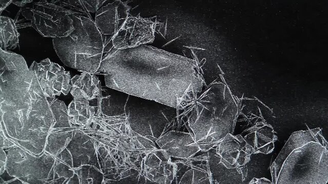
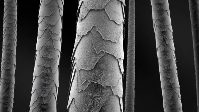
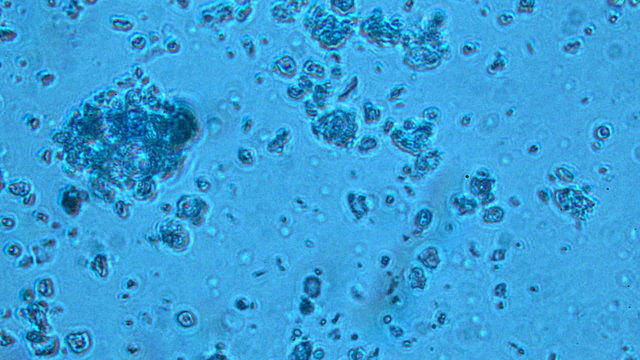
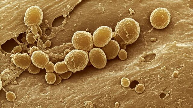
358 ผลลัพธ์สำหรับ
sem microscope
ใน วิดีโอ
00:13
4K
HD
00:20
4K
HD
00:13
4K
HD
00:05
4K
HD
00:10
4K
HD
00:10
4K
HD
00:06
4K
HD
00:20
4K
HD
00:05
4K
HD
00:05
4K
HD
00:05
4K
HD
00:08
4K
HD
00:08
4K
HD
00:11
4K
HD
00:08
4K
HD
00:05
4K
HD
00:05
4K
HD
00:06
4K
HD
00:07
4K
HD
00:10
4K
HD
00:26
4K
HD
00:10
4K
HD
00:18
4K
HD
00:10
4K
HD
00:10
4K
HD
00:08
4K
HD
00:10
4K
HD
00:08
4K
HD
00:07
4K
HD
00:10
4K
HD
00:06
4K
HD
00:10
4K
HD
00:16
4K
HD
00:05
4K
HD
00:10
4K
HD
00:08
4K
HD
00:05
4K
HD
00:14
4K
HD
00:07
4K
HD
00:05
4K
HD
00:36
4K
HD
00:10
4K
HD
00:12
4K
HD
00:07
4K
HD
00:10
4K
HD
00:09
4K
HD
00:31
4K
HD
00:10
4K
HD
00:08
4K
HD
00:08
4K
HD
00:10
4K
HD
00:07
4K
HD
00:05
4K
HD
00:08
4K
HD
00:08
4K
HD
00:08
4K
HD
00:08
4K
HD
00:08
4K
HD
00:07
4K
HD
00:10
4K
HD
00:24
4K
HD
00:10
4K
HD
00:14
4K
HD
00:05
4K
HD
00:10
4K
HD
00:10
4K
HD
00:20
4K
HD
00:10
4K
HD
00:12
4K
HD
00:10
4K
HD
00:08
4K
HD
00:05
4K
HD
00:10
4K
HD
00:05
4K
HD
00:05
4K
HD
00:20
4K
HD
00:11
4K
HD
00:10
4K
HD
00:07
4K
HD
00:10
4K
HD
00:22
4K
HD
00:04
4K
HD
00:10
4K
HD
00:05
4K
HD
00:05
4K
HD
00:20
4K
HD
00:20
4K
HD
00:10
4K
HD
00:12
4K
HD
00:10
4K
HD
00:05
4K
HD
00:10
4K
HD
00:10
4K
HD
00:20
4K
HD
00:14
4K
HD
00:15
4K
HD
00:07
4K
HD
00:20
4K
HD
00:07
4K
HD
00:10
4K
HD
ตกลง
ตกลง
ตกลง
เลือกภูมิภาคของคุณ
การเลือกภูมิภาคอาจเปลี่ยนภาษาและเนื้อหาส่งเสริมการขายที่คุณเห็นบนเว็บไซต์ Adobe Stock
อเมริกาเหนือ
Canada - English
Canada - Français
El Salvador
México
República Dominicana
United States
อเมริกาใต้
Argentina
Bahamas
Barbados
Bolivia
Brasil
Chile
Colombia
Costa Rica
Ecuador
Guatemala
Honduras
Jamaica
Nicaragua - English
Panamá
Paraguay
Perú
Trinidad and Tobago
Uruguay
Venezuela
ยุโรป ตะวันออกกลาง และแอฟริกา
Algeria - English
Armenia - English
Azerbaijan - English
Bahrain - English
Belgium - English
Belgique - Français
België - Nederlands
Česká republika
Croatia - English
Cyprus - English
Danmark
Georgia - English
Deutschland
Eesti
Egypt - English
España
France
Greece - English
Iceland - English
Ireland
Israel - English
Italia
Jordan - English
Казахстан
Kenya - English
Kuwait - English
Киргизия
Latvija
Lebanon - English
Lietuva
Luxembourg - Deutsch
Luxembourg - English
Luxembourg - Français
Mauritius - English
Moldova - English
Hungary - English
Malta - English
Morocco - English
Nederland
Nigeria
Norge
Oman - English
Österreich
Россия
Polska
Portugal
Qatar - English
România
Saudi Arabia - English
Schweiz
Slovenija
Slovensko
Suisse
Suomi
Sverige
Svizzera
Таджикистан
Turkey - English
Turkmenistan - English
UAE - English
Україна
United Kingdom
Узбекистан
България
เอเชียแปซิฟิก
Australia
Indonesia - English
中國香港特別行政區
India
日本
한국
中華人民共和國澳門特別行政區
Malaysia - English
New Zealand
ไทย
Philippines
Singapore
Sri Lanka - English
台灣地區
Vietnam - English