A link to set your password has been sent to: To access your purchases in the future you will need a password.
Create without limits — unlimited downloads, plus powerful AI editing
New filters added
Sort by

97 results for tibial in videos

Did you really mean: tibal
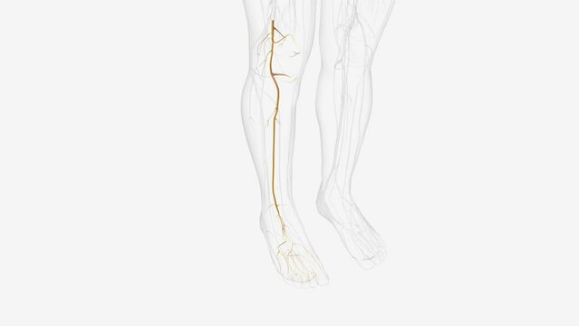
tibial nerve and terminal branches .
00:11
HD
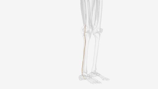
The tibial nerve branches off from the sciatic nerve .
00:19
HD
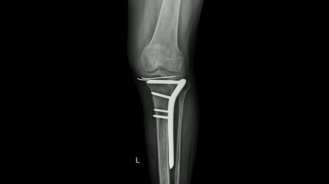
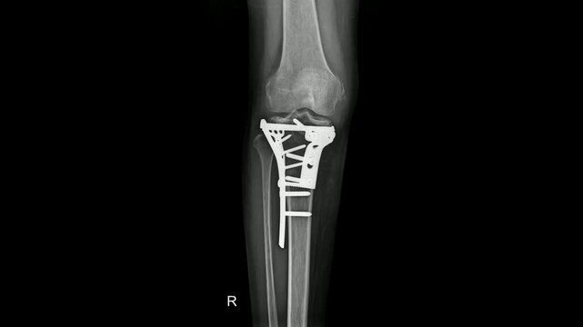
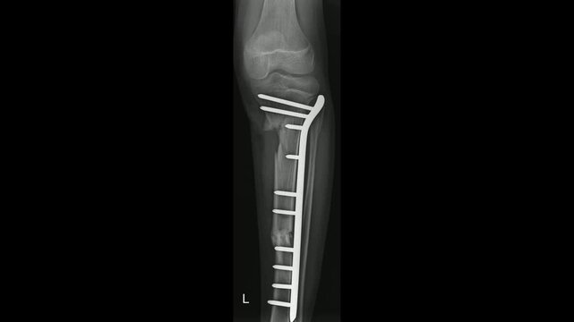
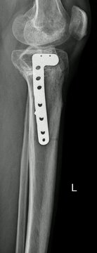
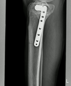
Post-operative X-ray showing internal fixation of a proximal tibial fracture with plate and screws
00:15
HD
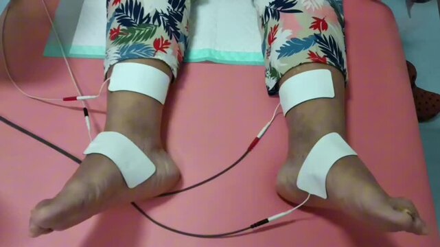
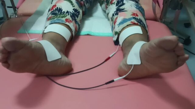
Close up of elderly woman's feet receiving Percutaneous Tibial Nerve Stimulation (PTNS) physiotherapy treatment for overactive bladder and bowel function in the hospital.
00:07
HD
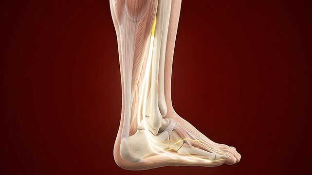
3D medical animation of tibial nerve pathway through ankle and foot showing tarsal tunnel anatomy on dark red background
00:05
HD
Nervous system , Doctor Writing On Transparent Screen
00:09
HD
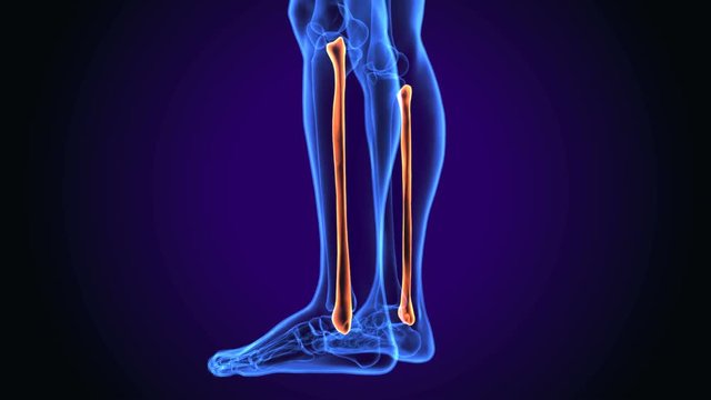
Fibula Bone Anatomy | Bone and Spine anatomy 3d illustration
00:15
HD
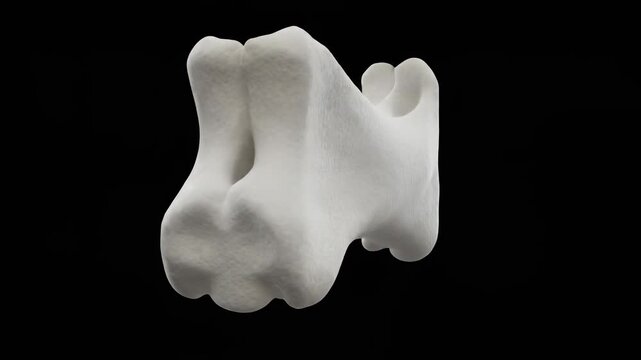
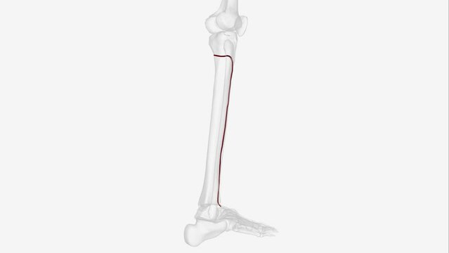
3D model animation of a tibial tuberosity bone, shin bone landmark
00:10
HD
It is like Siberian pine (Pinus sibirica)) from tibial base plate, Shangri La. cones with large healthy nuts
00:11
HD
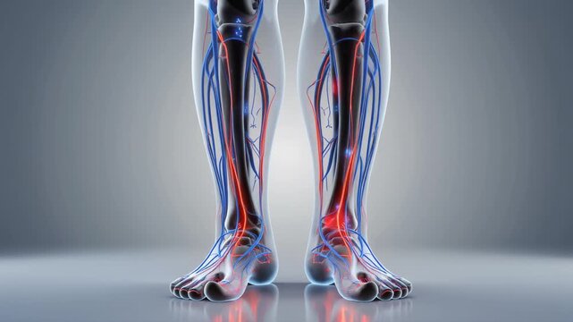
Human Leg Anatomy with Visible Veins and Arteries Medical 3D Animation.
00:05
HD
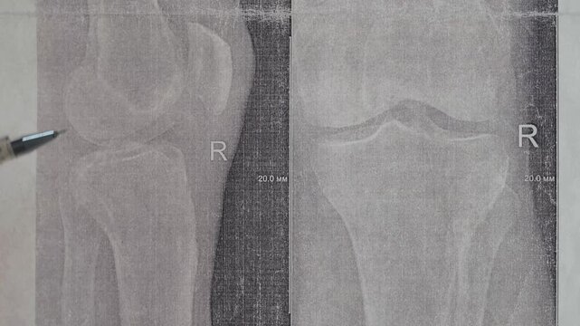
Doctor learn X-ray with narrowed joint space, compacted tibial endplate, exacerbation of intercondylar elevation of the tibia. Osteoarthritis of the knee joint.
00:23
HD
German shepherds first hydrotherapy session after Tibial Plateau Levelling Osteotomy surgery
00:14
HD
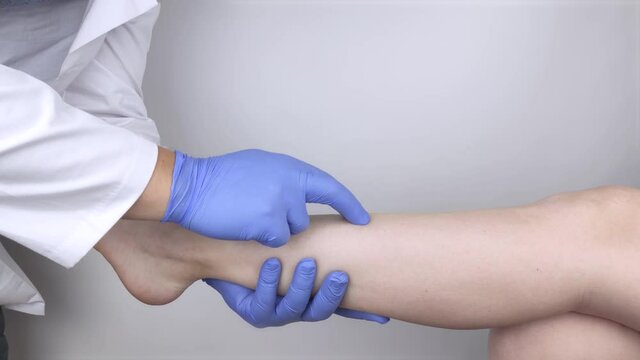
The podiatrist examines the tibia of a patient who complains of pain in it. The concept of diagnostics of bone microcracks and latent course of diseases
00:20
HD
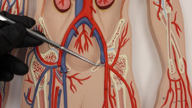
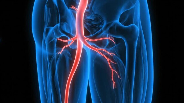
Following Femoral Artery Down Left Leg To Posterior Tibial
00:19
HD
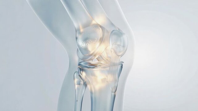
Transparent human knee joint bending and extending, showing bones, cartilage, ligaments and patella movement
00:17
HD
Blood vessels, Vessel health, Vein structure. Vascular health. Capillaries constrict under low pressure. Vein elasticity enhances oxygen delivery. Cardiovascular efficiency supports organ function.
00:10
HD
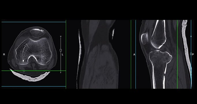
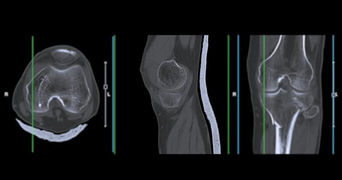
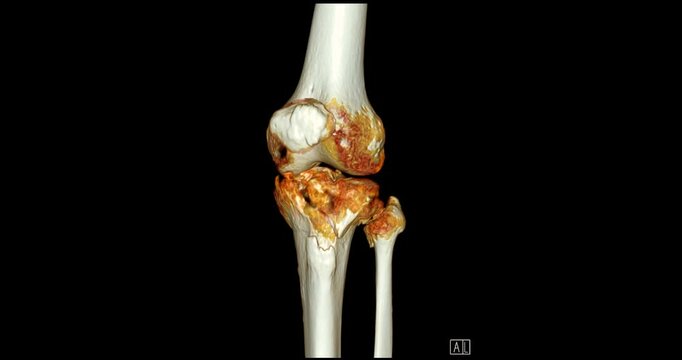
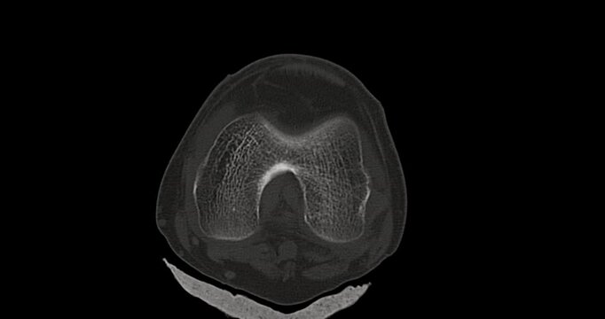
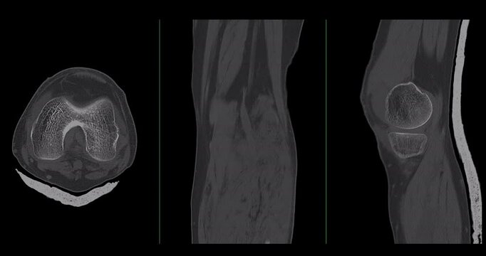
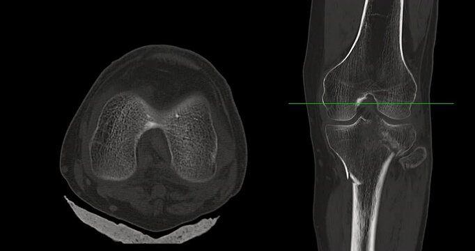
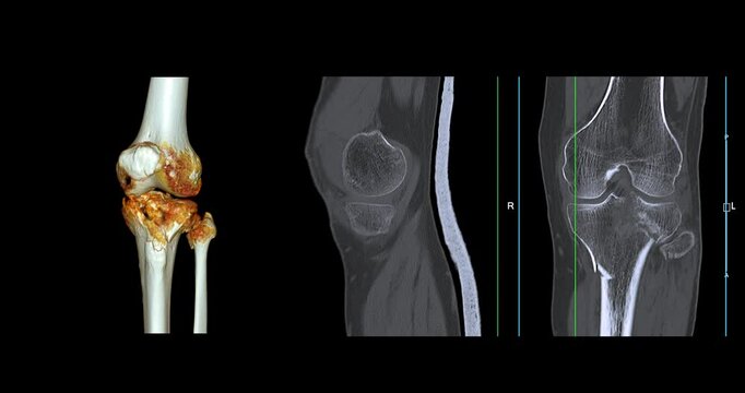
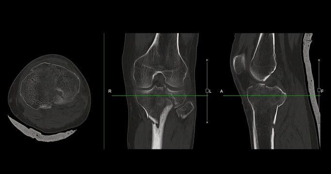
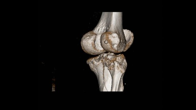
Knee Joint CT-Scan Revealing Tibial Fracture:The scan highlights a distinct fracture in the tibia, located beneath the knee joint. The break is visible as a crack.
00:13
HD
Vertical video: Lifting model, Dr in coat turning knee joint near camera showing tibial detail
00:05
HD
Knee Joint Anatomy Visualization Bone Structure Illustration
00:07
HD
Knee Joint Anatomy Visualization for Medical Explainer Videos
00:07
HD
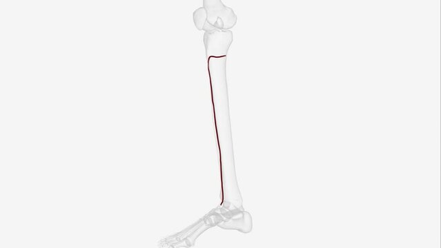
The anterior tibial artery is one of the terminal branches of the popliteal artery. It arises below the popliteal fossa, in the posterior .
00:10
HD
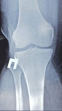
X-ray of Right Tibial Plateau Fracture with Surgical Fixation Using Plates and Screws
00:15
HD
Close up of elderly woman's feet receiving Percutaneous Tibial Nerve Stimulation (PTNS) physiotherapy treatment for overactive bladder and bowel function in the hospital.
00:07
HD
Examining Ankle Anatomy Model Showing Bones and Ligaments for Medical Education
00:08
HD
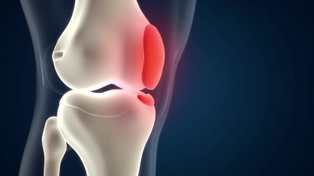
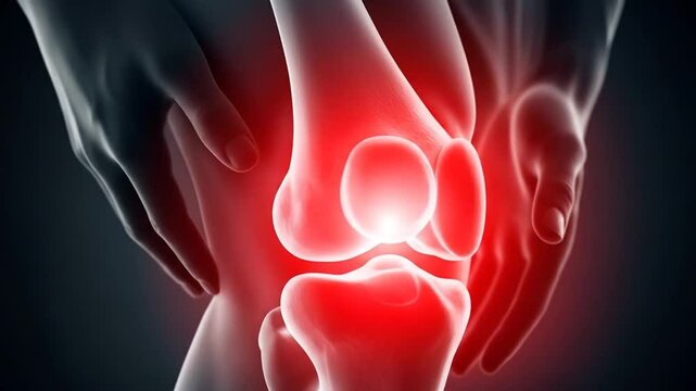
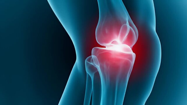
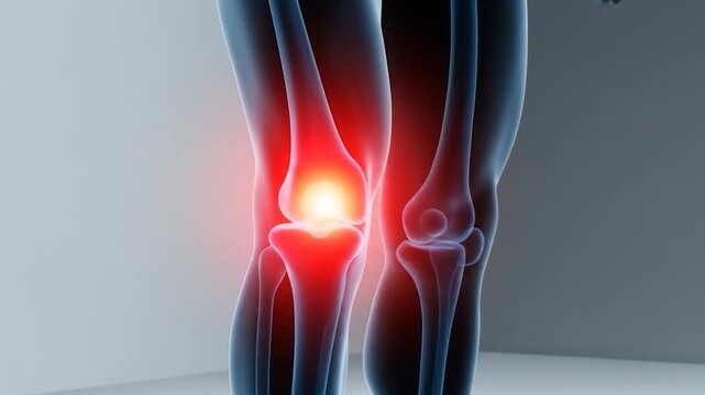
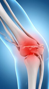
Knee Pain Animation with Red Hot Spot Medical Visualization Concept
00:08
HD
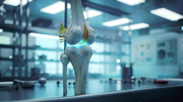
Examining Knee Joint Anatomy Model in Medical Lab
00:08
HD
Medical Animation of Tibial Tuberosity and Knee Joint Anatomy
00:07
HD
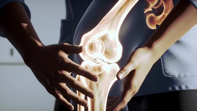
Detailed Anatomy of a Human Knee Bone Structure in White Coat with Warm Lighting
00:05
HD
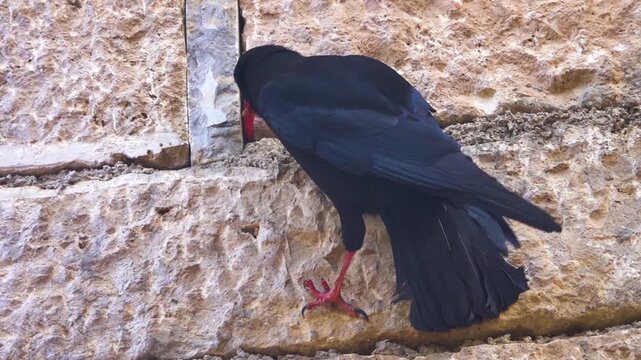
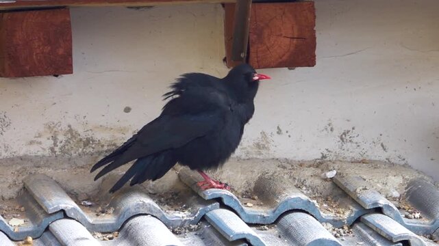
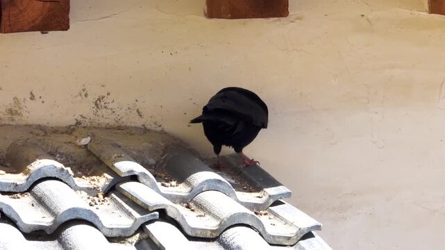
The amazing highest mountain bird chough (Pyrrhocorax pyrrhocorax brachypus) in the villages of an unknown country Shangri la. It's as ordinary as a commonplace crow there. tibial base plate
00:15
HD
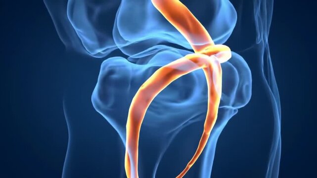
Animated Knee Joint with Orange Artery Visualization on Blue Background
00:08
HD
Doctor learn X-ray of Knee joint dislocation after accident. Patellar joint instability. Tibial dislocation in relation to the femur. 4k footage.
00:26
HD
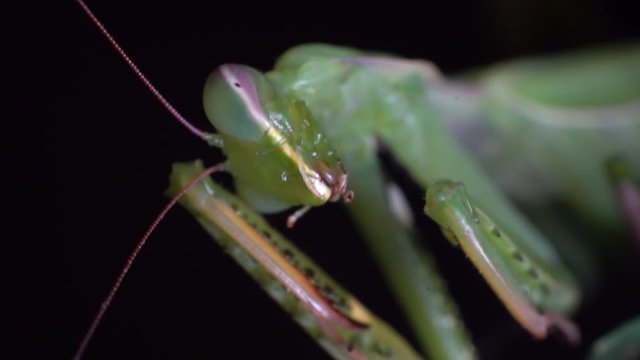
Extreme macro praying mantis cleaning her raptorial leg tibial spine
00:06
HD
Rotating Knee Joint Animation on Dark Blue Background Anatomy Study for Medical Education Knee Ligaments Bones
00:08
HD
Anatomical View Of A Knee Joint With Visible Ligaments And Bone Structure In Warm Orange Hues
00:08
HD
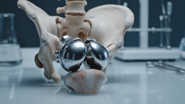
Anatomical Model Displaying Total Knee Replacement with Bone Structure and Surgical Tools in the Background on a Grey Table with Medical Implants and Orthopedic Research Showing Hip Bone
00:08
HD
Knee Joint CT-Scan Revealing Tibial Fracture:The scan highlights a distinct fracture in the tibia, located beneath the knee joint. The break is visible as a crack.
00:18
HD
CT Scan knee of the patient periarticular injuries fracture of the proximal tibia frequently associated with soft tissue injuries.Medical footage concept.
00:09
HD
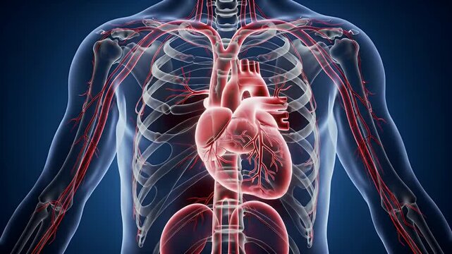
Detailed 3D Medical Animation of Human Torso Cardiovascular System Arteries Veins and Skeleton on Deep Blue Background Perfect for Medical Education and Healthcare Promotions
00:08
HD
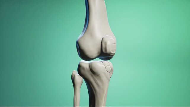
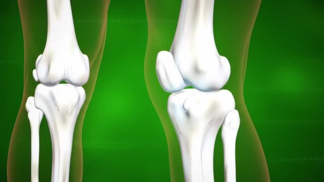
Knee Joint Anatomy Visual on Green Background
00:08
HD
The anterior tibial artery is one of the terminal branches of the popliteal artery. It arises below the popliteal fossa, in the posterior .
00:15
HD
Postoperative tibial fracture fixation with plate and screws – medical X ray video
00:15
HD
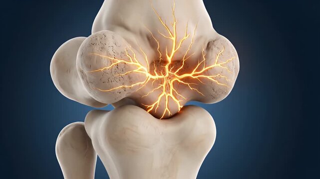
Knee Bone Anatomy Inflammation Animation On Blue Background Skeletal System Joint Pain Injury Medical Visualization
00:05
HD
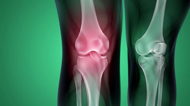
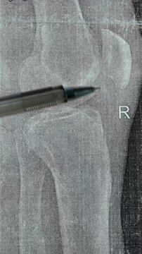
Knee Joint X Ray View with Red Highlight on Pain Area on Dark Background
00:05
HD
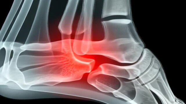
Illustration of Foot Bones in Medical Visualization with Red Spot on Dark Background
00:08
HD
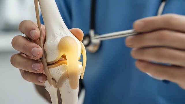
Orthopedic Surgeon Examining Knee Joint Model in Blue Scrubs with Stethoscope, Demonstrating Joint Function and Medical Assessment in Clinic Setting
00:08
HD
Femur, bone structure, fracture healing. Leg anatomy. Bones glow under X-ray examination. Calcium deposition strengthens skeletal stability. Tibial stress impacts walking endurance.
00:10
HD
Viewing Knee Joint Anatomy in X-Ray Style Animation
00:08
HD
Chinese pine (Pincus yunnanensis) is long-needle pines from tibial base plate, Shangri La
00:11
HD
3D Rendered Fracture of Femur Bone Medical Visualization Isolated on Dark Gradient Background
00:08
HD
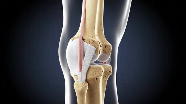
Human knee joint anatomy illustration medical diagram showing bone ligaments and tendons
00:10
HD
Girl in black sport suit does jumping jacks in a white Studio
00:50
HD
Human Knee Joint Illustration with Red Highlighted Pain Area on Blue Background Anatomical Visualization of Knee Bones and Structure for Medical Purposes
00:08
HD
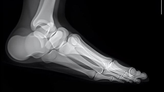
Anatomical Foot X-Ray Scan Showing Bone Structure in Black Background Diagnostic Radiology and Medical Visualization
00:05
HD
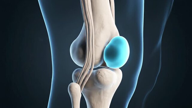
Detailed 3D Animation of Human Knee Joint Anatomy, Showing Tibial Tuberosity and Patella
00:07
HD
X-Ray of Human Knee Showing Bone Structure and Skeletal Anatomy Against a Dark Background in Blue Tones for Medical Visualization and Analysis
00:07
HD
Detailed Anatomical Rendering of Human Legs Featuring Muscles Bones and Ligaments on Dark Background
00:05
HD
Viewing Bone Injury X-Ray of Foot with Area of Pain Highlighted
00:10
HD
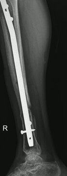
Tibial IM Nail X-Ray Imaging
00:15
HD
Shin Splints with medical digital technology concept
00:18
HD
Knee Joint CT-Scan Revealing Tibial Fracture:The scan highlights a distinct fracture in the tibia, located beneath the knee joint. The break is visible as a crack.
00:21
HD
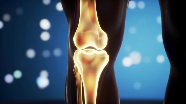
Illuminated knee joint anatomy with femur tibia patella and fibula against bokeh background in dark setting highlighting skeletal structure for medical concepts.
00:07
HD
Knee Pain Illustration of Young Woman with Visible Bones on Gray Background Suffering from Joint Issues Health Concerns and Soreness
00:08
HD
Human knee joint pain. Redness highlights discomfort against a plain backdrop. For medical use
00:08
HD
Doctor learn vertical X-ray with narrowed joint space, compacted tibial endplate, exacerbation of intercondylar elevation of the tibia. Osteoarthritis of the knee joint.
00:19
HD
The amazing highest mountain bird chough (Pyrrhocorax pyrrhocorax brachypus) in the villages of an unknown country Shangri la. It's as ordinary as a commonplace crow there. tibial base plate
00:11
HD
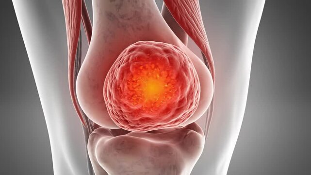
Inflamed Knee Joint Illustration - Close-up 3D video illustrating inflammation and pain within the knee joint. The visualization highlights the swelling and irritation affecting the joint.
00:08
HD
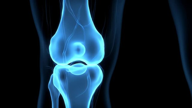
Detailed 3D Medical Visualization of a Translucent Blue Human Knee Joint Against a Black Background Revealing Complex Internal Bone and Ligament Structure
00:08
HD
Human body vascular system anatomy.
00:08
HD
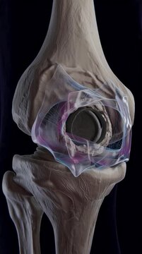
Knee Joint Replacement Anatomy Illustration on Black Background Depicting Bone Structure And Implant Visualization with Surgical Solution for Medical Education
00:08
HD
Medical illustration of a knee joint showing skeletal structure and highlights
00:05
HD
Knee Joint Anatomy Visualization on Blue Gradient Background Medical Demonstration of Skeletal Structure and Ligaments
00:08
HD
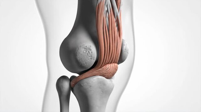
Medical Animation: Detailed Anatomy of Human Patellar Ligament and Tibial Tuberosity
00:07
HD
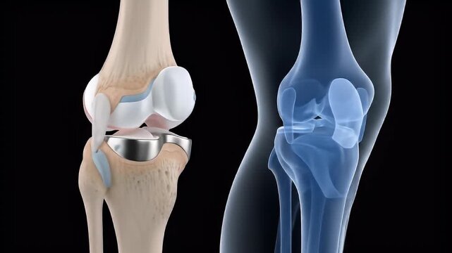
Knee joint model showing a total knee replacement implant, with bone structure and transparent cartilage.
00:05
HD
It is like Bhutan pine (Pinus wallichiana) from tibial base plate, Shangri La
00:11
HD
Chinese pine (Pincus yunnanensis) is long-needle pines from tibial base plate, Shangri La
00:17
HD
The amazing highest mountain bird chough (Pyrrhocorax pyrrhocorax brachypus) in the villages of an unknown country Shangri la. It's as ordinary as a commonplace crow there. tibial base plate
00:07
HD
Tibial Plateau Fracture Fixation X-Ray Imaging
00:15
HD
A bird rosefinch (Carpodacus) sits on an evergreen bush on a mountain stream. Tibial base plate, Shangri la.
00:05
HD
Tibial Plateau Fracture Fixation X-Ray
00:15
HD
Detailed anatomical illustration of knee joints on a teal background, showcasing bone structure, skeletal system for medical purpose.
00:07
HD
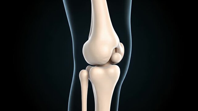
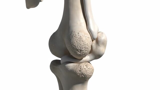
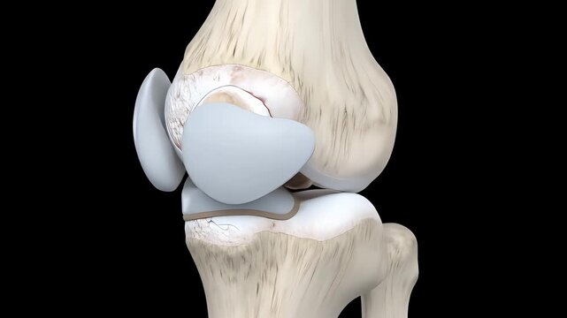
Detailed 3D Illustration of Human Knee Joint Anatomy in White and Beige on Black Background for Medical Visualization
00:08
HD
A corner of a mountain meadow over river on Tibial base plate. Probably orange flowers of Pilosella sp.. Asteraceae. Tibet, Shangri la.
00:10
HD
Shrubby bushclover (Lespedeza bicolor) in Tibial base plate, Shangri la. Cheegrass (jiji grass) backgroud
00:08
HD
Knee Joint CT-Scan Revealing Tibial Fracture:The scan highlights a distinct fracture in the tibia, located beneath the knee joint. The break is visible as a crack.
00:21
HD
Vertical video of doctor learning X-ray of Knee joint dislocation after accident. Patellar joint instability. Tibial dislocation in relation to the femur. 4k footage.
00:24
HD
Knee Joint CT-Scan Revealing Tibial Fracture:The scan highlights a distinct fracture in the tibia, located beneath the knee joint. The break is visible as a crack.
00:12
HD
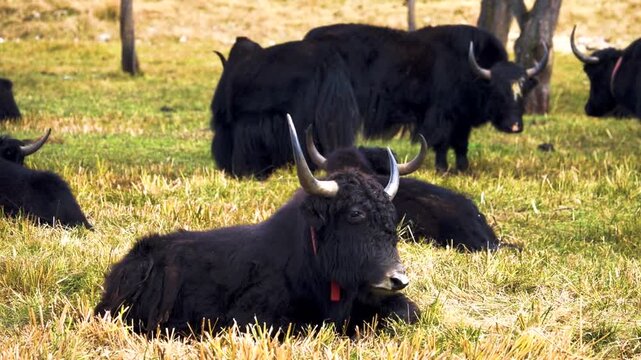
Black Domestic yaks (Bos grunniens), probably a Kyrgyz Yak breed in the mountain meadows of Shangri La, Tibet, China, Tibial base plate
00:06
HD
Knee Joint CT-Scan Revealing Tibial Fracture:The scan highlights a distinct fracture in the tibia, located beneath the knee joint. The break is visible as a crack.
00:15
HD
Knee Joint CT-Scan Revealing Tibial Fracture:The scan highlights a distinct fracture in the tibia, located beneath the knee joint. The break is visible as a crack.
00:24
HD
Knee Joint CT-Scan Revealing Tibial Fracture:The scan highlights a distinct fracture in the tibia, located beneath the knee joint. The break is visible as a crack.
00:21
HD
Knee Joint CT-Scan Revealing Tibial Fracture:The scan highlights a distinct fracture in the tibia, located beneath the knee joint. The break is visible as a crack.
00:12
HD
Detailed Anatomy of Human Knee Joint Bones and Ligaments on Neutral Background for Medical Educational
00:08
HD