A link to set your password has been sent to: To access your purchases in the future you will need a password.
Create without limits — unlimited downloads, plus powerful AI editing
New filters added
Sort by

358 results for sem microscope in videos

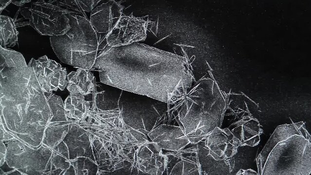
Scientific Research Sample Under Scanning Electron Microscope (SEM) - 4K
00:13
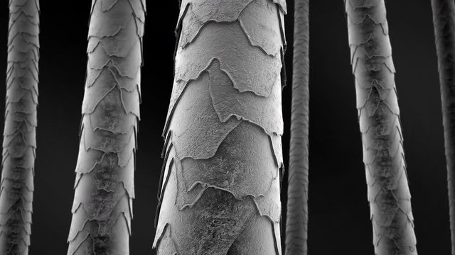
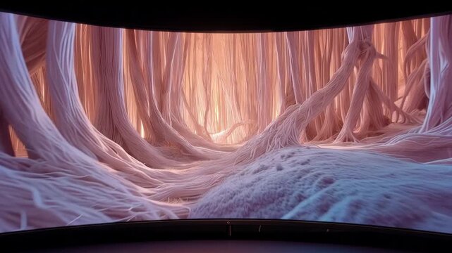
Hair microscope scan. Inside damaged strands of hair. Microscopic hair visualization
00:20
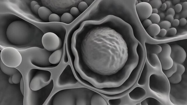
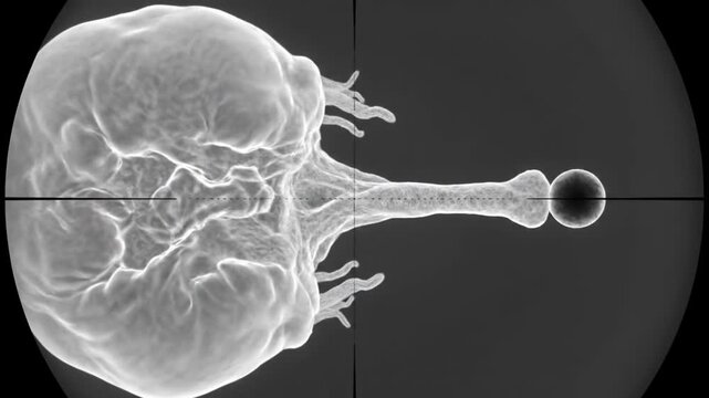
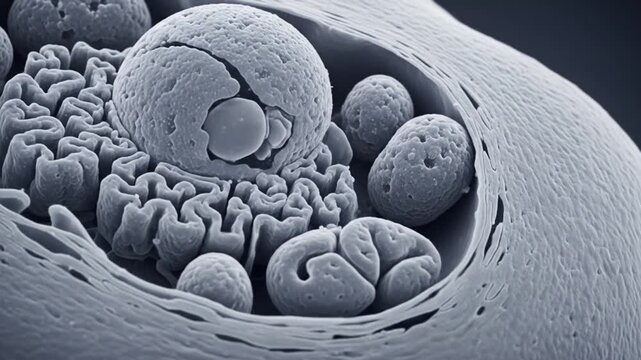
"Highly magnified grayscale microscopic image showing a complex biological structure with a central spherical formation resembling a nucleus or vesicle, surrounded by rounded and tubular components. T
00:05
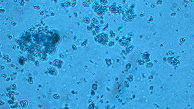
virus and bacteria from a microscope
00:13
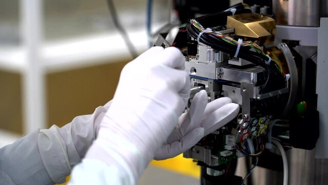
Gloved hands removing a sample from an electron microscope
00:14
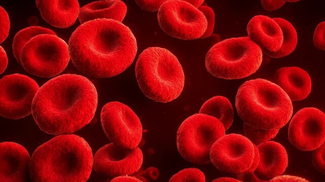
Electron Microscope Style View of Blood Cells Greyscale to Color
00:05
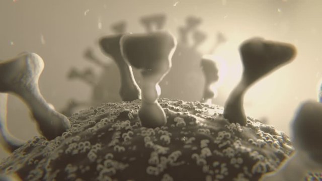
Animation of the microscopic view of the infectious Covid-19 Corona Virus with moving cells
00:16
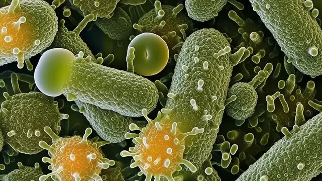
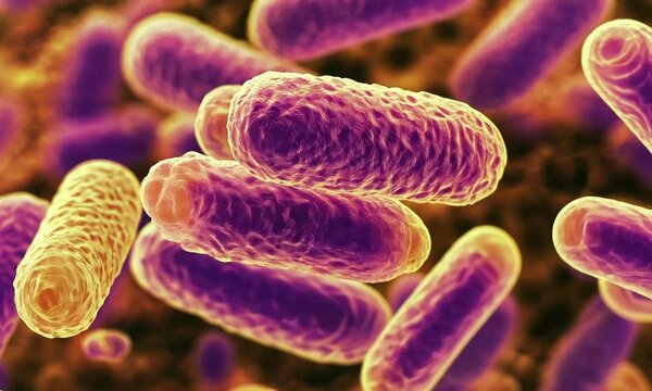
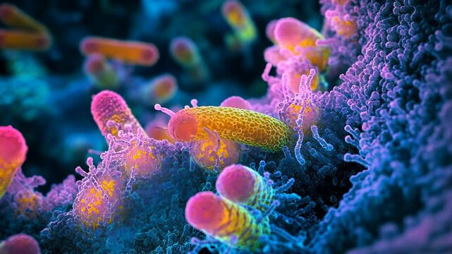
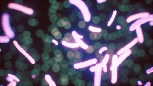
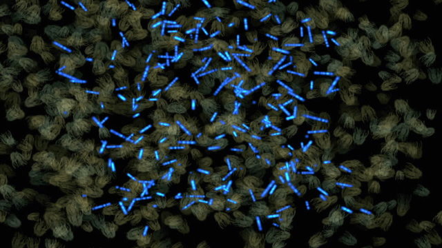
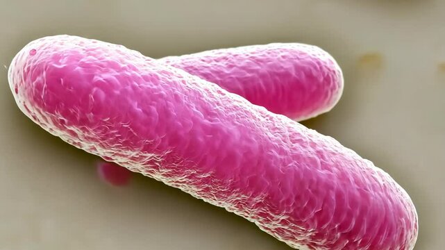
Colorful bacteria under a scanning electron microscope. 3D render of rod-shaped microbes, pathogens and microorganisms in a microscopic macro view.
00:08
Organic structure. SEM, scanning electrone microscope image.
00:06
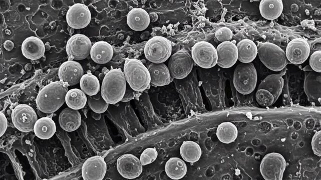
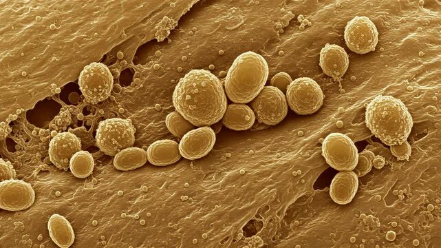
Electron microscope rendering of yeast cells (Saccharomyces cerevisiae) budding on the surface of fermenting dough, with realistic textures
00:10
Bacteriophage virus at microscope. Bacterial viruses landing over bacterias seen under a scanning electron microscope.
00:11
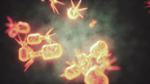
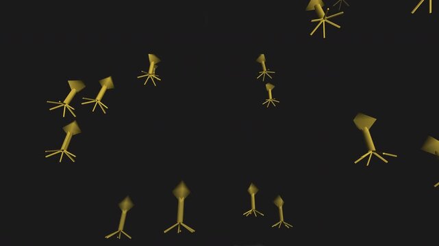
Procedural 3D animation of bacteriophage viruses with articulated legs drifting in fluid. SEM electron microscope aesthetic with film grain. Seamless loop for medical and science backgrounds.
00:12
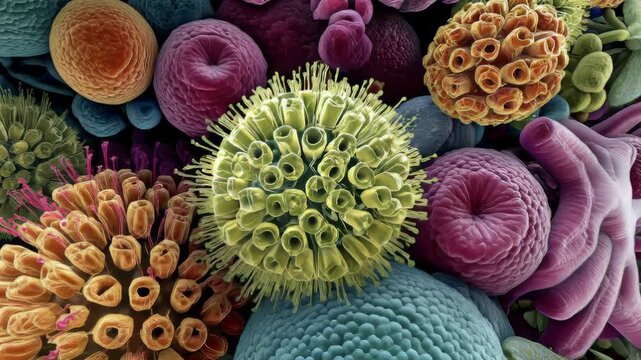
Microscopic view of diverse pollen grains and plant spores in vibrant colors
00:05
Carbon nanotube forest structure imaged under field emission scanning electron microscope, false-color rendering showing vertical alignment of billions of tubes, researcher reviewi
00:10
3D medical illustration of biological microorganisms. Abstract scientific background of germs, viruses or nematodes moving in gray monochrome.
00:08
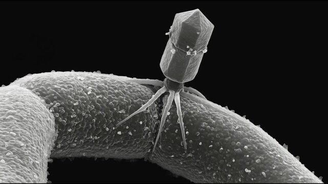
T4 Bacteriophage Virus Attaching to E. Coli Bacterial Cell Surface in Scanning Electron Micrograph
00:05
A close-up image of a fly trapped in a spider web, taken with a scanning electron microscope
00:10
Colorful Scanning Electron Microscope Image of Cell.
00:08
Microscopic View of Bacteria and Viruses.
00:05
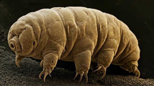
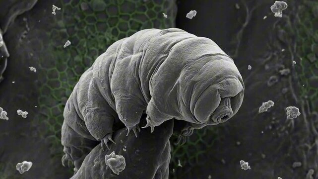
Tardigrade water bear microscopy macro, DNA protection, concept for radiation resistance
00:10
Scanning Electron Microscope (SEM) Microstructure Image, Laboratory Analysis
00:36
A microscopic view of a bacterium, showing the cell wall, capsule, flagellum, pili, and ribosomes. This is an electron micrograph, highlighting the bacteria's structure and features.
00:10
Hair damage animation. Single hair strand transition from Low to High porosity
00:20
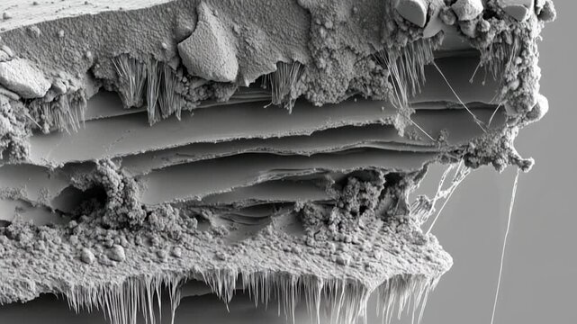
Microscopic layered structure
00:10
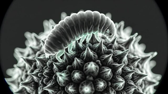
Microscopic Tardigrade Crawling on Spiked Surface
00:08
Electron Microscope Style View of Blood Cells Greyscale to Color
00:05
Corona Virus COVID-19 Animation Loop
00:19
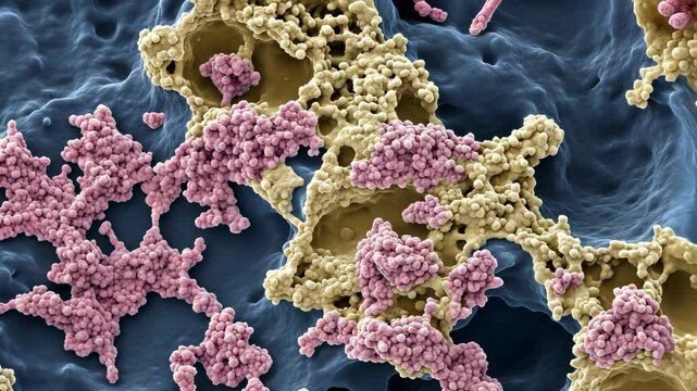
This is a scanning electron microscope image of a biofilm, a community of bacteria that adhere to a surface
00:10
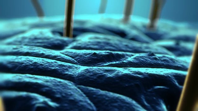
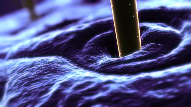
SEM  Scanning Electron Microscope photo of human hair and skin. Blue and yellow.
00:20
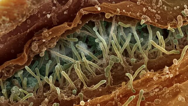
Electron microscope view of Cryptosporidium oocysts clinging to intestinal walls in early infection stage
00:10
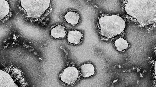
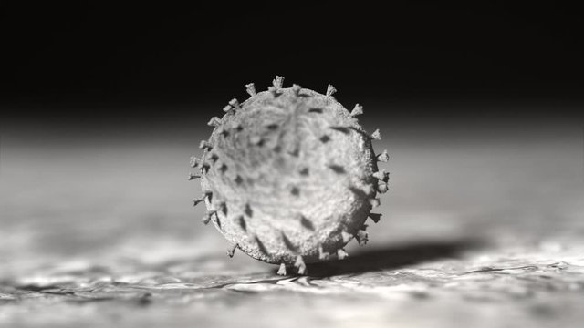
SARS-CoV-2 coronavirus at microscope.Representation of the Covid-19-causing coronavirus seen under a scanning electron microscope.
00:14
Scanning Electron Microscope Texture of Macrophage Cell
00:07
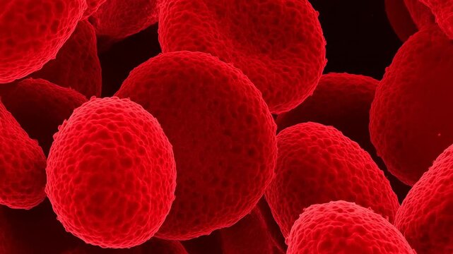
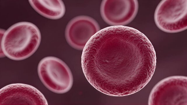
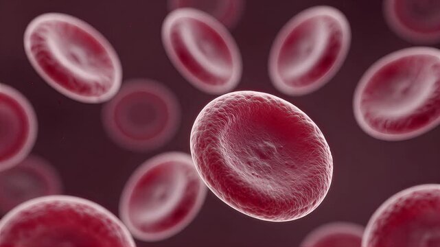
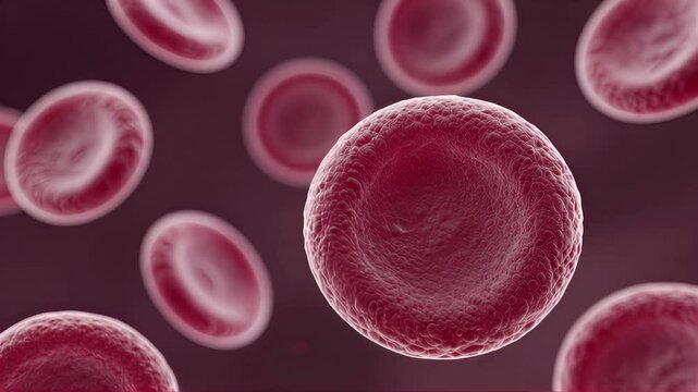
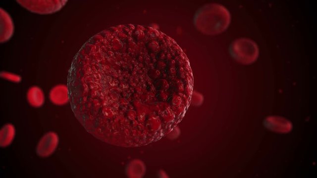
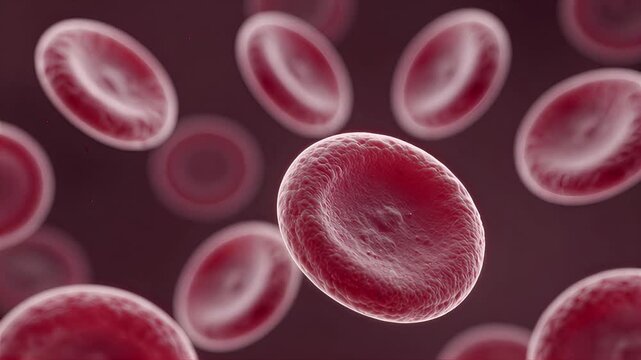
AI Generation: Red blood cells under an ultra-high magnification microscope.
00:07
virus and bacteria from a microscope
00:14
Extreme close-up of pollen grains seen through a scanning electron microscope. Grayscale SEM imagery reveals alien-like textures, spikes, and intricate microstructures during a slow, scientific pan.
00:05
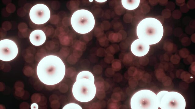
Glowing red cells forming a chain on a dark background
00:05
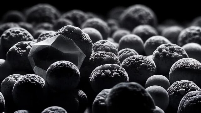
Abstract SEM Nanoparticles Background in Black and White
00:08
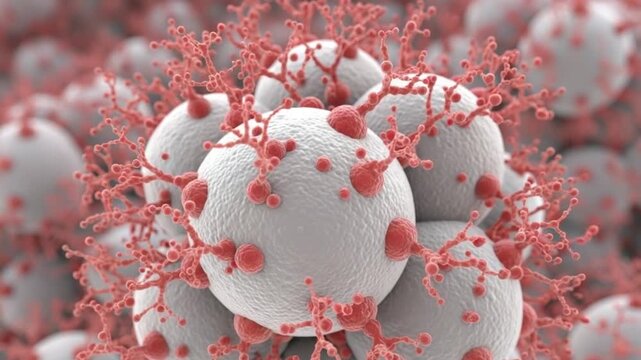
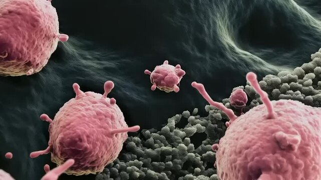
Microscopic view of white spherical cells with red protrusions on surface in closeup
00:10
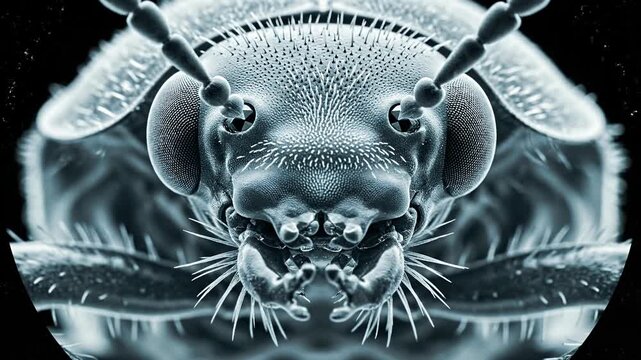
Detailed monochrome scanning electron micrograph of insect head.
00:08
Whipped cream under microscope, macro food science textures and bubbles close up
00:08
Scanning Electron Microscope (SEM) Image
00:26
3D medical illustration of biological microorganisms. Abstract scientific background of germs, viruses or nematodes moving in gray monochrome.
00:08
Cancer cell and T lymphocytes SEM image faceless microscopic visualization defocused scanning electron microscope background immune system interaction oncology research
00:10
Close up view of coral reef microstructure at a micron level. Scanning electron microscope videory.
00:10
A hyperdetailed microscopic view of cancer cells growing in a biological environment
00:07
Microscopic view of pollen grains inside an anther, showcasing intricate biological structures and textures at high magnification.
00:08
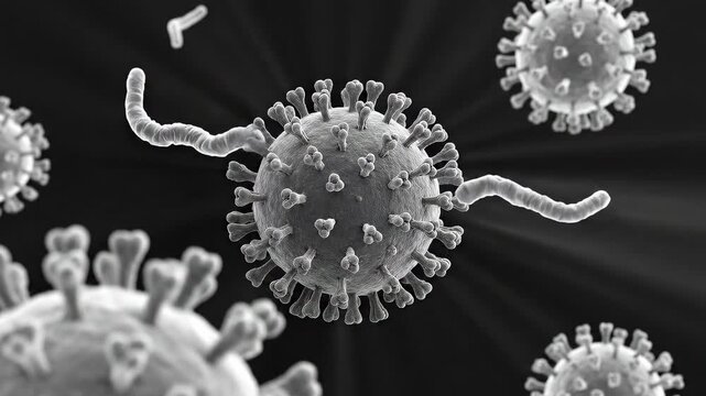
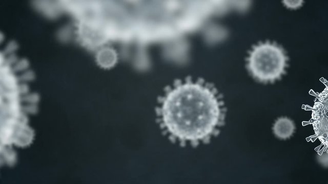
Coronavirus COVID-19 virus molecules spreading, scanning electron microscope view
00:14
Viewing the navirus under a digital microscope
00:10
Macrophage under the microscope.
00:30
Visualizing Nanoparticles: Atomic-Scale Particles in Nanotechnology and Nanomaterials Research
00:09
Pollen grain with white particles on surface, possibly fungus or mold, against blue background of branching tubes or fibers
00:08
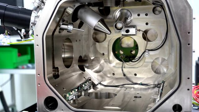
The inside of an electron microscope using accelerated electrons as illumination source
00:06
AI Generation: Red blood cells under an ultra-high magnification microscope.
00:07
Tardigrade water bear microscopy macro, DNA protection, concept for radiation resistance
00:10
Extreme close-up of pollen grains seen through a scanning electron microscope. Grayscale SEM imagery reveals alien-like textures, spikes, and intricate microstructures during a slow, scientific pan.
00:05
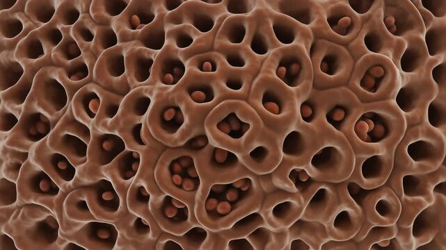
Microscopic View of Porous Structure Material.
00:05
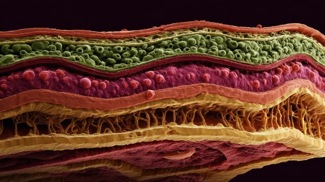
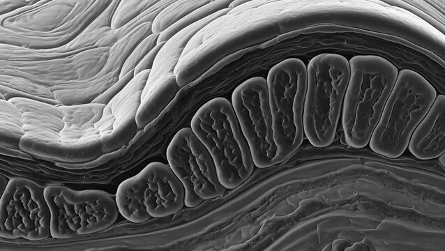
Ultra HD Detailed microscopic crosssection revealing complex layered biological tissue structure with distinct cellular formations and intricate organic patterns video
00:10
Microscopic Scanning Electron Microscopy View of Stratified Epithelial Cells
00:05
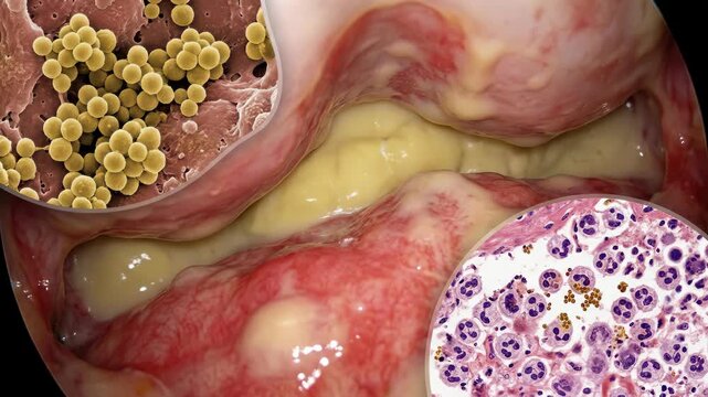
Pathogen disease manifestation: Staphylococcus bacteria, body infection, tissue inflammation, purulent exudate, immune cell, microscopic view, germ invade, host reaction, cellular defense.
00:08
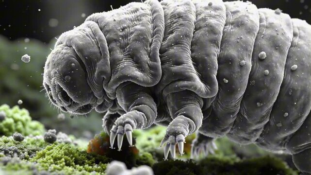
Tardigrade Water Bear Under Microscope Walking on Moss, Microscopic Life Biology
00:08
Scanning Electron Microscope (SEM) Image in a Chemical Lab.
00:36
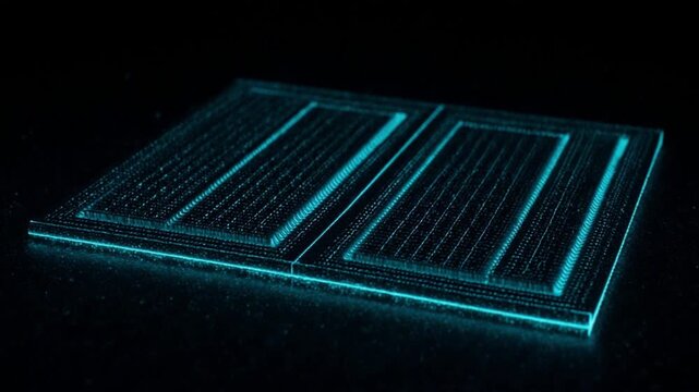
走査型電子顕微鏡 半導体ウェーハダイ ナノスケール シアン擬似カラー
00:05
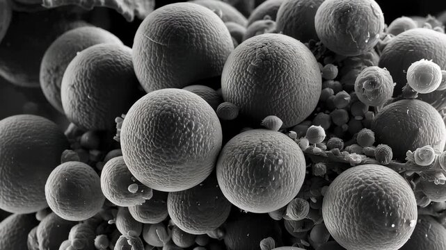
Black and White Microscopic View of Spherical Particles or Cells
00:10
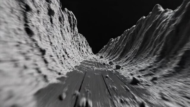
Electron Microscope Style Flyover of Vinyl Record Groove Texture
00:07
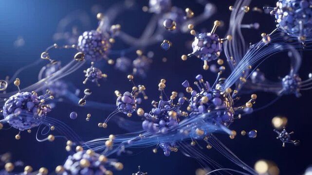
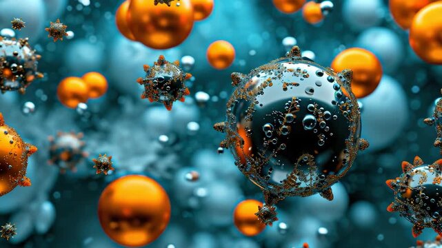
False-color SEM visualization of nanoparticles swarming a pulsing viral pathogen. Golden immune particles encapsulate a beating grey threat. Seamless loop for medical and science usage.
00:12
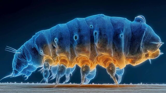
Microscopic Water Bear Tardigrade Close-up in Vibrant Blue and Orange Hues
00:05
Hair damage and repair concept. Low and High porosity hair strands animation.
00:20
Electron microscope rendering of yeast cells (Saccharomyces cerevisiae) budding on the surface of fermenting dough, with realistic textures
00:10
Virus closeup revealing another virus in the background. 3D animation. Coronavirus COVID-19. SEM electron microscope image. Series of clips.
00:10
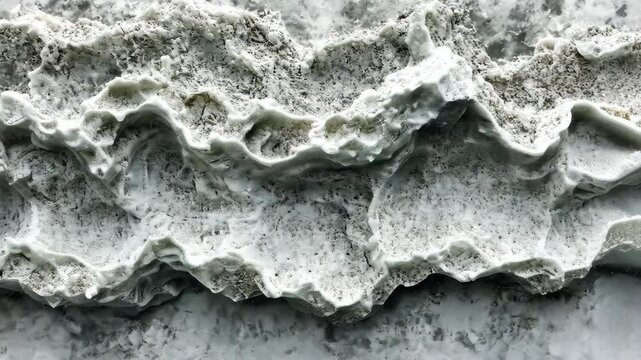
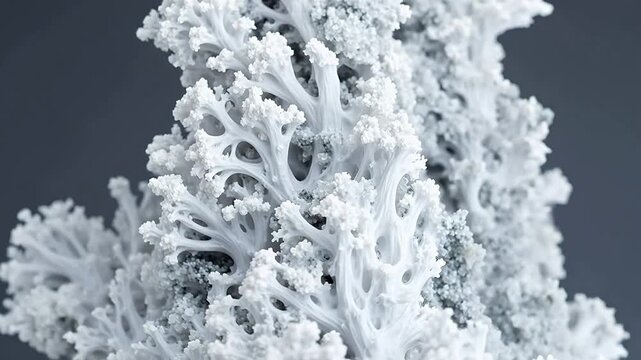
A close-up of a rough, white surface resembling a mountain range, taken with a microscope
00:10
Viewing the navirus under a digital microscope
00:10
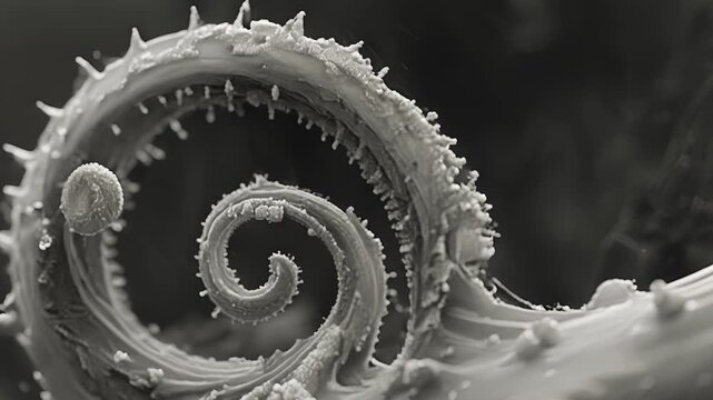
An SEM image revealing the delicate spiral shape of a cells tail used for propulsion.
00:08
AI-generated: Close-up of red blood cells that are visible under extreme microscope zoom.
00:07
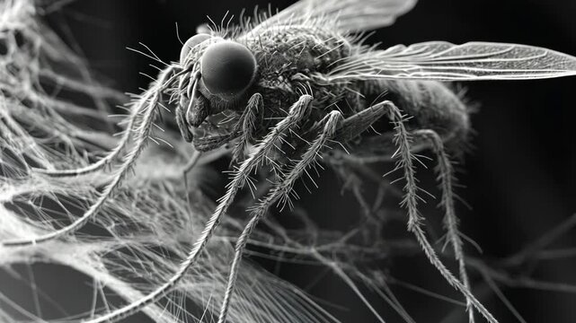
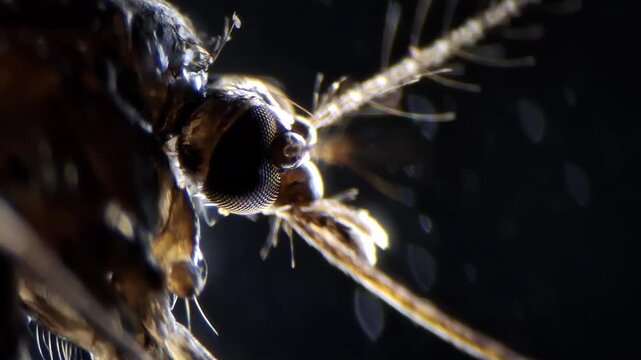
Close up of mosquito head under the microscope showing compound eyes and antennae
00:11
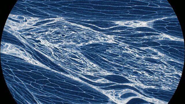
Colorized Scanning Electron Microscope View of Muscle Tissue Highlighting Intricate Structures and Patterns for Medical and Biological Research
00:08
Intricate fractal patterns, 4K Video
00:10
Microscopic View of Cancer Cells: A Detailed Look into the Cellular Structure and Morphology of Malignant Neoplasms
00:05
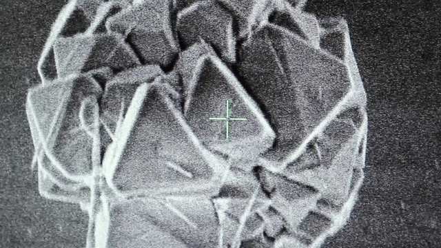
Electron Microscope View of Carbonate Crystals from CO2 Mineralization
00:05
A microscopic view of viruses and bacteria in a scientific setting.
00:10
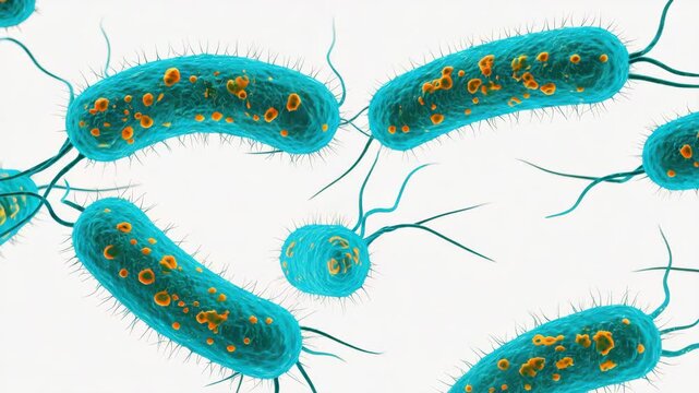
Microscopic Close-up of Rod-Shaped Bacteria, Possibly Escherichia coli
00:10
Scanning Electron Microscope (SEM) Image in a Chemical Lab.
00:22
virus and bacteria from a microscope
00:18
Black and White Microscopic View of Spherical Particles or Cells
00:10
3D Animation of bacterias
00:15
Computed 3D visualization of geometric bacteriophages landing on an organic cell surface. Seamless loop with SEM aesthetic. Schematic representation of viral binding for medical and scientific.
00:12
This is a microscopic image of bacteria attaching to a cell
00:10
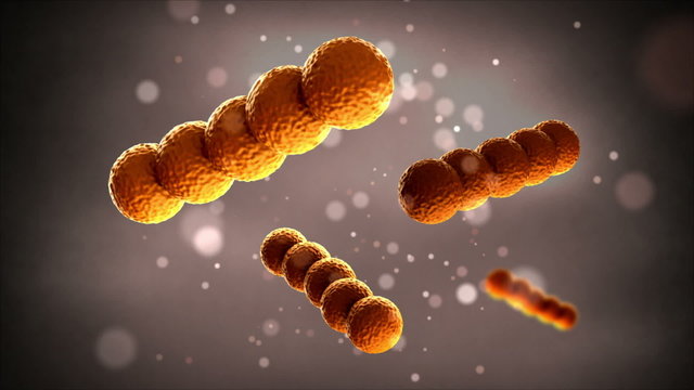
Bacteria - Streptococcus
00:20
High-magnification image of Aspergillus fungi colonies on spoiled fruit, with radiating conidiophores and spore chains under soft lighting
00:10
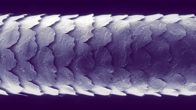
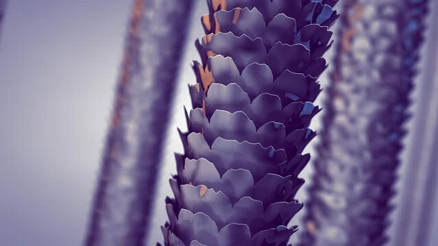
SEM  Scanning Electron Microscope photo of  single human hair and skin. Purple and yellow.
00:20
Realistic rendering of bacteria - in red colors
00:20
Viewing the navirus under a digital microscope
00:10
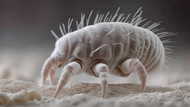
Detailed microscopic view of a dust mite, showcasing its unique features and texture. Highlighting the intricate details of these microscopic creatures.
00:05
AI-generated: Red blood cells are seen under extreme magnification.
00:07
Spermatozon fast movement microscopic image, dark field technique
00:14
Biological Animation of Cancer Cell Division
00:27
Bacteria
00:15
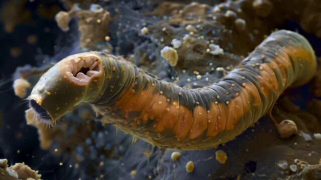
A colorized SEM scanning electron microscope image of a soil nematode allowing for a detailed examination of its rugged exterior and . AI generation.
00:06
Tumour
00:10
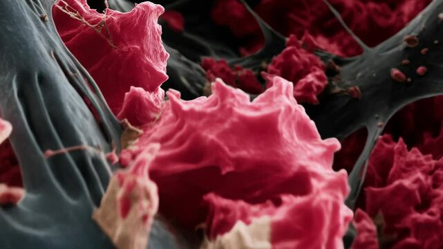
Scanning electron microscope view of fibrin meshwork and red blood cells involved in clot formation, blood components detail
00:05
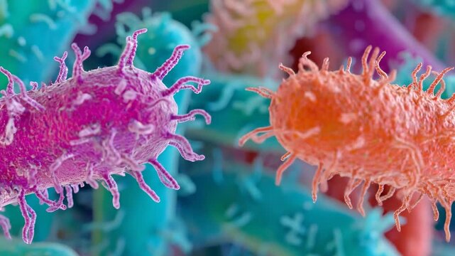
A scanning electron microscope image shows two pink bacteria
00:10