A link to set your password has been sent to: To access your purchases in the future you will need a password.
Create without limits — unlimited downloads, plus powerful AI editing
New filters added
Sort by

3,199 results for mitral in videos

Did you really mean: mitra
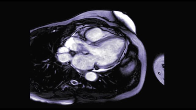
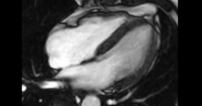
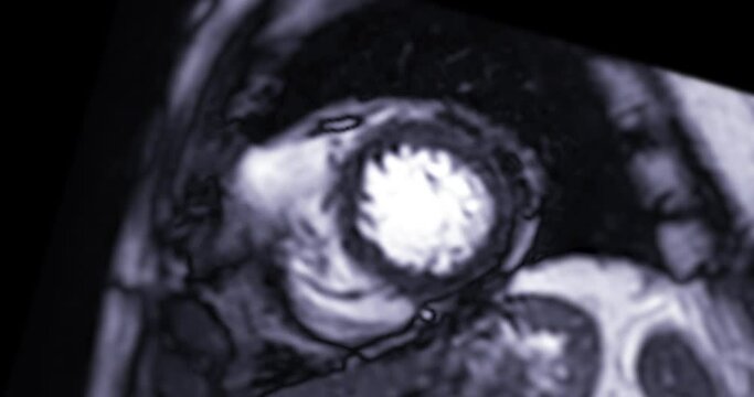
MRI heart or Cardiac MRI  magnetic resonance imaging  of heart in 3 chamber view showing left atrium ,left ventricle, mitral  and aortic valve  for detecting heart disease.
00:13
HD
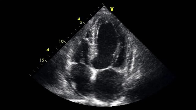
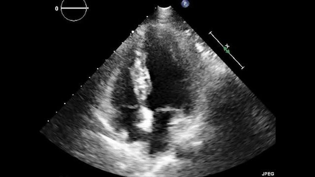
Transesophageal ultrasound video in gray-scale mode.
00:17
HD
Neural Network of Olfaction: Mitral Cell Activation in Bulb
00:05
HD
3d rendered medical animation of  a beating heart
00:12
HD
heart valves, valve function, disease prevention. Cardiac health. Arteries widen during stress response. Aortic durability enhances blood flow. Heart performance supports systemic circulation.
00:10
HD
Mitral Valve Prolapse Animation: Heart Function and Flow
00:26
HD
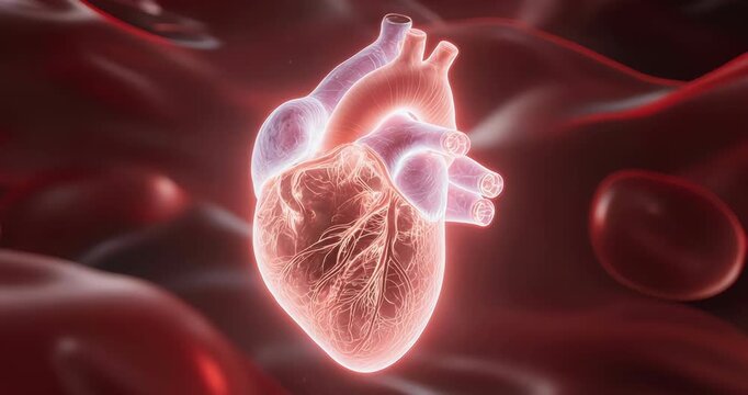
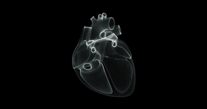
Hologram representation screen 3d of heart beating in the human body
00:19
HD
Mitral valve disease is a problem with the valve located between the left heart chambers .
00:12
HD
surgeon looking at a picture of a sonogram, tavi, transcatheter aortic valve implantation, surgery of the aortic valve. Coronary angioplasty and stent insertion. Stent Placement for the Heart
00:15
HD
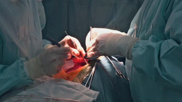
Doctors perform surgery is mitral valve replacement operation on open heart
00:21
HD
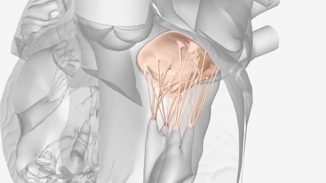
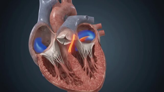
The mitral valve is located between the upper left heart chamber (left atrium) and the lower left heart chamber (left ventricle)
00:09
HD
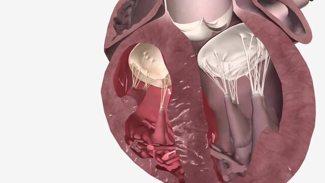
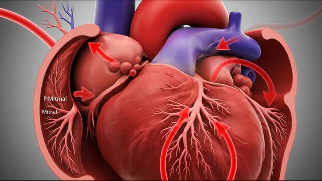
Animated cross-section of a human heart showing mitral valve leaflets in motion
00:10
HD
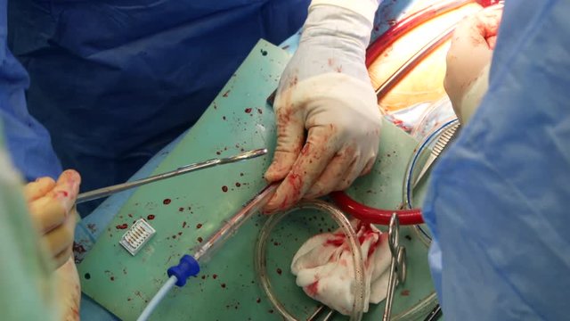
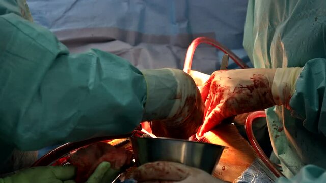
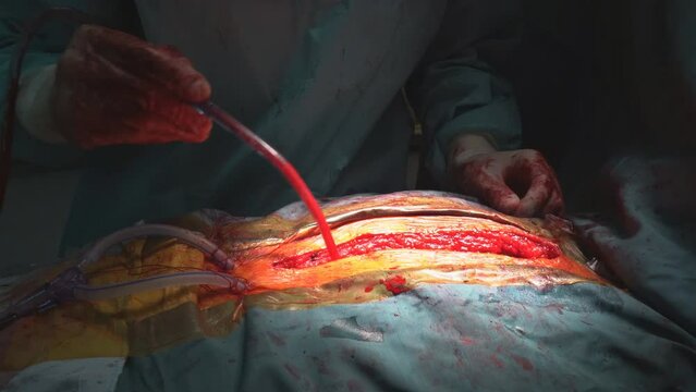
Close up of open heart surgery cardiopulmonary bypass, minimal invasive cardiac surgery, cannulation techniques. Minimally invasive mitral surgery through mini-thoracotomy with cannula tube
00:12
HD
Vibrant Heart Valve Animation with Doppler Flow
00:05
HD
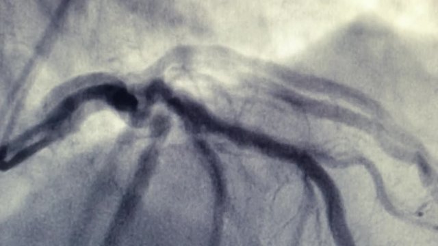
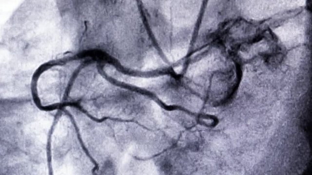
Catheterization. Cardiac ventriculography is a medical imaging test used to determine a patient cardiac function in the right or left ventricle
00:11
HD
Senior experiencing severe chest pain, indicative of cardiovascular distress, heart attack symptoms, and potential heart disease requiring immediate medical intervention and prevention strategies
00:08
HD
Doctor holding in hand Billowing Mitral Valve
00:15
HD
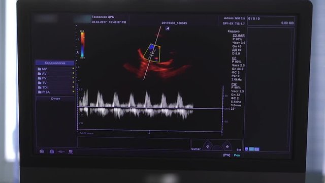
Echocardiography
00:06
HD
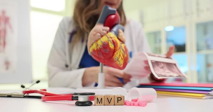
Wooden blocks on cardiological table spell MVP while female cardiologist holds scan images. Doctor reviews echocardiogram printout about mitral valve
00:16
HD
Computer-generated ry of human heart with red blood circulation.
00:10
HD
MRI heart or Cardiac MRI ( magnetic resonance imaging ) of heart  showing heart beating for detecting heart disease.
00:16
HD
Transesophageal Ultrasound Echocardiography.
00:12
HD
Close-up 3D Animation of Human Heart Mitral Valve Opening and Closing.
00:08
HD
A 10-second 16:9 3D medical animation video loop of a realistic human mitral heart valve opening and closing in a smooth, rhythmic 60 BPM heartbeat motion. Camera is fixed at a 45-degree angle inside
00:08
HD
heart valves, valve replacement, cardiac functioning. Valve structure. Arteries expand during exercise stimulation. Aorta flexibility aids blood flow. Heart efficiency supports vital activity.
00:10
HD
A 3D animation of a Mitral Valve Prolapse in the human heart
00:10
HD
Human heart beating gyrating 360 degree in loop
00:24
HD
Mitral valve disease is a problem with the valve located between the left heart chambers .
00:09
HD
Anatomical illustration of the human olfactory bulb and mitral cells
00:07
HD
As result of malfunctioning the heart valve, patient undergoes open heart surgery, which involves a valve replacement in an operating room.
00:29
HD
The mitral valve is located between the upper left heart chamber (left atrium) and the lower left heart chamber (left ventricle)
00:10
HD
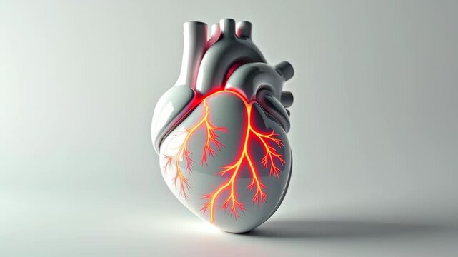
Delicate Luminous Mitral Valve Heart Anatomy
00:05
HD
Close up of open heart surgery cardiopulmonary bypass, minimal invasive cardiac surgery, cannulation techniques. Minimally invasive mitral surgery through mini-thoracotomy with cannula
00:11
HD
Takotsubo Syndrome with Apical Ballooning and Transient Ventricular Motion for Medical Education
00:08
HD
Catheterization. Cardiac ventriculography is a medical imaging test used to determine a patient cardiac function in the right or left ventricle
00:11
HD
Hand of doctor cardiologist with scalpel anatomy of heart
00:08
HD
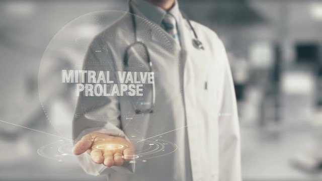
Doctor holding in hand Prolapsing Mitral Valve
00:15
HD
Echocardiography
00:20
HD
Side view of a man using a spirometer.
00:08
HD
3D anatomy man suffering from heart attack, coronary artery disease, atherosclerosis, myocardial infarction, angina, stroke and chest pain on a dramatic background. Video a great for medical purposes.
00:10
HD
MRI heart or Cardiac MRI ( magnetic resonance imaging ) of heart  showing heart beating for detecting heart disease.
00:13
HD
Transesophageal Ultrasound Echocardiography.
00:13
HD
The doctor makes an ultrasound in the hospital. Conducting an ultrasound procedure in a patient. Heart ultrasound image on computer screen.
00:09
HD
Interior view of a beating human heart showing the mitral valve as blood cells surge through and valve flaps close to stop backflow
00:10
HD
Vascular, vascular health, arterial pressure. Blood wellness. Capillaries constrict under low pressure. Vein elasticity enhances oxygen delivery. Cardiovascular efficiency supports organ function
00:10
HD
Close up detail of a surgery on male urogenital system. Action. Surgeons hands cutting out testicle malignant tumor with professional tools, concept of cancer disease and medicine.
00:05
HD
Animation of a human heart gyrating on black background
00:30
HD
The human heart has four valves, aortic, mitral, pulmonary and tricuspid that control blood flow.
00:20
HD
Mitral regurgitation diagnosis document panning shot. Heart disease medical concept
00:12
HD
Valve replacement is performed in operating room when a malfunctioning valve the heart causes an open heart surgery
00:29
HD
Left atrioventricular valve (bicuspid valve, mitral valve) .
00:16
HD
Delicate Luminous Mitral Valve Heart Anatomy
00:05
HD
Mitral valve prolapse.Doctor using stethoscope on medicine background. Futuristic technology. data hologram healthy concept.
00:14
HD
Mitral Valve Prolapse During Ventricular Systole in Beating Heart for Cardiology Education
00:08
HD
Catheterization. Cardiac ventriculography is a medical imaging test used to determine a patient cardiac function in the right or left ventricle
00:08
HD
Image on the monitor of a medical ultrasound machine.
00:20
HD
Doctor holding in hand Mitral Valve Prolapse
00:15
HD
Echocardiography
00:10
HD
3d illustration of human circulatory system heart anatomy
00:15
HD
Detailed 3D Anatomical Model of a Healthy Canine Mitral Valve Functioning During Cardiac Cycle
00:08
HD
MRI heart or Cardiac MRI ( magnetic resonance imaging ) of heart  showing heart beating for detecting heart disease.
00:15
HD
Transesophageal ultrasound video in gray-scale mode.
00:14
HD
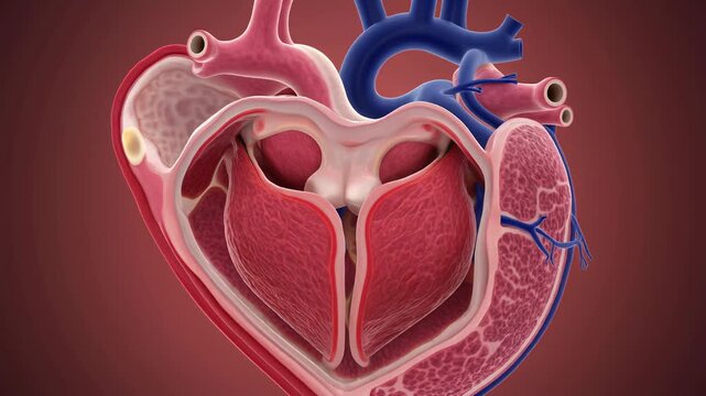
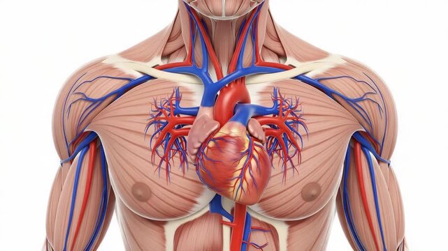
Anatomical Heart Illustration with Visible Muscles and Blood Vessels
00:05
HD
Heart pain: individual clutches chest, symbolizing cardiovascular distress, cardiac arrest, chest discomfort, heart condition, ischemic illness, circulatory system issues, myocardial infarction.
00:10
HD
Blood vessels show intricate branching.Red cells travel capillaries.Medical illustration depicts cardiovascular health.3D rendering highlights vascular system.Educational tool studying anatomy.
00:10
HD
Human Heart structure animation illustration  Shallow Depth of F
00:11
HD
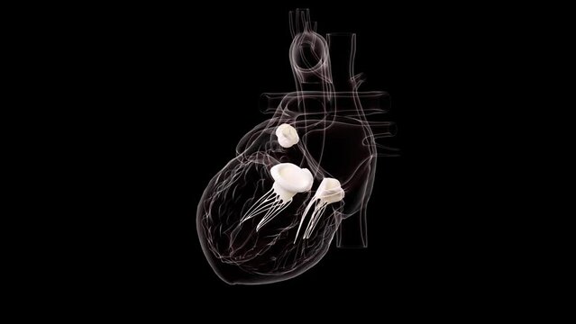
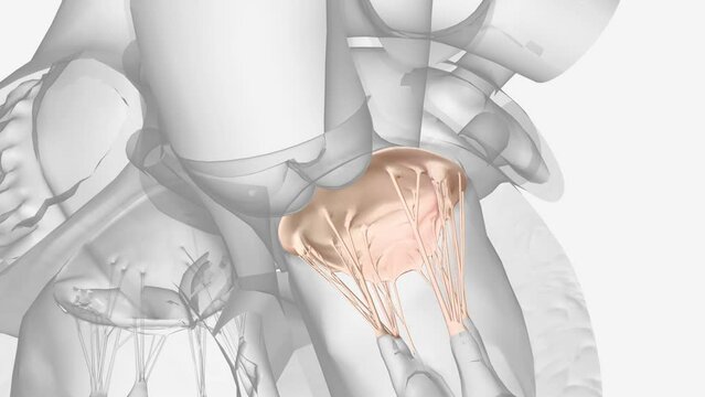
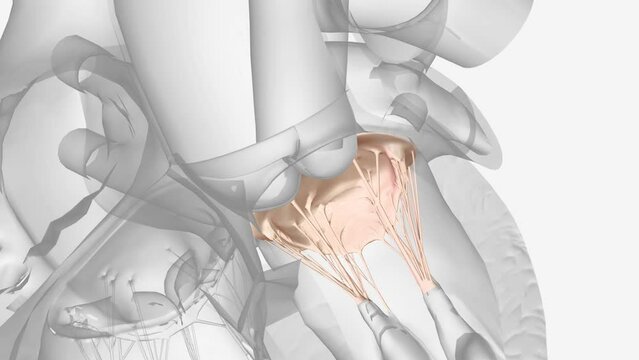
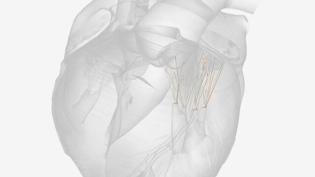
Mitral valve chordae tendinease 3D MED CAL
00:09
HD
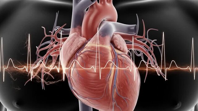
Anatomical heart render superimposed on a heartbeat graph and a silhouette of a torso
00:08
HD
Surgeon doctors perform on open heart valve replacement surgery.
00:20
HD
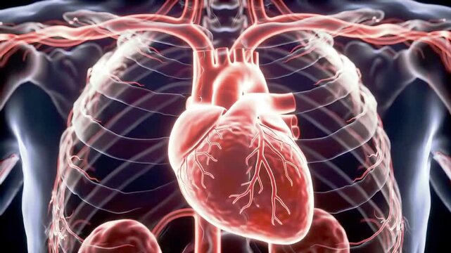
Anatomical View Showcasing The Human Heart And Rib Cage With Visible Blood Vessels System Clearly
00:08
HD
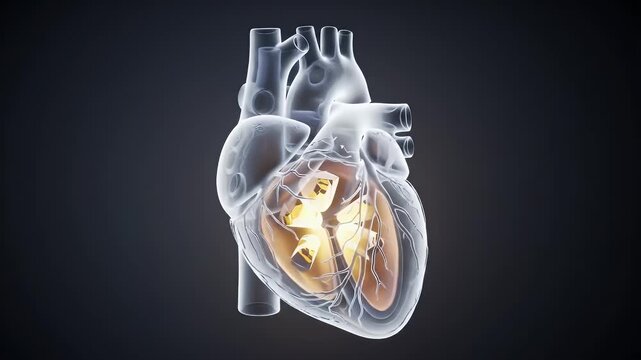
Detailed Translucent Anatomical Heart Model on Dark Background Cardiovascular System Study
00:08
HD
Cardiologist with scalpel anatomy of heart
00:11
HD
Human heart anatomy model cardiovascular system illustration 4k video
00:08
HD
Catheterization. Cardiac ventriculography is a medical imaging test used to determine a patient cardiac function in the right or left ventricle
00:11
HD
Mitral regurgitation.Doctor using stethoscope on medicine background. Futuristic technology. data hologram healthy concept.
00:14
HD
Myxomatous Mitral Valve with medical digital technology concept
00:18
HD
Echocardiography
00:21
HD
3d illustration of human circulatory system heart anatomy
00:15
HD
Ilustration Human Heart Anatomy 3D Model, Vessels, Chambers, Valves, Blood
00:08
HD
Doctor using the ECHO
00:08
HD
High quality transesophageal ultrasound video in gray-scale mode.
00:11
HD
Hologram representation screen 3d of heart beating in the human body - loop
00:19
HD
Chest pressure: figure holds heart region, depicting circulatory disorder, myocardial disturbance, heart failure, vascular problem, blood circulation issue, infarction risk, cardiac emergency.
00:10
HD
cardiac organ: heart anatomy, medical illustration, anatomical model, heart disease, blood flow, heart surgery, vascular health, heart function, heart muscle tissue, heart valves
00:10
HD
Human Heart structure animation illustration  Shallow Depth of F
00:13
HD
Animated human heart shows blood flow through chambers. Detailed 3D model illustrates cardiac circulation and anatomy.
00:08
HD
Mitral valve chordae tendinease 3D MED CAL
00:08
HD
MRI heart or Cardiac MRI ( magnetic resonance imaging ) of heart  showing heart beating for detecting heart disease.
00:20
HD
During open heart surgery, if heart valve malfunctions, valve replacement is performed in the operating room to resolve this issue
00:29
HD
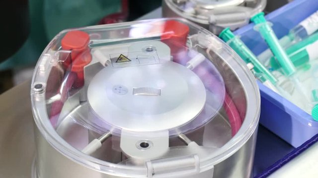
Heart-lung bypass machine, Close-up
00:20
HD
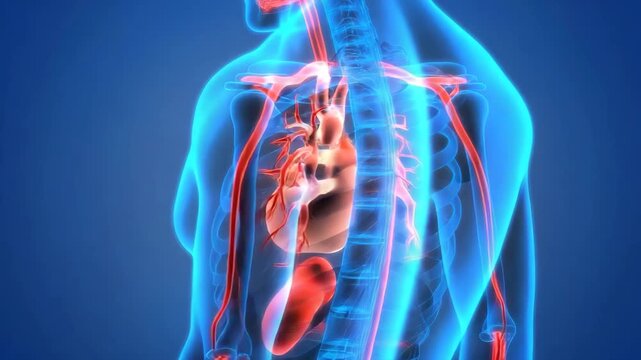
Human body anatomy with heart and blood vessels on blue background
00:10
HD
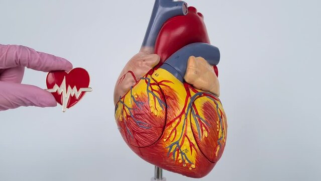
The model is made of plastic and is painted to show the different structures of the heart, including the chambers, valves, and blood vessels
00:05
HD
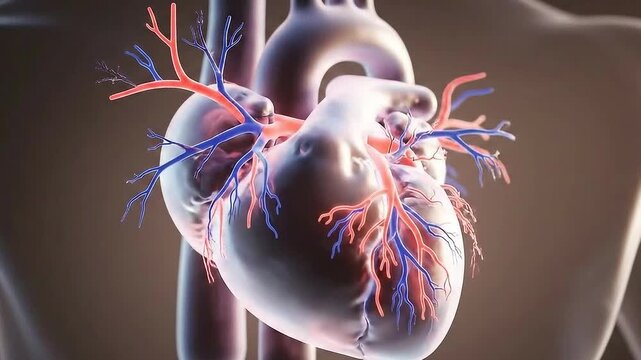
3D Rendering of a Human Heart with Arteries and Veins on Dark Brown Background
00:07
HD
Catheterization. Cardiac ventriculography is a medical imaging test used to determine a patient cardiac function in the right or left ventricle
00:24
HD
Transesophageal ultrasound video in Doppler mode.
00:17
HD
Transesophageal Ultrasound Echocardiography.
00:15
HD
Transesophageal ultrasound video in gray-scale mode.
00:16
HD
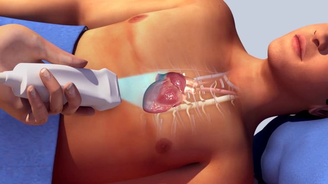
Echocardiography (ultrasound examination of the heart)
00:14
HD
Transesophageal ultrasound video in gray-scale mode.
00:21
HD