A link to set your password has been sent to: To access your purchases in the future you will need a password.
Create without limits — unlimited downloads, plus powerful AI editing
New filters added
Sort by

33,078 results for microscopy in videos

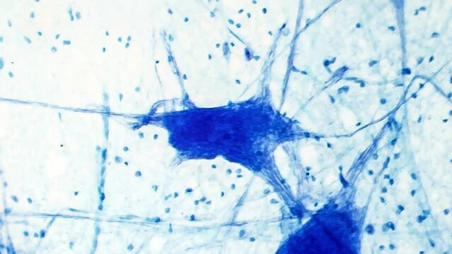
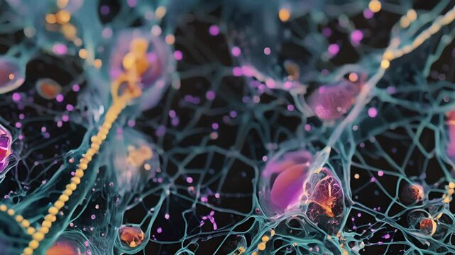
An intricate view of neurons showcasing synaptic connections and electrical activity, highlighting the beauty and complexity of the nervous system in amazing detail.
00:05
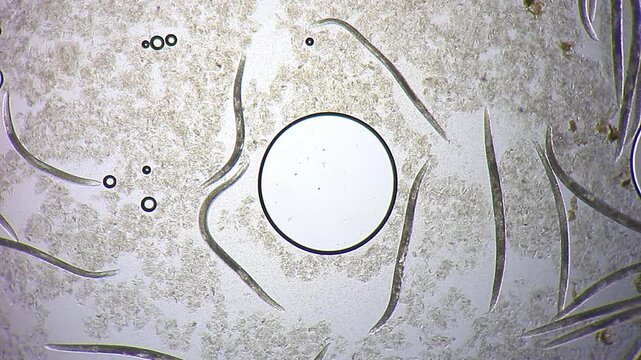
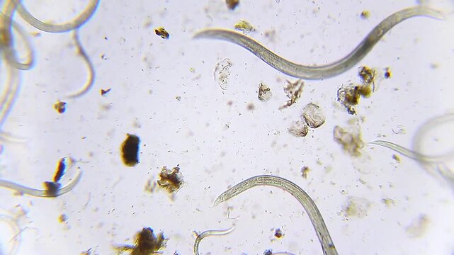
Nematodes actively moving in blue water droplet under 100x magnification brightfield microscopy. 4K scientific microscopy footage.
00:27
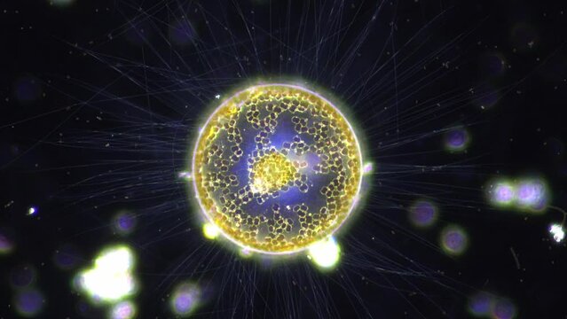
fresh water micro organism, Heliozoa, with its beautiful filaments spreading out like sun rays. 400x magnification, dark field microscopy
00:49
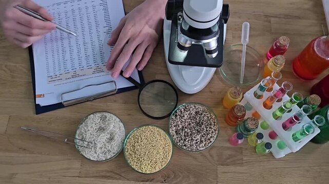
Scientific analysis of food samples in a laboratory with detailed observations and microscopy
00:13
Scientist using microscopy glass setup in laboratory
00:07
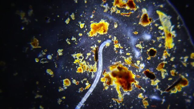
3D animation: Green glowing plankton, algae, and diatoms float in dark water. Microscopic aquatic life, darkfield microscopy style. Science, biology visualization.
00:10
A brightfield microscopy image of hepatocytes revealing their dual function in detoxification and the production of essential proteins such as albumin and clotting factors.
00:07
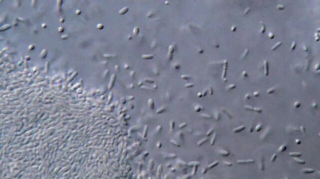
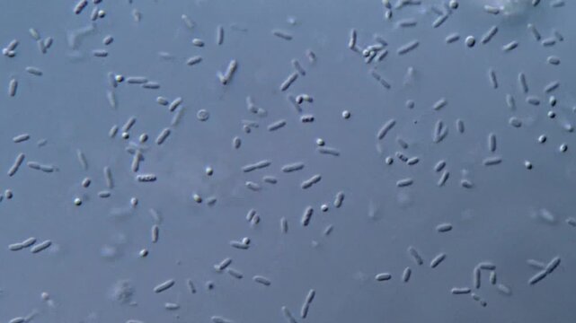
High resolution microscopy of bacteria colonies
00:44
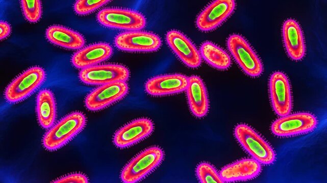
Bacterial infection Yersinia pestis microscopy fluorescent microscopic animation
00:10
Microscopic view of paramecium and amoeba actively swimming in a drop of pond water. Dark-field microscopy lighting makes the microorganisms glow vividly against a deep black background.
00:05
isolated neuron with red nucleus and blue mesh envelope, slow approach revealing membrane tension and surrounding microfilaments, scientific visualization for microscopy and virology
00:10
Resistance to antibacterial agents, growth of bacteria in the intestines, microscopy of bacteria
00:10
Abstract microscopic view of cells with glowing nucleus and soft texture. Concept for biology, life, medicine, science, health, and innovation
00:06
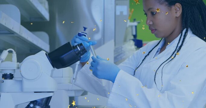
Female lab tech picking up pipette, aspirating sample for microscopy with gold stars overlaying
00:10
Red blood cells move in human blood stream during a close-up view under a microscope showing various shapes and sizes of the cells in a dark background
00:10
Molecule chemistry science cell biotechnology molecular nad longevity research laboratory featuring glowing molecular structure near cell surface and membrane DNA microscopy genetics medical biology
00:08
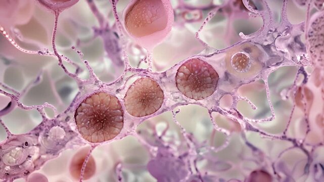
Microscopic digital scene of transparent cytoplasm and filamentous actin structures forming networks around a nucleus, representing cellular mechanics, tissue biology, and modern biotechnological
00:10
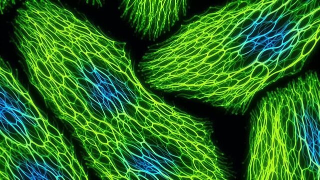
Neuron Network Green Light Patterns.
00:10
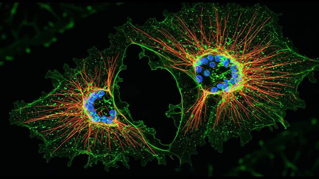
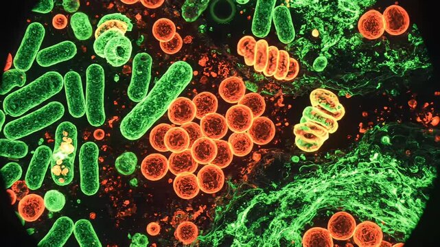
High-resolution fluorescence microscopy animation showing microorganisms stained with fluorescent dyes, emitting vivid green and red light under ultraviolet excitation in a dark laboratory environment
00:08
Rotifer microorganism swimming under darkfield microscopy representing ciliary corona light scattering and aquatic zooplankton biokinetics in 4K Ultra HD footage
00:25
Tardigrade water bear microscopy macro, DNA protection, concept for radiation resistance
00:10
Nematodes moving slowly in blue water droplet under 400x magnification brightfield microscopy. 4K scientific microscopy footage.
00:17
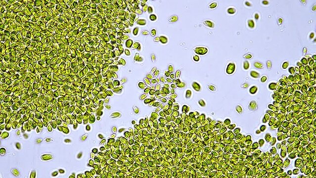
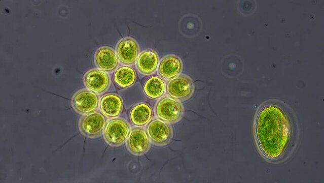
Beautiful and very active single-cell marine microalgae found in the bucket of a fisherman. Microscope footage, time lapse, 200x magnification, bright field microscopy
00:43
Human cerebellar cortex, light microscopy showing Purkinje cells, granular cells, and molecular layer involved in motor coordination and balance.
00:16
Bright laboratory-style abstract 3D microscopy visualization, multiple types of viruses and bacteria in vivid contrasting colors, fine structural details on surfaces, floating dyna
00:10
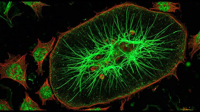
Fluorescence microscopy of actin filaments v1
00:10
A microscopy image of a thyroid follicle showing the unique shape of thyroid cells resembling tiny sacs filled with hormoneproducing machinery.
00:07
Movement of protozoa under microscopy  view
00:18
Lassa virus microscopy video showing cellular degeneration vacuoles and membrane rupture
00:10
Fibroblast cell neuron network microscopic biology glowing membrane organic structure molecular tissue science research connective extracellular matrix scientific visualization cellular anatomy
00:05
Macro cinematic movement through plant spores, scientific discovery concept
00:05
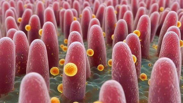
Glowing green particles drift above red intestinal villi in a microscopic view of gut absorption and microbiome
00:10
Abstract scientific background showing isolated stem cell under magnification, surrounded by soft blue microscopy textures
00:10
Microscopic intestinal villi covered in bacteria, showing nutrient absorption on mucosal surface, yellow spherical microbes attaching to pink villi, closeup microscopy style, highdetail
00:10
Examining Microscope View of Cell Sample in Laboratory Imaging
00:10
Female scientist microscope laboratory focusing on moving sample through eyepiece; gloved hands adjust controls in blue lit clinical research closeup.
00:21
Highdetail depiction of mitochondria in cellular energy production
00:10
Woman lifting pipette and drawing into tube in lab, molecules animating on pipette for microscopy
00:10
Pathogens infecting and replicating within human cells, representing an abstract biological concept of viral infection, disease, and medical research in microscopy and science
00:05
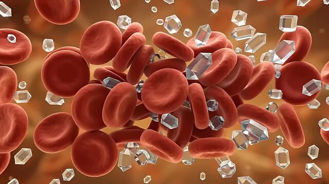
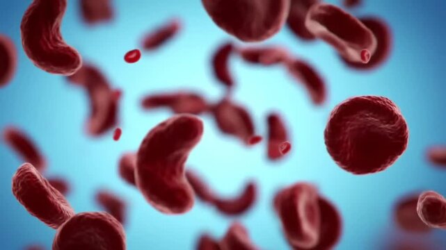
sickle cell anemia red blood cells microscopy
00:08
Darkfield Microscopy of Live Microorganisms Moving Fast across Slide with Stationary Algae for Microbiological Research and Exploration of Hidden Ecosystems in Water Samples and Environmental Study
00:18
Demodex brevis mite moving legs with visible body structure under 100x bright field microscopy. 4K scientific microscopy footage.
00:24
Detailed illustration of a cell with various organelles and structures highlighted.
00:07
Vivid Neon Microscopic View of Cells with Glowing Circular Structures
00:10
Green fluorescent microscopy showing cluster of cells under high magnification view
00:05
Fluorescent microscopy of Caenorhabditis elegans, with glowing neurons and transparent body structure under lab lighting
00:10
Microscopic diatoms with translucent silica shells scattered across slide, varied shapes and sizes, delicate frustules and algae cells visible, brightfield microscopy for taxonomy and environmental
00:38
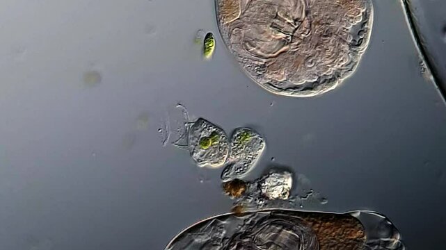
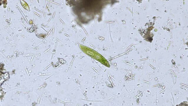
Movement of Euglena and rotifer under microscopy
00:18
Yersinia pestis bacteria animation microscopy fluorescent microbes research
00:10
Fluorescent microscopy animation of an activated B lymphocyte surrounded by a glowing ring of antigens.
00:08
Cytoplasmic streaming
00:10
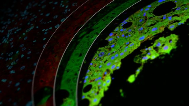
immunostained tissue (mouse liver), split up into single fluorescence channels, confocal microscopy
00:30
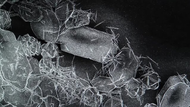
Scanning Electron Microscope (SEM) Image
00:26
A close-up view of vibrant red cellular structures, showcasing intricate details through advanced microscopy techniques.
00:05
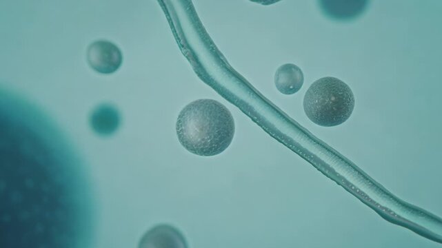
4k Blue Thick liquid chain,light,Magical microbes & splash mucilage under deep ocean.
00:23
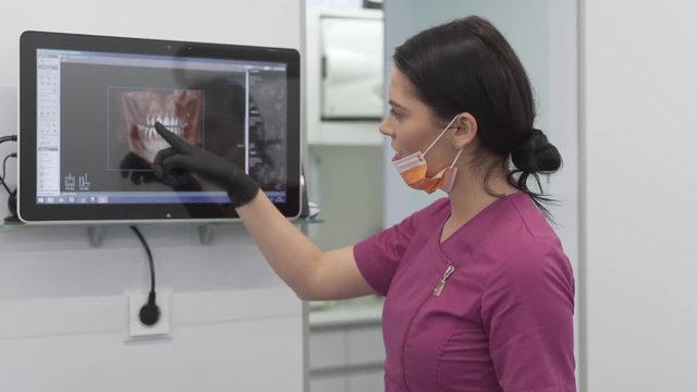
Female Dentist in Her Uniform and Gloves in Modern Dental Clinic Showing Scans Details on Monitor to Her Patient.
00:12
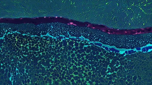
Histology Microscope Video of Mammal Cuboidal Epithelium in Kidney Medulla Tubules (Mouse, 400x)
00:11
Vibrant, neon-color microbes: Lepocinclis and Trachelomonas, two types of Euglenoid microorganisms, seen under the microscope in polarized light at 400x magnification.
00:24
Macro shot of green leaf - super close up - Zoom in
00:22
Combining cells into one. Laboratory research. The science. Blue glowing cells under a microscope. 3d renderer.
00:10
Amoeba at microscope
00:18
Nematodes aggregation moving rapidly under 40x magnification brightfield microscopy. 4K scientific microscopy footage.
00:34
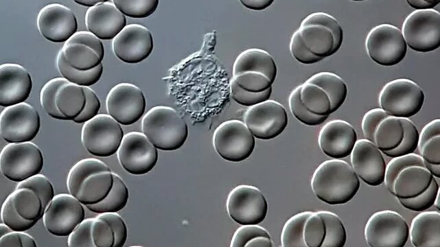
Human blood microscopy thin film at 600x in phase contrast light
00:18
Microscopic view of an amoeba with cellular structures.
00:08
microorganisms close up under the microscope. in a soil sample from a farm
00:11
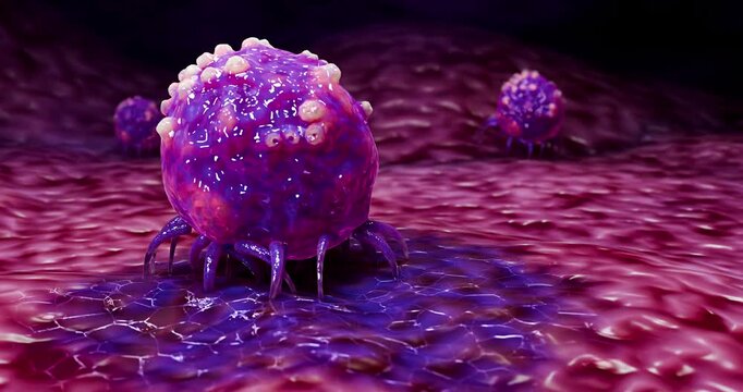
Cancer cell under fluorescence microscopy, showing highlighted mitochondria, nucleus, and surface receptors in bright neon colors
00:10
Toxic goitre of the thyroid, light microscopy
00:12
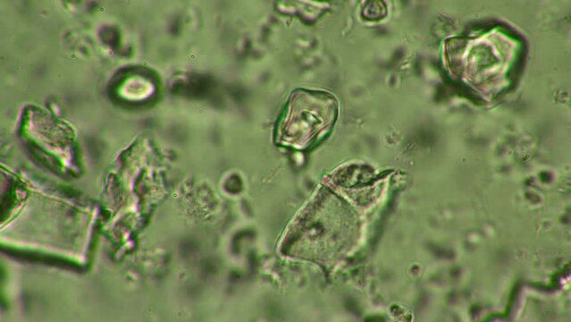
Stool sample under phase microscopy view, Trichuris egg
00:10
A Laboratory Scientist Uses Electron Microscopy to Inspect High-Contrast Biological Cell Structures
00:10
Lipid raft heterogeneity captured by atomic force microscopy depicting their varying size and shape within the cell membrane.
00:08
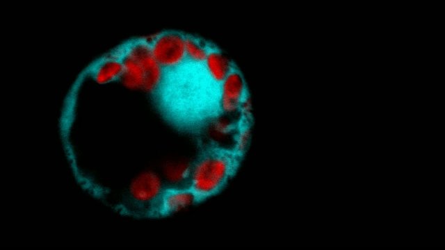
Protoplasts (plant cells without cell wall) expressing green fluorescent protein under fluorescence microscope.
00:12
Explore the Wonders of DNA Structure and Its Significance in Genetics and Biology Labs
00:10
Scanning Electron Microscope (SEM) Image in a Chemical Lab.
00:24
A lot of bacteria and bacilli in a drop of ferment for fermented milk in microscope. Theme of lactic acid foods is under 1000x magnification. Microcosmic color background.
00:18
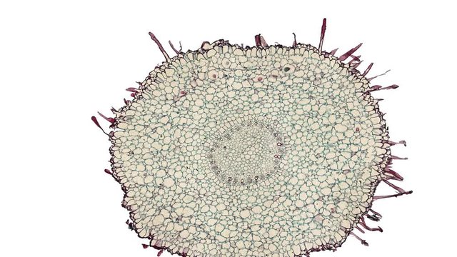
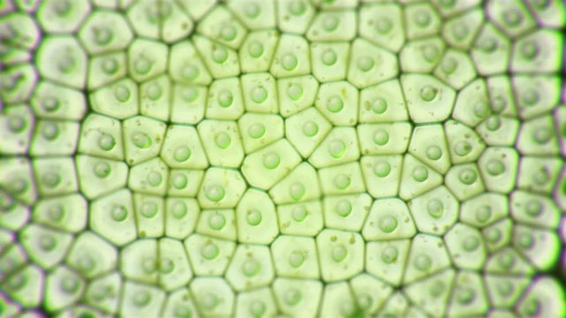
Plant stem cross section microscopy
00:08
Close-up shot of sample slide and microscope with metal lens at laboratory.
00:18
Fluorescence microscopy. Microscopic examination. Medical examination with contrast agent. Laboratory research, lab test. Biology science concept. Cyan orange acid colors abstract texture background
00:10
Cytoplasmic streaming and pulsation of slime mold plasmodium under microscope time lapse
00:10
Lobar pneumonia during the hemorrhagic edema period, light microscopy displaying lung tissue inflammation with hemorrhagic changes.
00:16
Microscopy reveals intricate details in a laboratory during advanced scientific research on materials
00:10
Instructor pointing, guiding researcher adjusting electron microscope settings for sample analysis
00:10
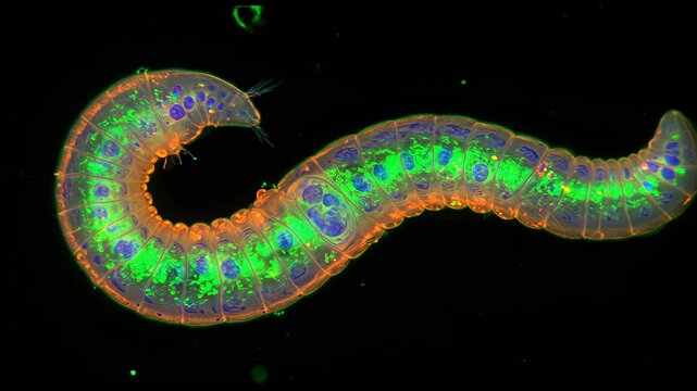
Annelids moving with rotifer colony in background under 40x magnification darkfield microscopy. 4K scientific microscopy footage.
00:27
Neuronen, fluoreszenzmarkiert
00:31
cross section cut of cymbidium root under the microscope
00:30
Molecular structure flow
00:11
3D animation: Green filamentous algae with smaller protists. Brightfield microscopy style. Aquatic life, biology, science visualization.
00:10
Swarm of navicula diatoms swimming dynamically sampled in Japan. Microscopy footage 400x magnification time lapse.
00:57
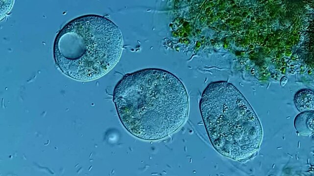
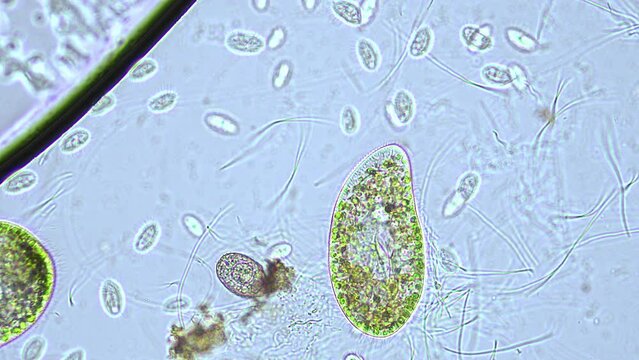
Blepharisma ciliate under microscopy view 400x magnification
00:21
Electron Microscopy Enables Precise Scientific Examination of Glowing Biological Cellular Material
00:10
A fluorescence microscopy image of liver cells marked with fluorescent stains to visualize the process of detoxification and identify the specific toxins being removed.
00:08
Arabidopsis protoplasts
00:07
Pleuronema coronatum under the microscope - unicellular ciliated organism
00:30
Close up on scientist hand with steriled gloves rotating and adjusting block of microscope lenses. Healthcare technology and pharmaceutical research and development concept.
00:08
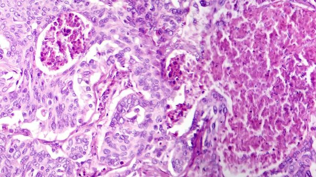
Lung adenocarcinoma, light microscopy
00:12
Biomedical animation of clostridium botulinum.
00:15
Researcher places sample under microscope in laboratory
00:12
Visual representation of mitochondria highlighting structure and function in cellular energy production during biological processes
00:10
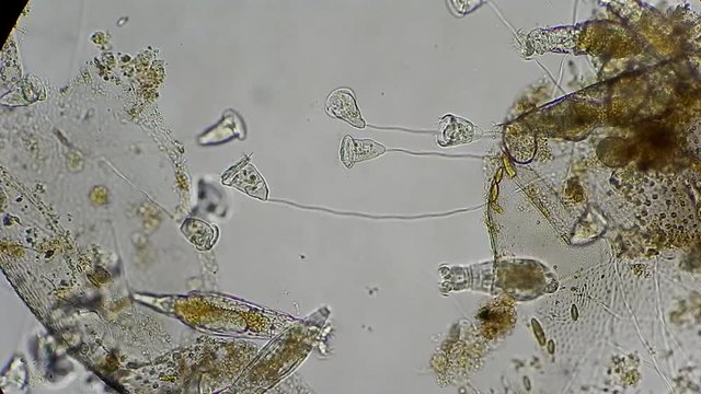
Ciliate swimming among vegetation
00:18
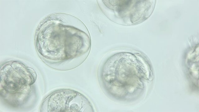
Movement of veliger mollusk larvae in laid eggs under a microscope, class Gastropoda. White Sea.
00:15
Confocal laser scanning microscopy in slow motion. Optical imaging technique
00:10