A link to set your password has been sent to: To access your purchases in the future you will need a password.
Create without limits — unlimited downloads, plus powerful AI editing
New filters added
Sort by

524 results for acls in videos

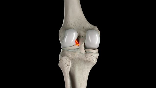
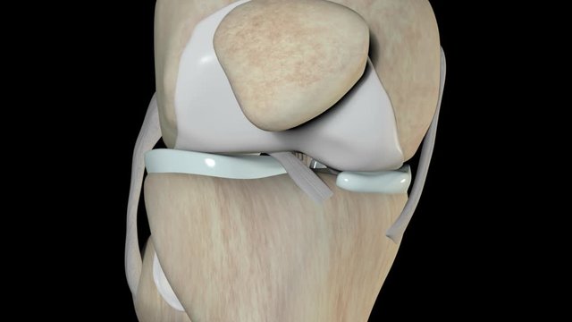
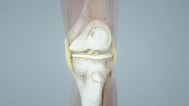
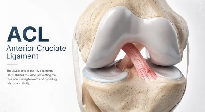
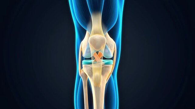
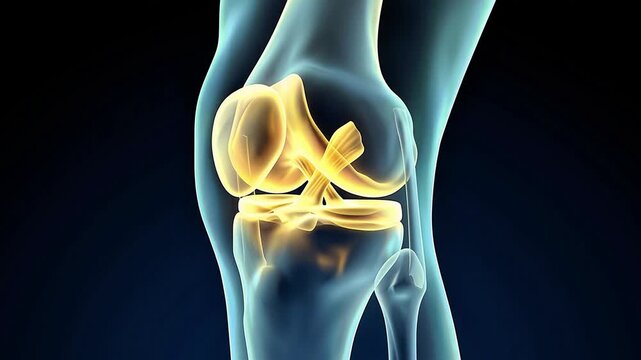
3D rendered medical animation of anterior cruciate ligament
00:13
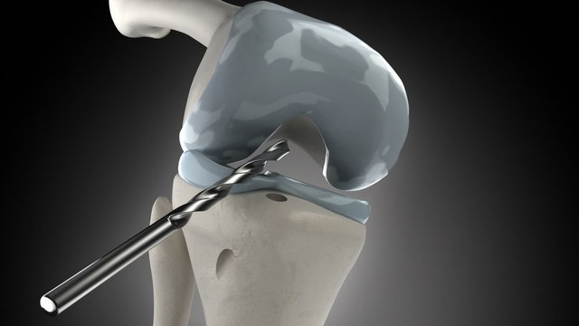
Knee arthroscopic cruciate ligament replacement stage
00:17
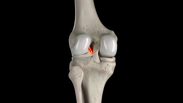
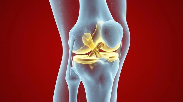
Animation of anterior cruciate ligament
00:13
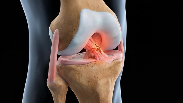
3D Illustration of ACL Injury and Knee Pain in Human Anatomy
00:08
Physical therapy knee acl meniscus rehab return sport quad isolation exercise ankle weight strap strengthening workout
00:05
Close-up of women touching her knee, depicting joint pain, stiffness, and inflammation in calm bedroom. Ideal for content about knee arthritis, injury, physiotherapy, recovery and pain relief.
00:10
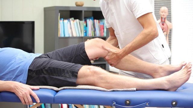
Doctor ot therapist performing Lachman Test
00:20
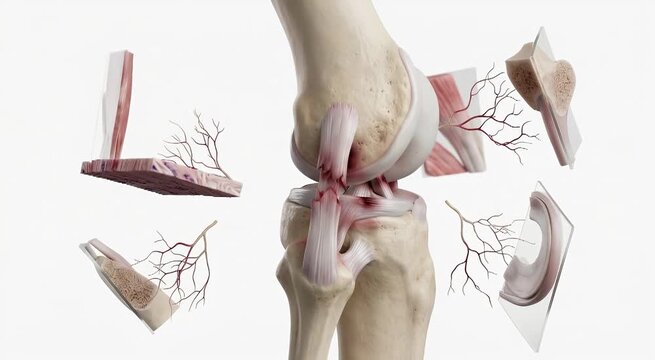
3d medical knee joint anatomy showing ligaments and nerves moving through flexion and extension motion
00:10
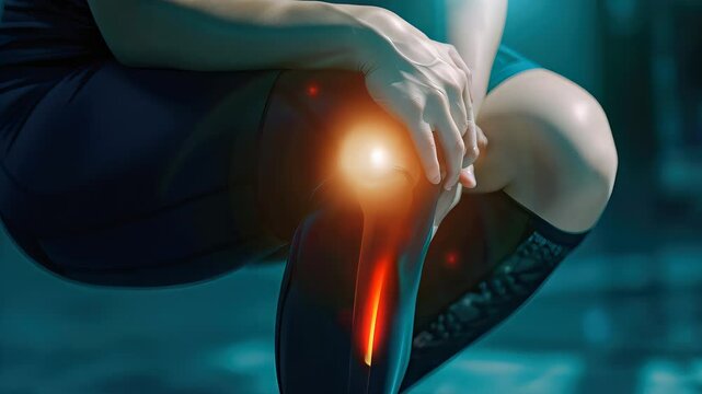
Man holding painful knee with red highlight showing joint pain and injury concept on gray background
00:05
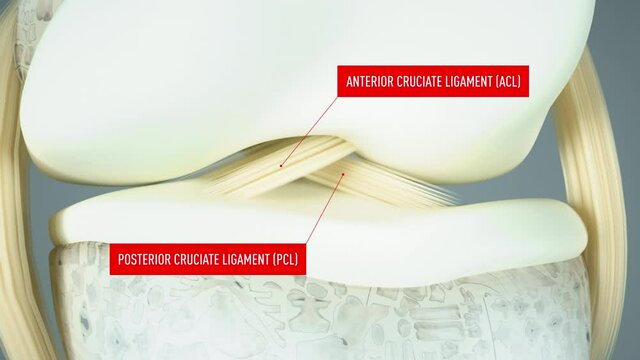
Athletic Knee Joint Anatomy ACL PCL Ligaments Explained with 3D X-ray Animation in Gym Setting
00:10

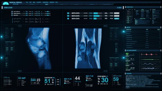
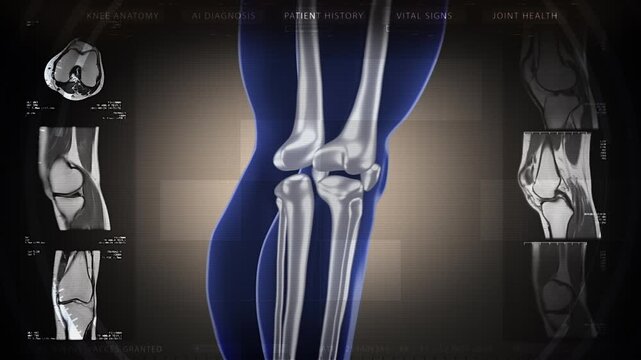
Futuristic Technological Interface Analyzing Human Anatomy. MRI Knee Joint or Magnetic Resonance Imaging of Knee for Diagnosis Sport Trauma. Damage of Ligaments (ACL). Several Healthcare Information
00:15
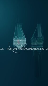
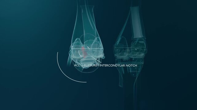
Anterior Cruciate Ligament Rupture
00:11
Complete Knee Anatomy, Medial Ligaments, Meniscus, Patella, knee joint, cruciate ligament of the knee, knee surgery, meniscus, hip, 3D render of human anatomy, 3d illustration
00:10
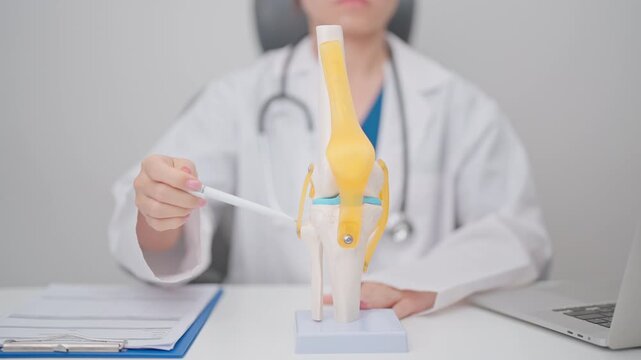
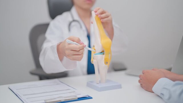
Doctor with Human Knee joint anatomy model. Osteoarthritis, Patellofemoral Pain Syndrome, Patellar Tendinitis, ACL, Anterior Cruciate Ligament, PCL, Posterior, MCL, Medial Collateral, LCL and Lateral
00:13
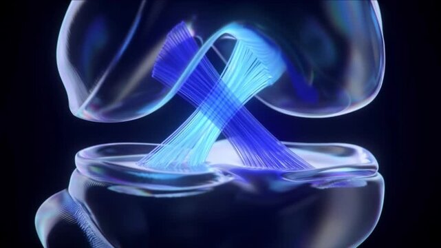
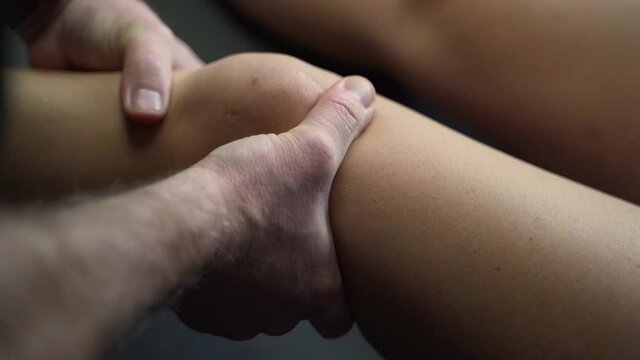
Knee pain, joint inflammation, pain relief. Chronic discomfort. Ligaments tear during excessive exercise. Cartilage degradation limits mobility. Arthritis symptoms increase joint pain.
00:10
Hands, knee and architect man with injury, joint and leg inflammation from arthritis. Construction worker, acl pain and tension from manual labour for male person and medical accident on job site
00:06
A 3D medical animation of an ACL tear in the human knee joint, showing the ligament rupture from a twisting motion in slow motion.
00:10
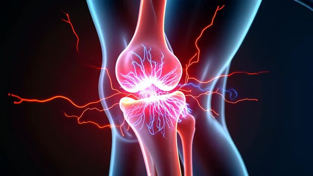
Female athlete suffering from a leg injury with a red painful joint inflammation after physical exercise. The concept of trauma, arthritis, and orthopedic problems in runners
00:05
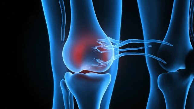
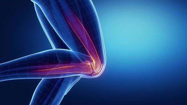
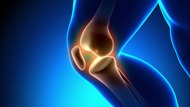
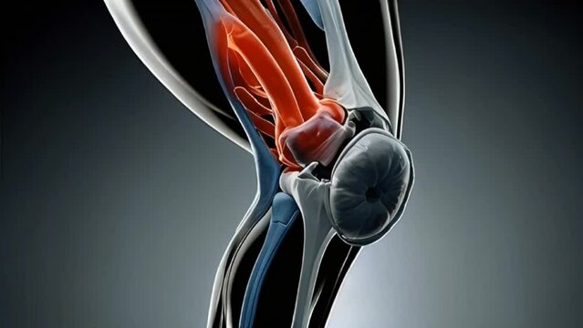
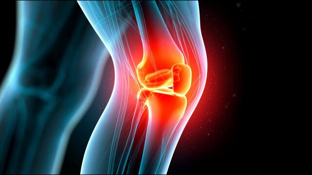
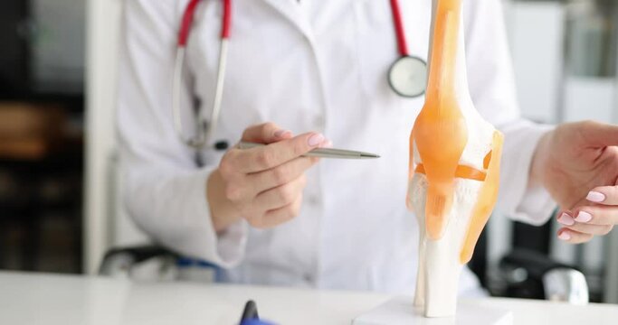
Detailed medical animation of knee joint and ligament pain. Red glow indicates inflammation in the patella and cruciate ligaments.
00:06
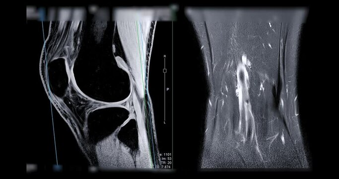
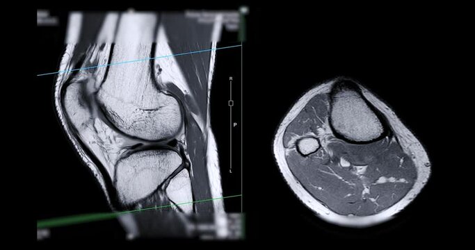
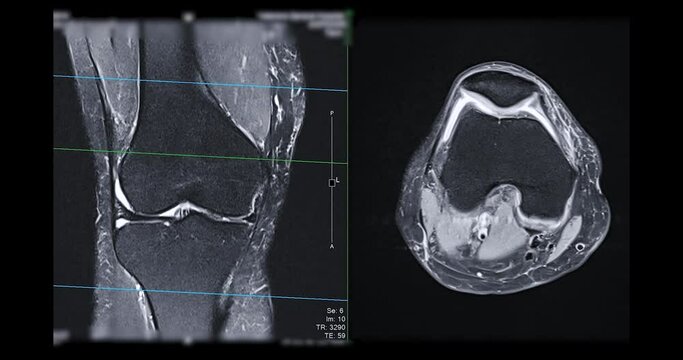
MRI knee or Magnetic resonance imaging of knee joint   for detect acl ligament tear.
00:18
Visualizing an ACL tear with emphasis on knee instability and swelling, providing a conceptual representation of common orthopedic injuries for educational purposes
00:05
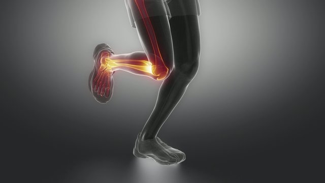
Running woman focused on knee left view
00:09
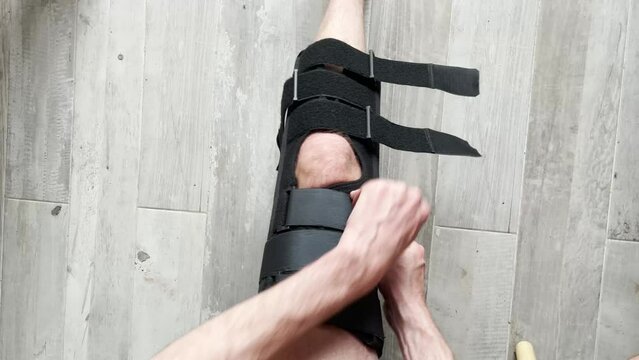
Closeup of knee brace to patient leg on background with copy space
00:13
Human knee joint anatomical illustration of an acl injury
00:08
Scar Skin Knee Physical Therapy Rehab Meniscus ACL Recovery Healing Return Sport Medical Injury Surgery Orthopedic Joint Healthcare Treatment Progress
00:05
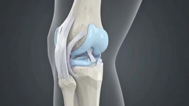
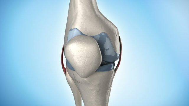
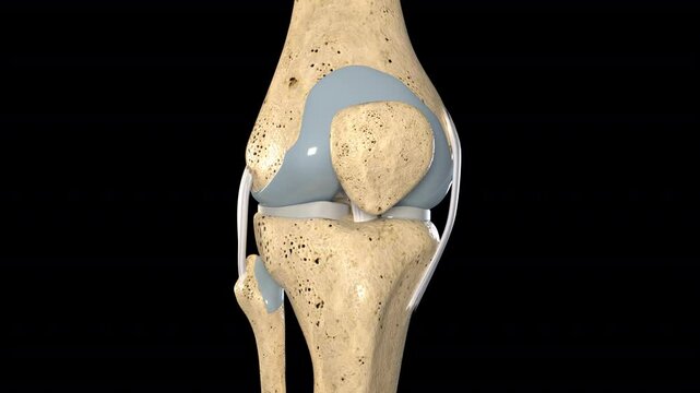
Detailed Medical Illustration of Human Knee Joint Anatomy, Depicting Bones, Cartilage, Ligaments, Including Femur, Tibia, Patella, with Articular Cartilage and Meniscus Highlighted in Blue
00:13
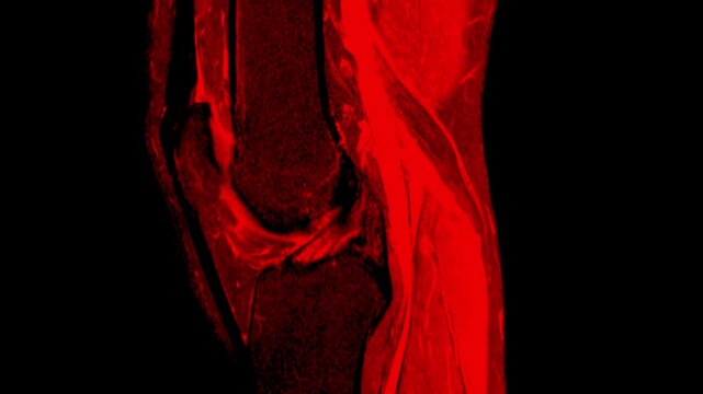
MRI Scan of Right Knee Joint in Red Color Medical Imaging Animation
00:09
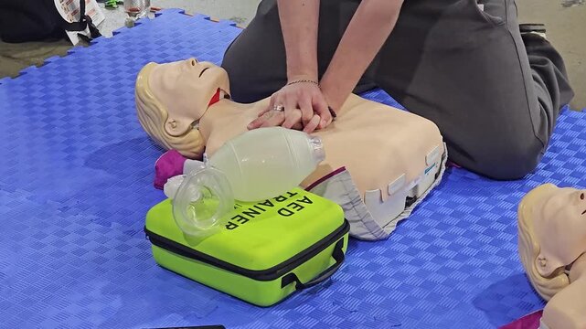
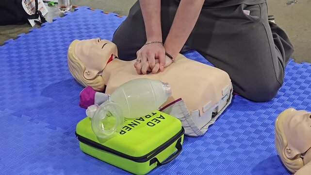
First Aid Training with CPR Manikin Chest Compression and AED Trainer Bag
00:17
Visual Representation of 'ACL P SANCTIONS' Emphasizing Regulatory Measures and Enforcement
00:08
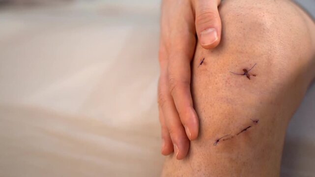
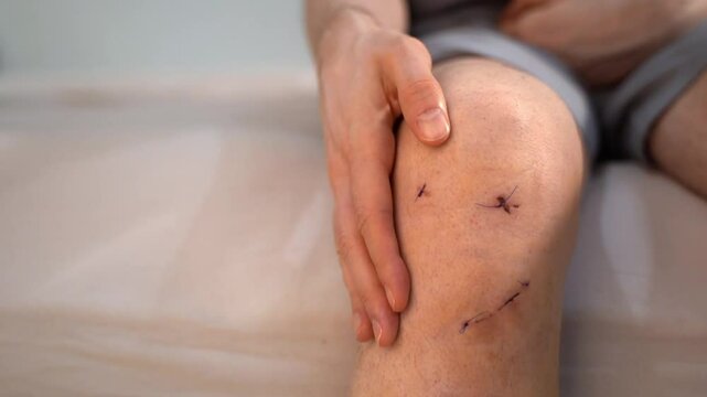
Male hand gently touches healing wound on knee with three stitched surgical incisions after ACL repair, realistic home setting. Concept of sports injury treatment, recovery and postoperative care
00:23
Capital view from Congress Ave in Austin, Texas 12/1/2024
00:28
Ligaments of the Knee Joint
00:38
Waving torn UNIA - ACL flag grunge brush burn light leaks, transparent background alpha channel loop full HD
00:10
Doctor with Human Knee joint anatomy model. Osteoarthritis, Patellofemoral Pain Syndrome, Patellar Tendinitis, ACL, Anterior Cruciate Ligament, PCL, Posterior, MCL, Medial Collateral, LCL and Lateral
00:14
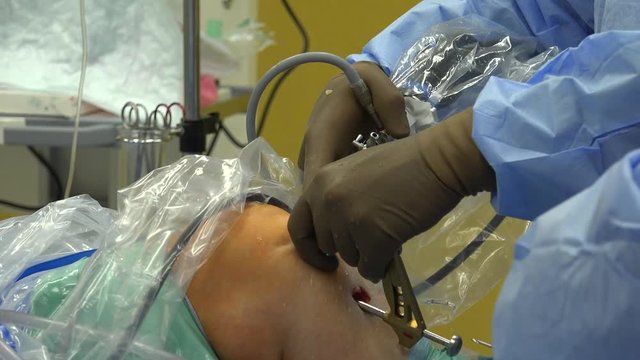
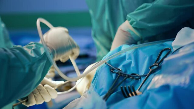
Unrecognized doctor in latex gloves performs operation on patient's knee. Some of the tools are on the patient.
00:09
Delving into the intricate world of the knee joint a vital intersection for mobility and health shown in detailed 3D
00:08
Knee Pain Animation - Understanding Causes and Treatment Options.
00:05
Access Control List concept with interconnected icons on a dark blue background network security authorization
00:10
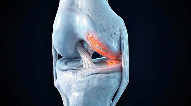
3D Medical Animation of Human Knee Joint Cross Section Showing Cartilage Damage and Glowing Red Orange Inflammation on Femur with Cruciate Ligaments Meniscus and Tibia on Dark Blue Background
00:06
MRI knee or Magnetic resonance imaging of knee joint   for detect acl ligament tear.
00:24
Knee Pain: Visual Effects Concept of Person Wincing with Hand on Knee, Indicating Knee Pain or Injury
00:08
ACL tear, knee injury, rehabilitation support. Joint recovery. Ligaments heal after severe stress. Therapy enhances stability improvement. Recovery process promotes long-term health.
00:10
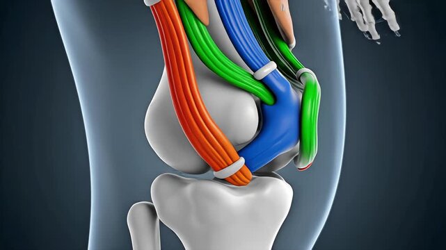
3D anatomical visualization of human knee joint showing color-coded ligaments and bones, rotating medical animation for orthopedic education
00:07
Medical 3D Illustration of Human Knee Joint and ACL Injury
00:08
Physical therapy knee acl meniscus rehab return sport with male hip joint alignment pain relief focus for safer lower body recovery and confident
00:05
Visual representation of ACL tear with knee instability and sweling highlighted. seamless overlay 4k virtual video animation background
00:04
3D medical animation of ACL ligament tear in the knee
00:09
膝関節の痛みと炎症を示す医療図
00:10
Detailed 3D Human Knee Joint Anatomy Showing Cartilage Bone Structure Visualization
00:13
Close-up of man calmly touching knee with stitches on wound after ACL surgery. Concept of gentle reassurance, body awareness, optimistic approach to recovery following orthopedic sports-related trauma
00:18
Close-up of middle-aged Asian male hands performing cardiopulmonary resuscitation (CPR) on a training dummy. Vital techniques include defibrillation, chest compressions, and maintaining the airway.
00:08
Anterior Cruciate Ligament Repair Showing Graft Placement Through Bone Tunnels and Restored Knee Stability in Detailed Anatomy
00:08
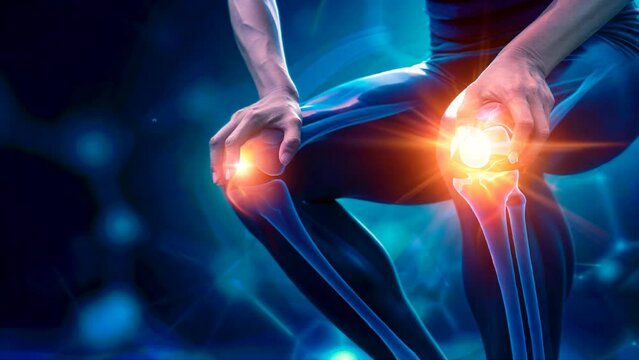
Joint discomfort: visualization of knee pain in patella area highlights challenges of mobility and importance of understanding symptoms for effective treatment, rehabilitation strategies.
00:10
Doctor with Human Knee joint anatomy model. Osteoarthritis, Patellofemoral Pain Syndrome, Patellar Tendinitis, ACL, Anterior Cruciate Ligament, PCL, Posterior, MCL, Medial Collateral, LCL and Lateral
00:13
Biomechanics of the knee joint- flexion and extension
00:36
Man clutching sore knee with red pain highlight showing joint injury and inflammation concept on dark background
00:05
3D Animation of Human Knee Joint Anatomy with Ligaments and Bones on Black Background.
00:08
3D medical animation of a rotating human knee joint. Features a glowing neon blue skeleton with highlighted bright red Medial and Lateral Collateral Ligaments (MCL & LCL) in a futuristic X-ray style.
00:06
3D Medical Animation Human Knee Joint Anatomy Showing Bones Ligaments Cartilage Meniscus Tendon Orthopedic Healthcare Concept Blue Background
00:05
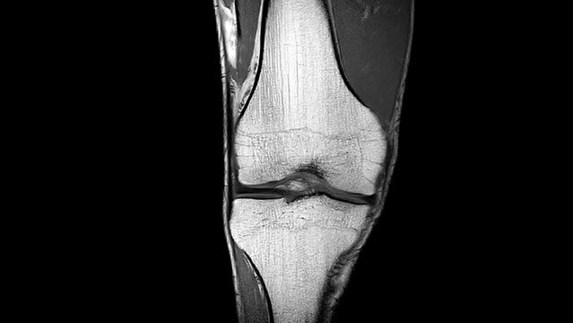
High-Resolution MRI Scan of the Human Knee Joint with Joint Effusion (Coronal  ACL View), showing fluid accumulation and related internal structures for orthopedic diagnosis.
00:11
ACL Tear Trauma: The Knee Instability and Swelling - Picture a person holding their knee, with highlighted instability and swelling, illustrating the knee instability and swelling of an ACL tear
00:12
Bempedoic acid, rotating 3D model of hypercholesterolemia medication, seamless looped video, molecular structure with selective focus
00:15
Detailed 3D Medical Render of Human Knee Joint Anatomy
00:08
3D Medical Animation of Human Knee Anatomy and ACL Injury
00:08
Physical therapy knee acl meniscus rehab return sport drill on turf for confident comeback training and fitness test session
00:05
Human cruciate ligament injury treatment concept. Orthopedist showing to cruciate ligament in a knee-joint medical teaching model, close-up
00:17
3D medical render of ACL ligament tear in the knee joint
00:10
Medical Imaging of Knee Joint Scan for Ligament and Meniscus Injury Assessment
00:13
3D medical animation of ACL ligament stretching and stress.
00:09
Meniscal root tears are less common than meniscal body tears and frequently go undetected
00:13
膝関節の炎症部位を学術的に表現した図
00:10
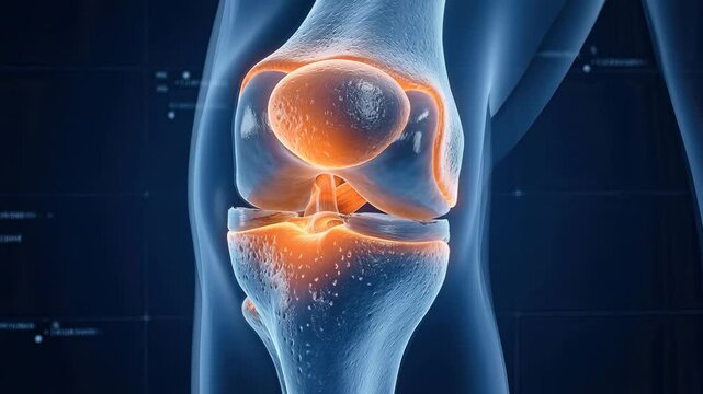
Xray view of human knee joint with inflammation red glow
00:10
Joint discomfort: visualization of knee pain in patella area highlights challenges of mobility and importance of understanding symptoms for effective treatment, rehabilitation strategies.
00:10
Doctor with Human Knee joint anatomy model. Osteoarthritis, Patellofemoral Pain Syndrome, Patellar Tendinitis, ACL, Anterior Cruciate Ligament, PCL, Posterior, MCL, Medial Collateral, LCL and Lateral
00:10
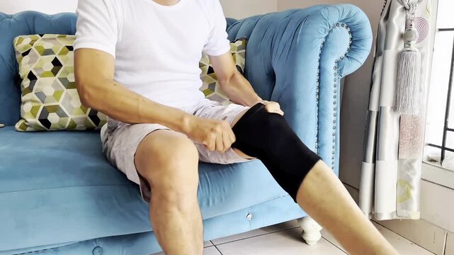
An adult man wears a knee support (knee brace or protector) which functions to stabilize the joint, reduce pain, prevent further injury, and help the recovery process.
00:14
Knee joint displays anatomy with ligament connections in xray view. Medical visualization shows knee bone and ligament structure. Anatomy of knee joint reveals ligament detail. Xray shows knee anatomy
00:07

Futuristic Technological Interface Analyzing Human Anatomy. MRI Knee Joint or Magnetic Resonance Imaging of Knee for Diagnosis Sport Trauma. Damage of Ligaments (ACL). Healthcare Information, EKG.
00:13
Unveiling the Human Knee Anatomy, Structure, and Function
00:08
3D Medical Animation of Human Knee Joint Anatomy with ACL Cruciate Ligament Tear, Meniscus, Cartilage and Inflammation Pain Highlight on Dark Background
00:05
High-Resolution MRI Scan of the Human Knee Joint with Joint Effusion (Coronal View), showing fluid accumulation and related internal structures for orthopedic diagnosis.
00:13
Treatment of human cruciate ligament injuries closeup
00:09
Doctor in surgery for ACL Reconstruction
00:13
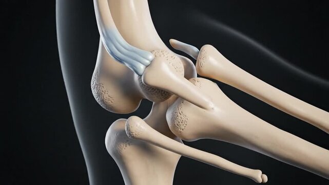
Human skeleton Knee and Foot joint ligaments anatomy 3D Animation
00:12
3D Illustration of ACL Injury and Knee Joint Ligament Inflammation
00:08
Physical therapy knee acl meniscus rehab return to sport concept focus on muscle recovery progress and tendon health during manual treatment
00:05
MRI knee joint. Dynamics of scrolling through MRI images. Magnetic resonance imaging of right knee - view from above. Diagnosis sport trauma and damage of ligaments.
00:09
Pain in the knee. Problems with joints.
00:13
Running woman skeleton scan
00:14
Male hand gently touches his knee wound with healing stitches after anterior cruciate ligament surgery. Concept of postoperative monitoring, injury management and personal care at home. Copy space
00:16
Meniscal root tears are less common than meniscal body tears and frequently go undetected
00:08
4K Austin Texas Skyline USA Time Lapse Drone Shot Cesar Chavez Frost Tower
00:13
Group of surgeons in protective suits and helmets work under the round lamps in surgery room. Team of doctors cooperate at work.
00:17
Joint discomfort: visualization of knee pain in patella area highlights challenges of mobility and importance of understanding symptoms for effective treatment, rehabilitation strategies.
00:10
Doctor with Human Knee joint anatomy model. Osteoarthritis, Patellofemoral Pain Syndrome, Patellar Tendinitis, ACL, Anterior Cruciate Ligament, PCL, Posterior, MCL, Medial Collateral, LCL and Lateral
00:13
knee medical scan footage
00:10
Tilt up of an elderly black woman using an ice pack on her knee to relieve pain
00:11
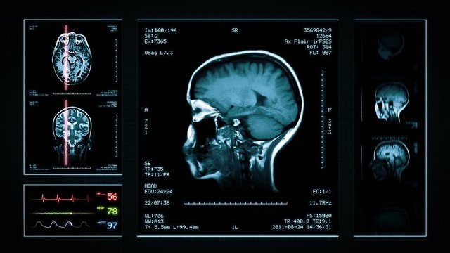
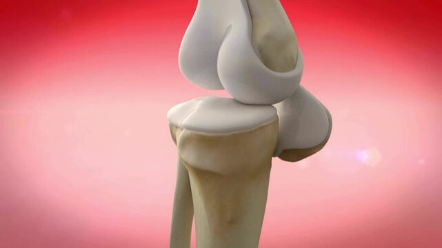
Knee MRI Scan. Blue. 4 videos in 1 file. Animation showing top, front, lateral view and ECG display. Each video is loopable. Medical Background. More options in my portfolio.
00:24
Close-up of middle-aged Asian male hands performing cardiopulmonary resuscitation (CPR) on a training dummy. Vital techniques include defibrillation, chest compressions, and maintaining the airway.
00:17
Front view of senior Golden Labrador Retriever dog walking and limping on a forest path beside a flowing creek.
00:20
High-Resolution MRI Scan of the Human Knee Joint with Joint Effusion (Coronal View), showing fluid accumulation and related internal structures for orthopedic diagnosis.
00:14