Электронное письмо с подтверждением и ссылкой для установки пароля отправлено по адресу Для доступа к покупкам в будущем вам понадобится пароль.
Получите 3 видео или 25 изображений в рамках бесплатной пробной версии
Добавлены новые фильтры
Сортировать по

198 результатов по запросу «proteus» в категории «видео»

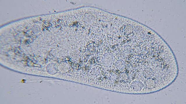
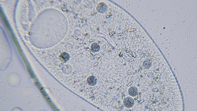
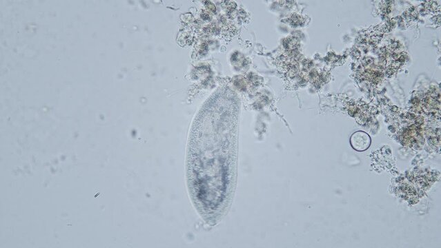
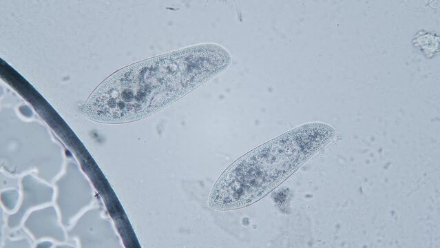
Paramecium large magnification inside organelle movement bright field microscopical view
00:19
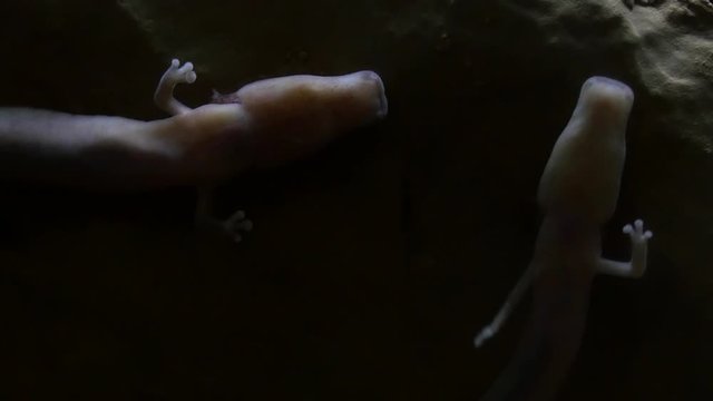
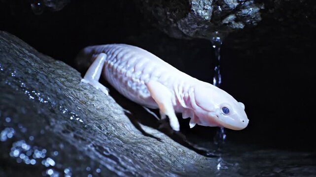
olm or proteus (Proteus anguinus) aquatic salamander in captivity
00:15
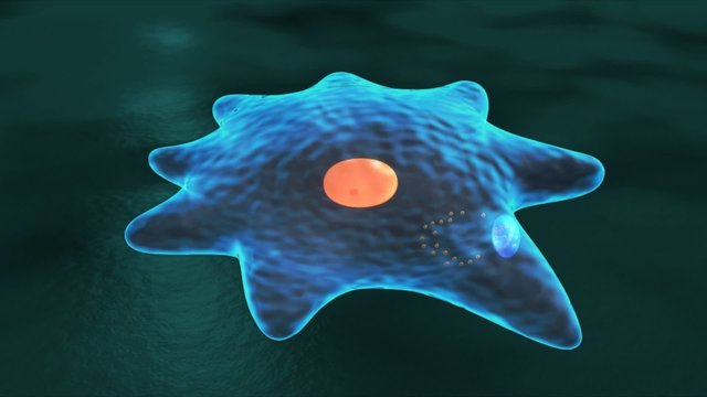
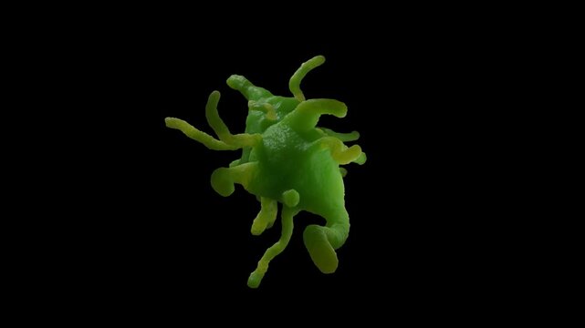
Amoeba
00:14
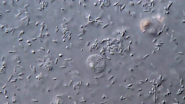
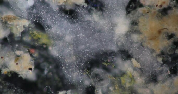
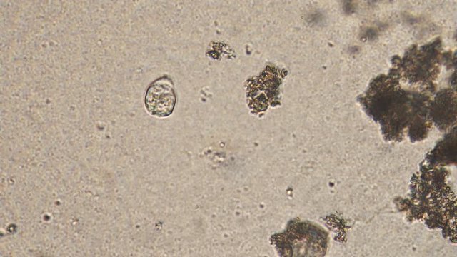
Bacteria and protozoa parasite in feces under microscope
00:23
Microscopic Examination of Bacterial Families in Healthcare Settings
00:09
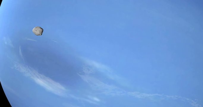
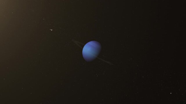
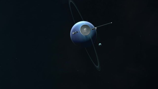
Satellites Proteus and Triton orbiting around Neptune planet
00:20
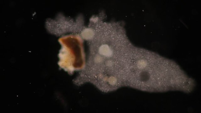
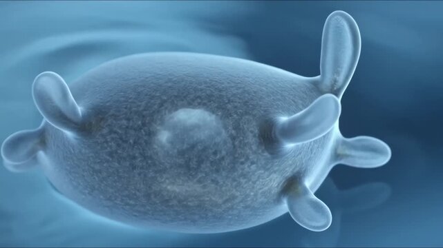
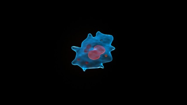
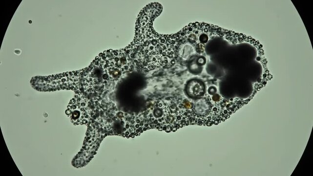
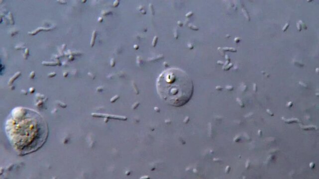
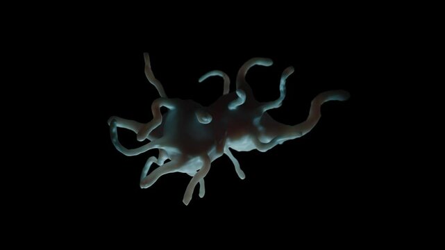
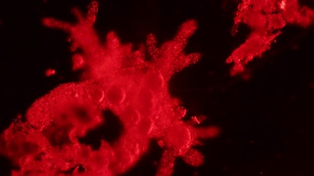
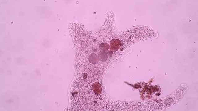
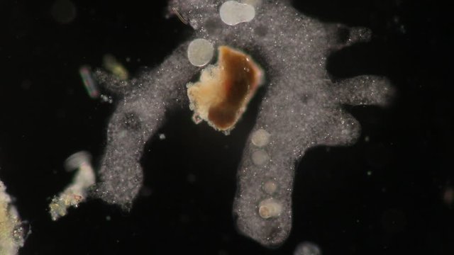
Amoeba proteus protozoan. This freshwater single-celled organism feeds on bacteria and smaller protozoa
00:10
Amoeba proteus / Chaos diffluens - Fortbewegung
01:00
Virus, bacteria, cell infected organism. Hepatitis viruses in infected organism. Gram negativas Proteobacteria, bacteria such as salmonella, escherichia coli, yersinia pestis, klebsiella. 3D animation
00:16
Amoebae move and feed by using pseudopods, which are bulges of cytoplasm formed by the coordinated action of actin microfilaments pushing out the plasma membrane that surrounds the cell.
00:24
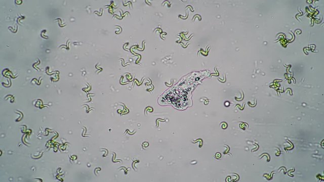
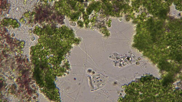
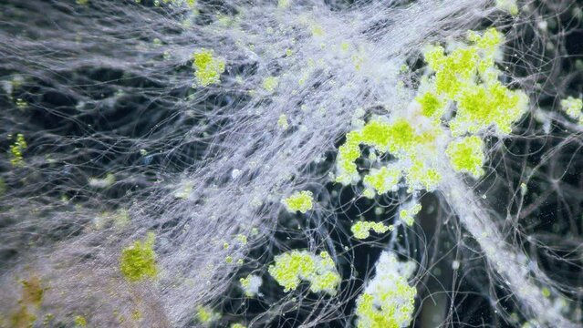
Amoeba moving in the water between the algae close-up. Theme of laboratory biological research under microscope. Microscopic protozoa in a drop of water under magnification. Microcosm close-up.
00:17
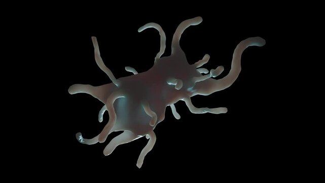
Amoeba proteus this small protozoan uses tentacular protuberances called pseudopodia to move and phagocytose smaller unicellular organisms.
00:24
Microscopic Amoeba Proteus Moving in Water Scientific Macro
00:06
Amoeba. Explain the nutrition process in an Amoeba.
00:12
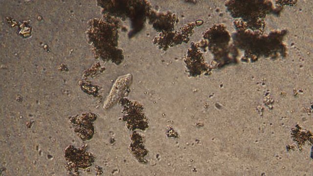
Microscopy of protozoa ciliate and small microorganisms in the water sample. Magnification 150x infusoria under microscope in active movement and feed process.
00:28
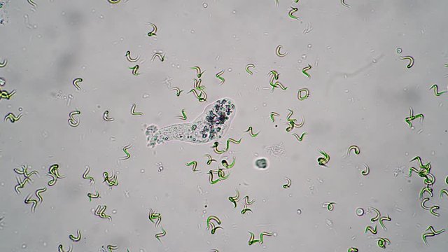
Amoeba and Vorticella hunt among unicellular algae. The incredible and amazing life of microorganisms under the microscope. Bright-field microscopy method
00:29
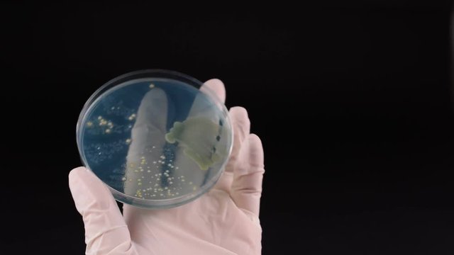
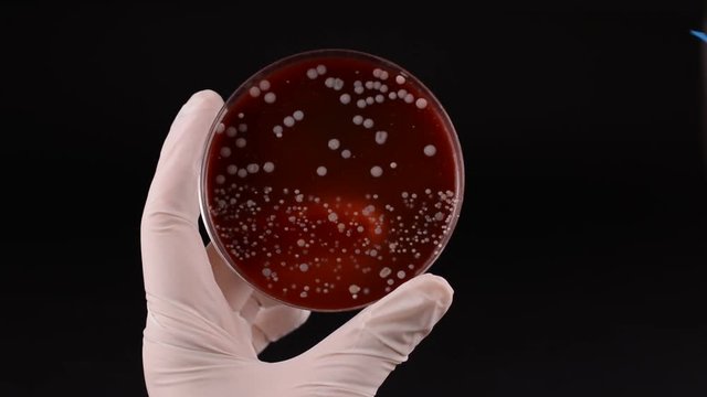
Footage, Scientist hand holding petri dish with colony of Proteus bacteria, a type of gram-negative bacteria that can cause a variety of infections, including UTI.
00:08
Scientist's hand checking petri dish and pointing with inoculation loop Proteus bacteria
00:17
Looped video of Kanamycin A, kanamycin, rotating molecular 3D ball-and-stick model
00:15
Cinematic microscopic time-lapse of bacteria activity A colorful biofilm structure forms. Precise view of Proteus bacteria's role
00:05
Paramecium large magnification inside organelle movement bright field microscopical view
00:22
animation amoeba protozoa unicellular biology. 2d motion
00:10
Amoeba
00:13
Bacteria under microscope showing cell structure
00:22
Neon Spiral Energy Rotating Precise Proteus Abstract Loop
00:05
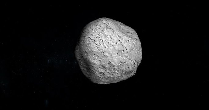
Proteus moon, satellite of Neptune. Loop
00:30
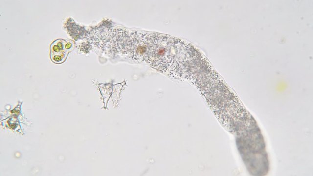
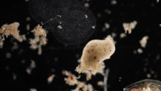
Amoeba proteus is a large, well-known species of single-celled amoeboid protozoan. 1  It is commonly found in freshwater environments like ponds and slow-moving streams
00:05
Amoeba Proteus: Microscopic Single-Celled Organism
00:07
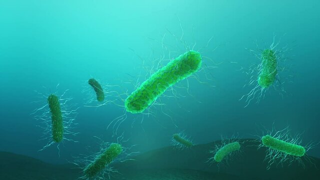
Microscopic view of bacteria showing flagella and pili growing in colonies
00:05
Amoebae move and feed by using pseudopods, which are bulges of cytoplasm formed by the coordinated action of actin microfilaments pushing out the plasma membrane that surrounds the cell.
00:56
Amoeba moving in the water between the algae timelapse. Theme of laboratory biological research under microscope. Microscopic protozoa in a drop of water under magnification. Microcosm close-up.
00:31
Amoeba proteus this small protozoan uses tentacular protuberances called pseudopodia to move and phagocytose smaller unicellular organisms.
00:43
Microscopic View of Bacteria Cells, Medical Science Research Animation Concept
00:10
proteus emerging from the water
00:09
Microscopy of protozoa in the water sample. Magnification 300x microorganism under microscope in active movement.
00:18
Ball-Shaped Lump Of Swarming Tadpoles Amoebas Spermatozoa
00:50
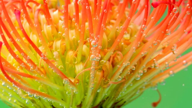
Beautiful flower with stamens under water. Stock footage. Exotic bright flower under water with bubbles. Beautiful flower stamens in clear water with bubbles. Refreshing water with proteus
00:20
Scientist's hand checking petri dish and pointing with inoculation loop Proteus bacteria
00:10
Kanamycin A, rotating 3D model of kanamycin, seamless looped video, molecular structure with selective focus
00:15
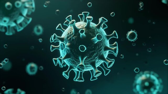
Microscopic view of a contagious virus particle with glowing structures, illustrating the threat of infectious disease and pandemic
00:05
Paramecium large magnification inside organelle movement bright field microscopical view
00:11
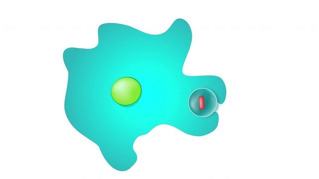
Amoeba Proteus garm bacteria text animation cartoon style background animated video
00:18
Golden lights floating in the air on a green background, creating a vibrant, festive atmosphere with bokeh effect.
00:08
Protozoa microorganism and bacteria under microscope, feces sample
00:42
Footage, Scientist hand holding petri dish with colony of Proteus bacteria, a type of gram-negative bacteria that can cause a variety of infections, including UTI.
00:13
Proteus moon, inner satellite of Neptune planet, orbiting around Neptune planet
00:20
Amoeba proteus protozoan. This freshwater single-celled organism feeds on bacteria and smaller protozoa
00:10
Doctor holding in hand Dysentery
00:15
Aerial sunset view of the town of Marathopoli in Messinia, Peloponese, Greece, with the island of Proti
00:34
Amoebae move and feed by using pseudopods, which are bulges of cytoplasm formed by the coordinated action of actin microfilaments pushing out the plasma membrane that surrounds the cell.
00:21
Albino Cave Salamander Clinging to Damp Rock Face
00:08
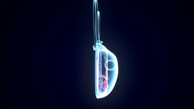
3d illustration of Chlamydomonas anatomy
00:15
The simplest single-celled organism. The amoeba infusoria is moving.
00:12
The microcosm of amoeba moves in water among algae close-up. Theme of laboratory biological research under microscope. Microscopic protozoa in a drop of water under magnification. Microcosm close-up.
00:26
Microscopy of Amoeba limax in the water (pond) sample. Magnification 300x microorganism in active movement.
00:28
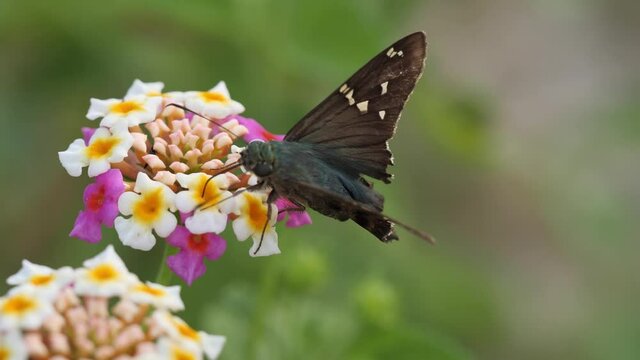
Long-tailed skipper butterfly with damaged tail feeding on lantana
00:14
Beautiful flower with stamens under water. Stock footage. Exotic bright flower under water with bubbles. Beautiful flower stamens in clear water with bubbles. Refreshing water with proteus
00:07
Scientist's hand checking petri dish infected with Staphylococcus aureus bacteria
00:13
Sea gate. The sea washed the entrance to the cliff
00:10
Neptune Moons Seamless Loop
00:15
Approaching Neptune and Moon Titania
00:15
Proteus Mirabilis garm bacteria text animation cartoon style background animated video
00:18
Aerial view of a winding road through dense forest with misty mountains in the background
00:08
White Olm Salamander 3D Render Isolated on Green Screen Backdrop for Chroma Key Visual Effects
00:08
Kanamycin A molecule, rotating 3D model of antibiotic, looped video on a black background
00:15
Satellite Proteus, inner moon of Neptune planet, orbiting around Neptune planet
00:20
Amoeba proteus is a large, well-known species of single-celled amoeboid protozoan. 1  It is commonly found in freshwater environments like ponds and slow-moving streams
00:05
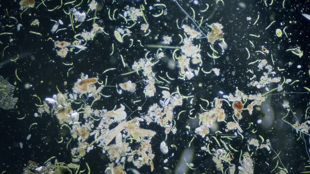
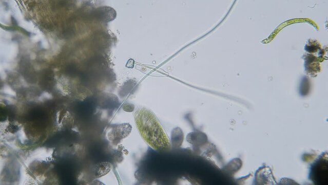
Different freshwater  species of protozoa single cell organisms as stentors, ciliates and algae movement under microscope bright filed view
00:14
Microorganisms ciliates and algae high concentration microscopic dark field view
00:10
Different freshwater  species of protozoa single cell organisms as stentors, ciliates and algae movement under microscope bright filed view
00:20
Microorganisms ciliates and algae high concentration microscopic dark field view
00:10
Different freshwater  species of protozoa single cell organisms as stentors, ciliates and algae movement under microscope bright filed view
00:15
Amoeba proteus is a large, well-known species of single-celled amoeboid protozoan. 1  It is commonly found in freshwater environments like ponds and slow-moving streams
00:05
Different freshwater  species of protozoa single cell organisms as stentors, ciliates and algae movement under microscope dark field view
00:20
Paramecium large magnification inside organelle movement bright field microscopical view
00:13
Amoebae move and feed by using pseudopods, which are bulges of cytoplasm formed by the coordinated action of actin microfilaments pushing out the plasma membrane that surrounds the cell.
00:23
Amoeba proteus is a large, well-known species of single-celled amoeboid protozoan. 1  It is commonly found in freshwater environments like ponds and slow-moving streams
00:05
Amoebae move and feed by using pseudopods, which are bulges of cytoplasm formed by the coordinated action of actin microfilaments pushing out the plasma membrane that surrounds the cell.
00:20
Amoebae move and feed by using pseudopods, which are bulges of cytoplasm formed by the coordinated action of actin microfilaments pushing out the plasma membrane that surrounds the cell.
00:18
Different freshwater  species of protozoa single cell organisms as stentors, ciliates and algae movement under microscope bright filed view
00:11
Amoebae move and feed by using pseudopods, which are bulges of cytoplasm formed by the coordinated action of actin microfilaments pushing out the plasma membrane that surrounds the cell.
00:28
Paramecium large magnification inside organelle movement bright field microscopical view
00:14
Different freshwater  species of protozoa single cell organisms as stentors, ciliates and algae movement under microscope bright filed view
00:12
Paramecium large magnification inside organelle movement bright field microscopical view
00:10
Eating typical German bockwurst sausage and homemade potato salad close up personal perspective
00:25
Amoebae move and feed by using pseudopods, which are bulges of cytoplasm formed by the coordinated action of actin microfilaments pushing out the plasma membrane that surrounds the cell.
00:40
Voyager passing Neptune and It's Moon
00:15
Amoebae move and feed by using pseudopods, which are bulges of cytoplasm formed by the coordinated action of actin microfilaments pushing out the plasma membrane that surrounds the cell.
00:29
Amoebae move and feed by using pseudopods, which are bulges of cytoplasm formed by the coordinated action of actin microfilaments pushing out the plasma membrane that surrounds the cell.
00:21
Amoebae move and feed by using pseudopods, which are bulges of cytoplasm formed by the coordinated action of actin microfilaments pushing out the plasma membrane that surrounds the cell.
00:27
Amoebae move and feed by using pseudopods, which are bulges of cytoplasm formed by the coordinated action of actin microfilaments pushing out the plasma membrane that surrounds the cell.
00:24
Amoebae move and feed by using pseudopods, which are bulges of cytoplasm formed by the coordinated action of actin microfilaments pushing out the plasma membrane that surrounds the cell.
01:06
Microscopy of protozoa ciliate and small microorganisms in the water sample. Magnification 300x infusoria under microscope in active movement and feed process.
00:12
Amoeba proteus protozoan. This freshwater single-celled organism feeds on bacteria and smaller protozoa
00:10
Amoebae move and feed by using pseudopods, which are bulges of cytoplasm formed by the coordinated action of actin microfilaments pushing out the plasma membrane that surrounds the cell.
00:23
Microscopy of protozoa ciliate and small microorganisms in the water sample. Magnification 300x infusoria under microscope in active movement and feed process.
00:16
Amoebae move and feed by using pseudopods, which are bulges of cytoplasm formed by the coordinated action of actin microfilaments pushing out the plasma membrane that surrounds the cell.
00:33
Amoebae move and feed by using pseudopods, which are bulges of cytoplasm formed by the coordinated action of actin microfilaments pushing out the plasma membrane that surrounds the cell.
00:29
Microorganisms ciliates and algae high concentration microscopic dark field view
00:10
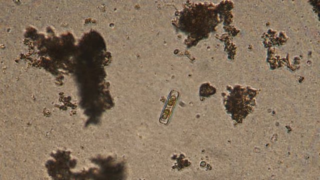
Microscopy of Diatoms. Magnification 150x of unicellular organism. Cell walls made of glass and pennales tend to be elongated.
00:20