판매
가격
암호 설정을 위한 링크가 다음으로 전송됨:
나중에 구매 기능을 이용하려면 암호가 필요합니다.
모두
이미지
비디오
오디오
템플릿
3D
무료
Premium
유사 항목 찾기:
시리즈:
모델:
문서:
내 라이브러리
유사 항목 찾기.
여기로 이미지 드래그
또는
검색
이미지 업로드 중...
무제한 다운로드와 강력한 AI 편집 기능으로 제약 없이 창작해 보세요
무제한으로 스톡 이용
무제한 다운로드와 강력한 AI 편집 기능으로 제약 없이 창작해 보세요
무제한으로 스톡 이용
패널 숨기기
패널 보기
새 필터 추가됨
정렬 기준
비디오에서
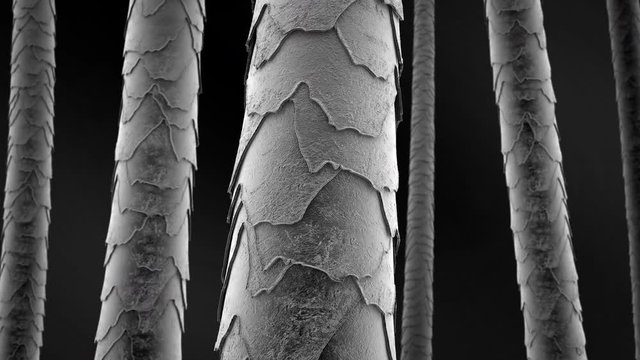
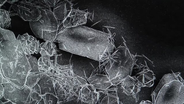
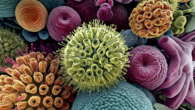
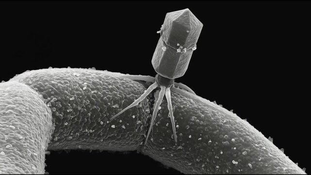
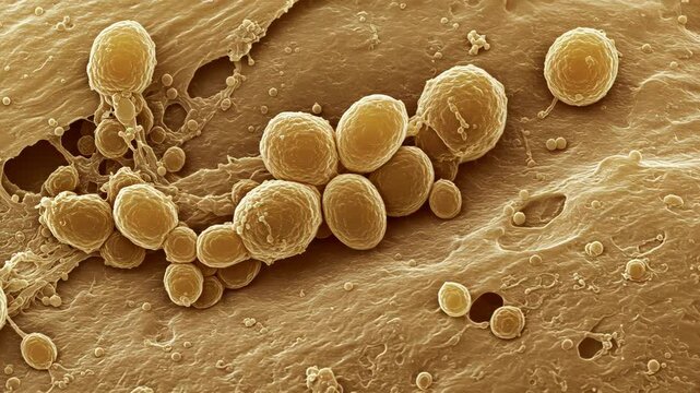
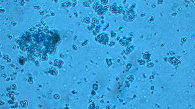
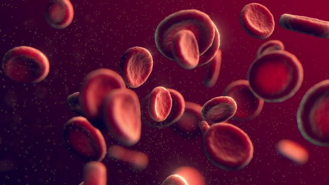
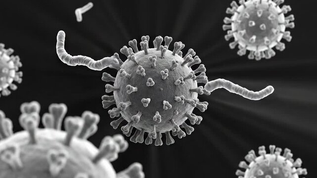
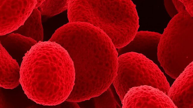
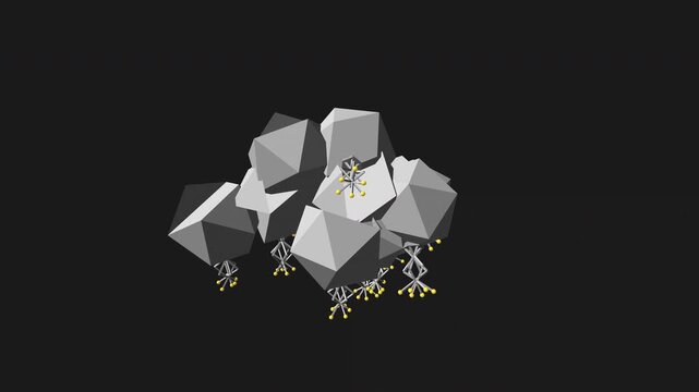
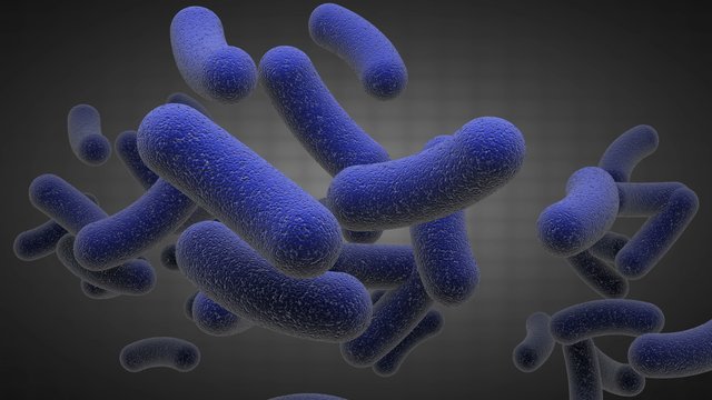
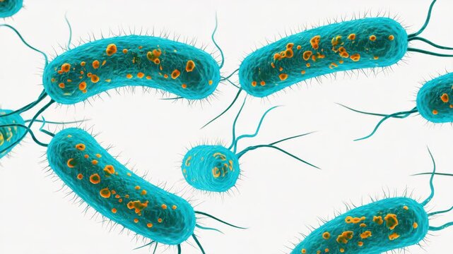
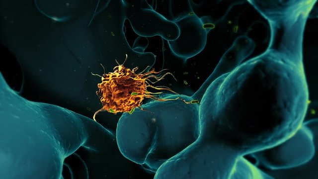
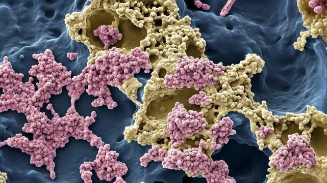
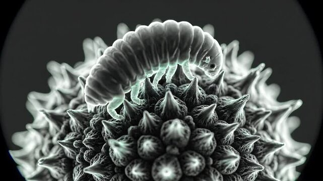
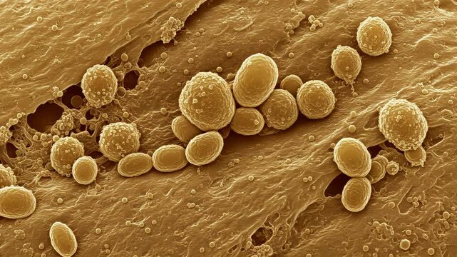
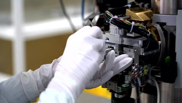
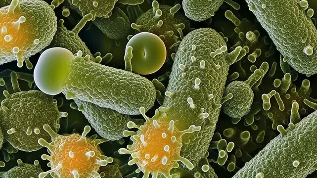
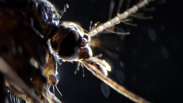
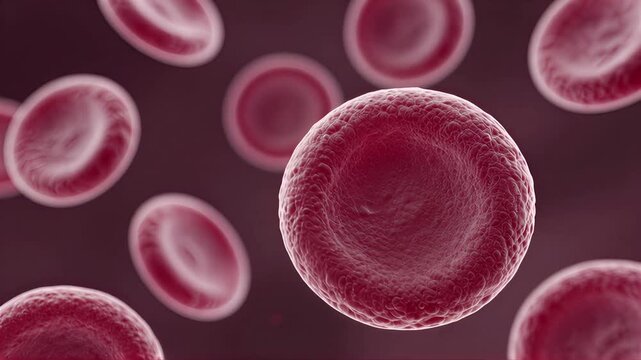
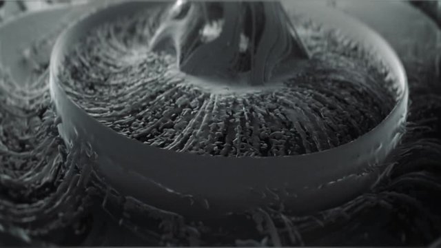
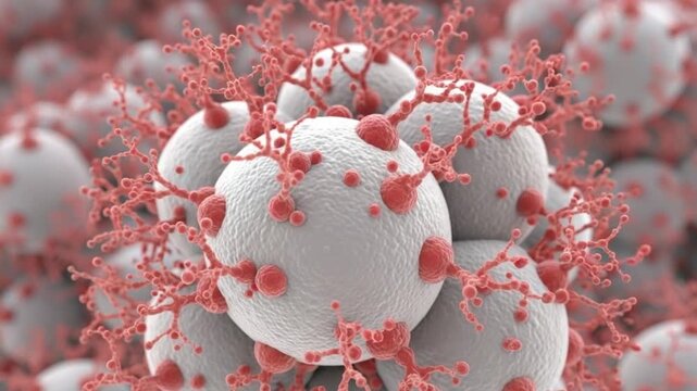
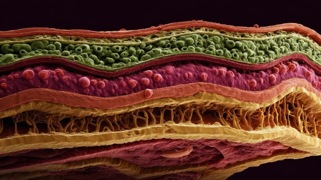
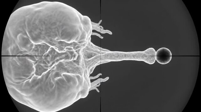
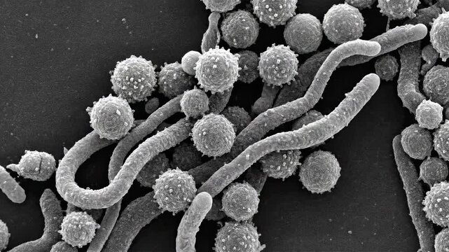
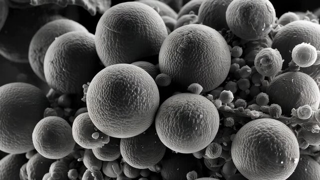
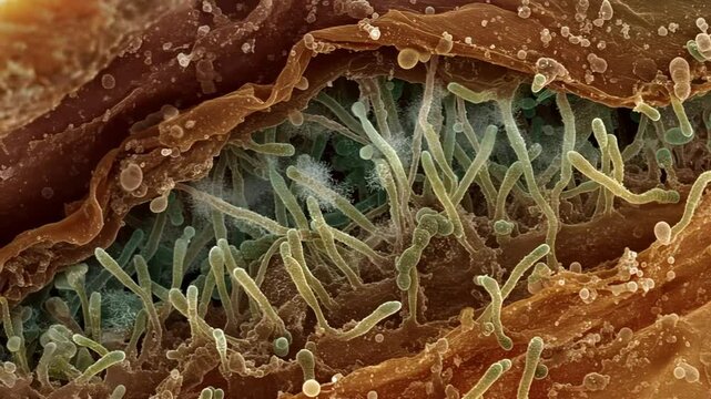
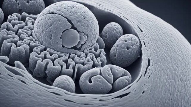
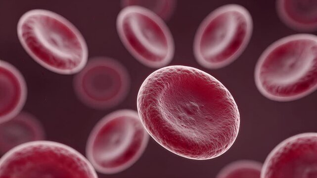
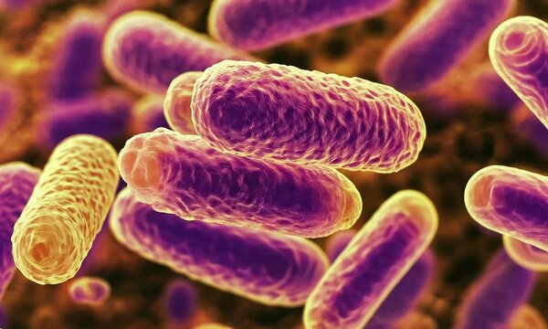
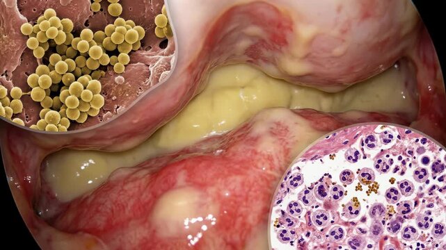
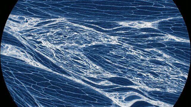
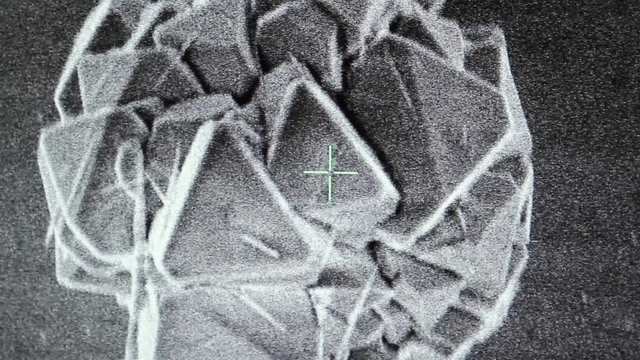
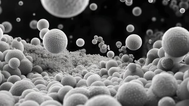
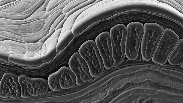
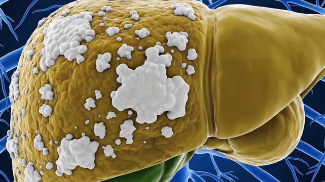
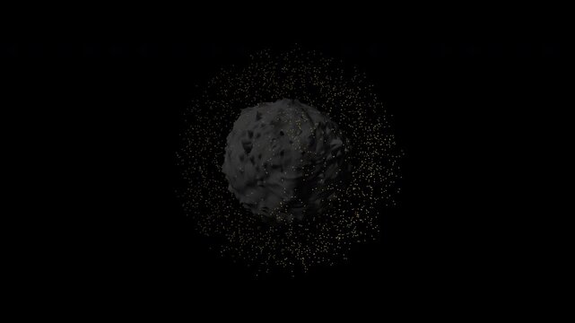
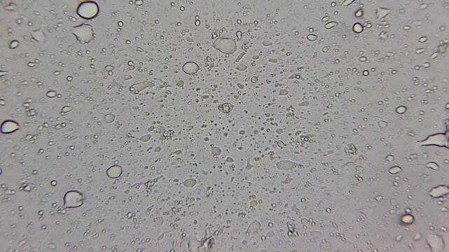
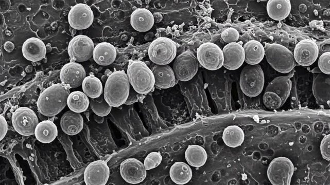
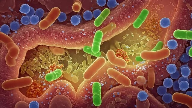
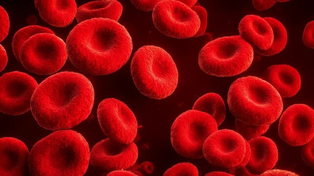
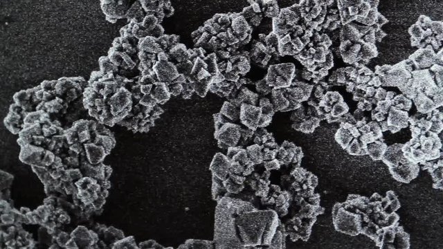
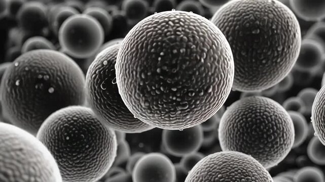
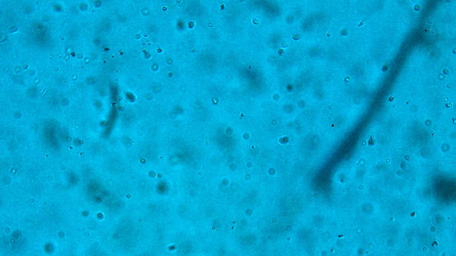
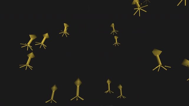
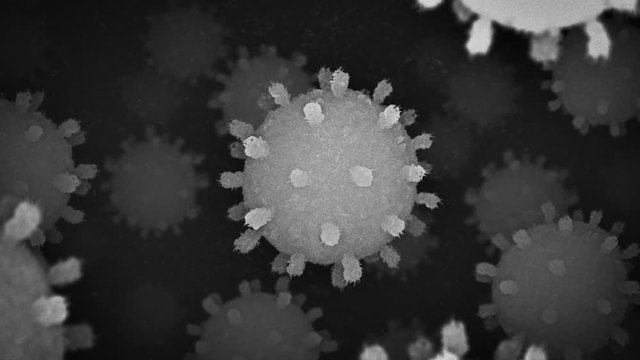
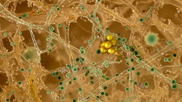
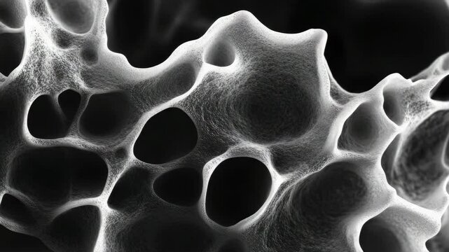
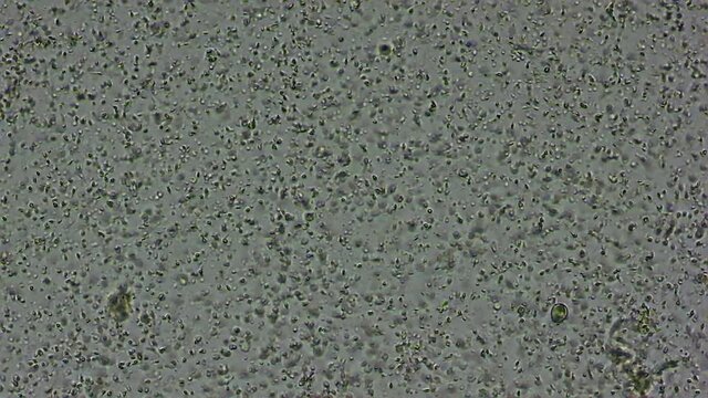
sem microscope
에 관한 358개 결과
00:20
4K
HD
00:13
4K
HD
00:20
4K
HD
00:05
4K
HD
00:05
4K
HD
00:10
4K
HD
00:05
4K
HD
00:13
4K
HD
00:10
4K
HD
00:06
4K
HD
00:08
4K
HD
00:08
4K
HD
00:10
4K
HD
00:05
4K
HD
00:12
4K
HD
00:20
4K
HD
00:10
4K
HD
00:30
4K
HD
00:07
4K
HD
00:10
4K
HD
00:10
4K
HD
00:24
4K
HD
00:20
4K
HD
00:10
4K
HD
00:08
4K
HD
00:10
4K
HD
00:14
4K
HD
00:08
4K
HD
00:05
4K
HD
00:09
4K
HD
00:05
4K
HD
00:05
4K
HD
00:05
4K
HD
00:11
4K
HD
00:05
4K
HD
00:07
4K
HD
00:10
4K
HD
00:06
4K
HD
00:10
4K
HD
00:05
4K
HD
00:10
4K
HD
00:22
4K
HD
00:07
4K
HD
00:07
4K
HD
00:08
4K
HD
00:10
4K
HD
00:06
4K
HD
00:08
4K
HD
00:05
4K
HD
00:10
4K
HD
00:10
4K
HD
00:19
4K
HD
00:08
4K
HD
00:10
4K
HD
00:05
4K
HD
00:07
4K
HD
00:10
4K
HD
00:20
4K
HD
00:08
4K
HD
00:10
4K
HD
00:08
4K
HD
00:36
4K
HD
00:04
4K
HD
00:05
4K
HD
00:14
4K
HD
00:10
4K
HD
00:05
4K
HD
00:08
4K
HD
00:08
4K
HD
00:12
4K
HD
00:08
4K
HD
00:05
4K
HD
00:10
4K
HD
00:07
4K
HD
00:08
4K
HD
00:07
4K
HD
00:10
4K
HD
00:31
4K
HD
00:10
4K
HD
00:05
4K
HD
00:08
4K
HD
00:32
4K
HD
00:10
4K
HD
00:08
4K
HD
00:10
4K
HD
00:10
4K
HD
00:06
4K
HD
00:12
4K
HD
00:13
4K
HD
00:12
4K
HD
00:14
4K
HD
00:10
4K
HD
00:10
4K
HD
00:15
4K
HD
00:15
4K
HD
00:07
4K
HD
00:20
4K
HD
00:20
4K
HD
00:05
4K
HD
00:50
4K
HD
확인
확인
확인
지역 선택
지역을 선택하면 Adobe Stock 웹 사이트에 표시되는 언어와 프로모션 내용이 변경될 수 있습니다.
북아메리카
Canada - English
Canada - Français
El Salvador
México
República Dominicana
United States
남아메리카
Argentina
Bahamas
Barbados
Bolivia
Brasil
Chile
Colombia
Costa Rica
Ecuador
Guatemala
Honduras
Jamaica
Nicaragua - English
Panamá
Paraguay
Perú
Trinidad and Tobago
Uruguay
Venezuela
유럽, 중동 및 아프리카
Algeria - English
Armenia - English
Azerbaijan - English
Bahrain - English
Belgium - English
Belgique - Français
België - Nederlands
Česká republika
Croatia - English
Cyprus - English
Danmark
Georgia - English
Deutschland
Eesti
Egypt - English
España
France
Greece - English
Iceland - English
Ireland
Israel - English
Italia
Jordan - English
Казахстан
Kenya - English
Kuwait - English
Киргизия
Latvija
Lebanon - English
Lietuva
Luxembourg - Deutsch
Luxembourg - English
Luxembourg - Français
Mauritius - English
Moldova - English
Hungary - English
Malta - English
Morocco - English
Nederland
Nigeria
Norge
Oman - English
Österreich
Россия
Polska
Portugal
Qatar - English
România
Saudi Arabia - English
Schweiz
Slovenija
Slovensko
Suisse
Suomi
Sverige
Svizzera
Таджикистан
Turkey - English
Turkmenistan - English
UAE - English
Україна
United Kingdom
Узбекистан
България
아시아 태평양
Australia
Indonesia - English
中國香港特別行政區
India
日本
한국
中華人民共和國澳門特別行政區
Malaysia - English
New Zealand
ไทย
Philippines
Singapore
Sri Lanka - English
台灣地區
Vietnam - English