암호 설정을 위한 링크가 다음으로 전송됨: 나중에 구매 기능을 이용하려면 암호가 필요합니다.
무제한 다운로드와 강력한 AI 편집 기능으로 제약 없이 창작해 보세요
새 필터 추가됨
정렬 기준

비디오에서 microscopy에 관한 31,090개 결과

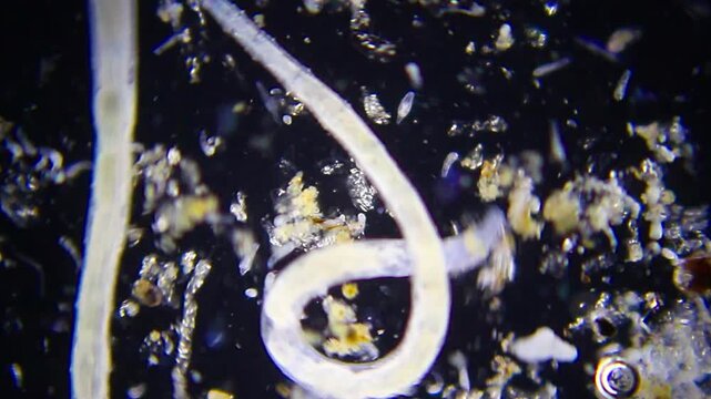
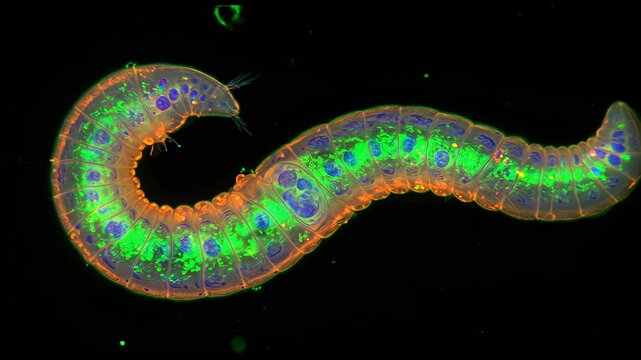
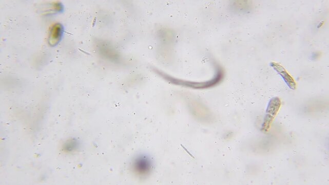
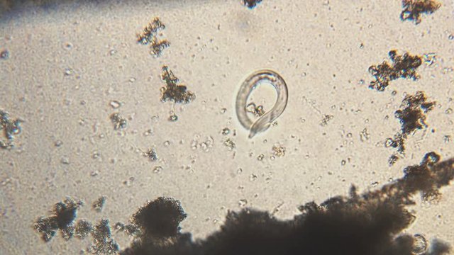
Annelids moving with rotifer colony in background under 40x magnification darkfield microscopy. 4K scientific microscopy footage.
00:27
HD
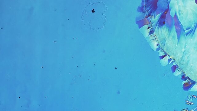
Real time swift crystal growth under polarized light microscopy showing energetic fluid movement and expanding birefringent structures filling space completely
00:20
HD
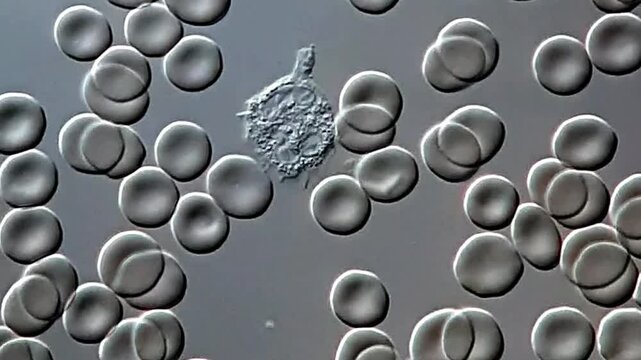
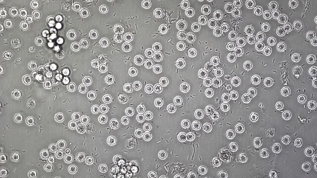
White blood cells under microscopy view, WBC under microscope
00:11
HD
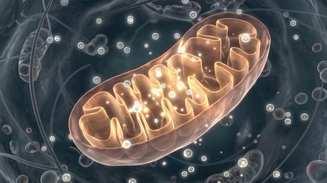
Cinematic 3D microscopic human cell animation, glowing mitochondria generating energy particles, detailed organelles, vibrant biological activity, metabolism and biotechnology concept, 4K video
00:10
HD
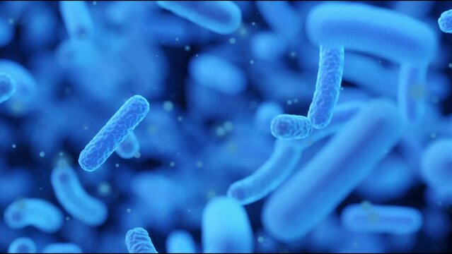
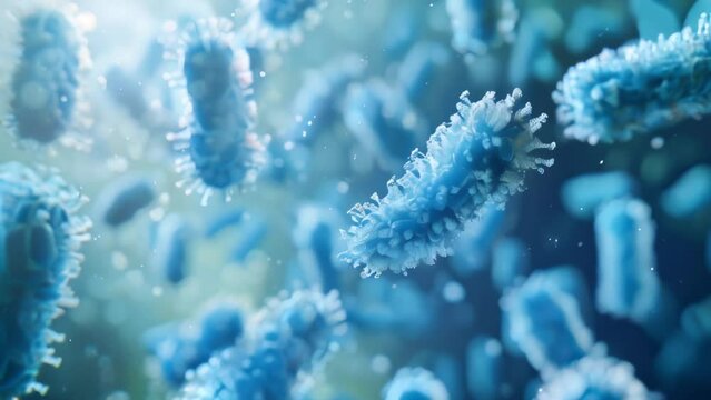
Resistance to antibacterial agents, growth of bacteria in the intestines, microscopy of bacteria
00:10
HD
A brightfield microscopy image of hepatocytes revealing their dual function in detoxification and the production of essential proteins such as albumin and clotting factors.
00:07
HD
Closeup of microscope with lens and laboratory glass setup
00:14
HD
Fluorescent microscopy of Caenorhabditis elegans, with glowing neurons and transparent body structure under lab lighting
00:10
HD
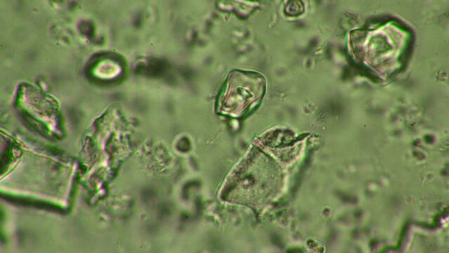
Microscopic examination of a probiotic at 1000x zoom, revealing dynamic crystals and bacteria aiding digestion.
00:22
HD
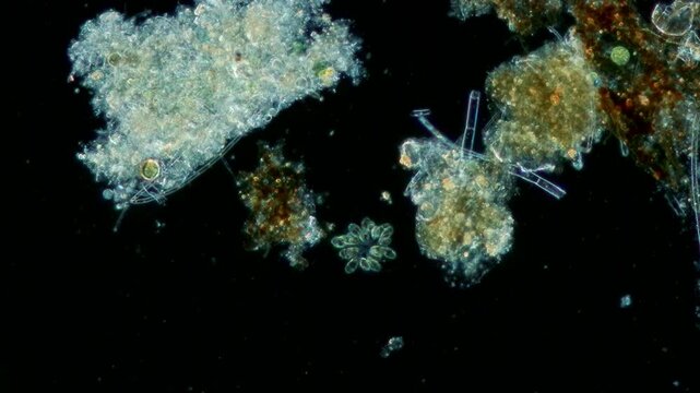
Synura colony at 10x under darkfield microscopy
00:16
HD
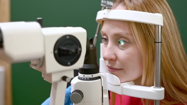
Examination of fundus of woman by ophthalmologist using retinal scanner. Patient during visit to optometrist for microscopic analysis of eyelids, sclera, conjunctiva, iris, lens, cornea. Eye exam
00:22
HD
A close-up image of green algae under a microscope. The algae is visible in various shapes and sizes
00:10
HD
4K Glowing Neuron Brain Cell Neural Network Medical Science
00:06
HD
Exploring cellular structures with confocal microscopy in a laboratory setting
00:10
HD
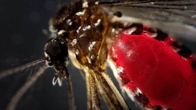
Darkfield microscopy of a mosquito filled with human blood after feeding
00:14
HD
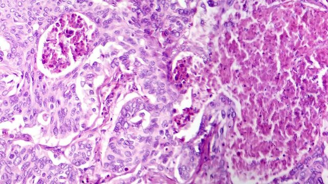
Light microscopy of lung adenocarcinoma, a malignant tumor originating from glandular cells in the lung. Histologically, it shows atypical glandular structures, and increased mitotic activity.
00:16
HD
Research professional conducting experiments with laboratory equipment and team
00:10
HD
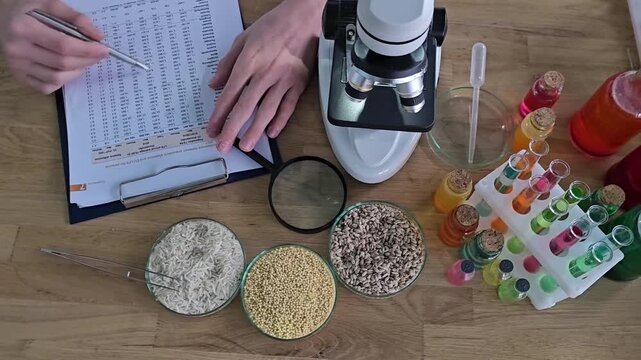
Scientific analysis of food samples in a laboratory with detailed observations and microscopy
00:13
HD
Two female scientists conducting chemical experiments and microscopy in a modern laboratory setting
00:21
HD
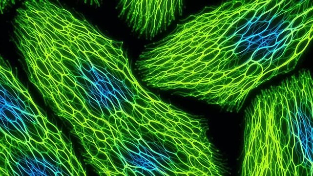
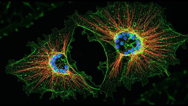
Fibroblast cell cytoskeleton fluorescence microscopy green filament network blue nucleus closeup scientific cell biology tissue sample pattern texture microscopic research
00:05
HD
Nematode worm wriggling among swimming rotifers in liquid medium under 100x brightfield microscopy
00:18
HD
Real time opposing crystal growth under polarized light microscopy showing rapid deceleration and leaving empty liquid gap between two beautiful birefringent structures
00:14
HD
Movement of Euglena and rotifer under microscopy
00:18
HD
Microscopic view of biological cells multiplying and dividing in organism
00:11
HD
Laboratory slide revealing cells top view under fluorescent microscopy green and blue structures for biomedical research, pathology analysis, microbiology education, diagnostic imaging, tissue study
00:05
HD
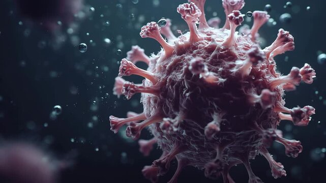
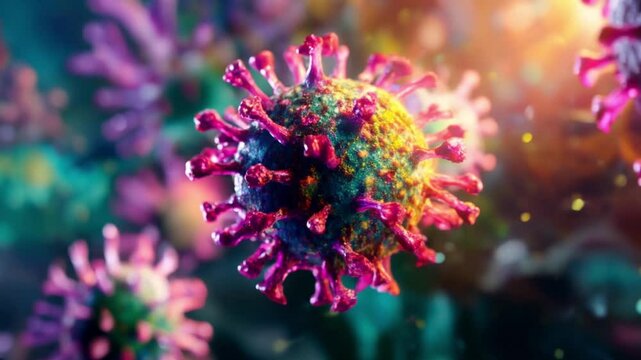
4K Ultra HD 3d render of a coronavirus sarscov2 virus cell, microscopic view of a pathogen causing respiratory illness, medical science concept video
00:10
HD
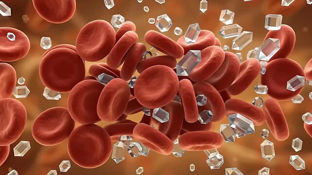
3D Medical Animation of Glucose Crystals Accumulating Among Red Blood Cells in Hyperglycemia Onset
00:06
HD
Fluorescence microscopy of actin filaments v1
00:10
HD
Molecule chemistry science cell biotechnology molecular nad longevity research laboratory featuring glowing molecular structure near cell surface and membrane DNA microscopy genetics medical biology
00:08
HD
Scientist using a pipette near a microscope with test tubes and blue liquid in a modern laboratory
00:10
HD
A fluorescent microscopy image of melanocytes responsible for producing melanin to protect the skin from UV radiation.
00:08
HD
Female Scientist Compares Microscope Slides With AI Infographics on Lab Computer in a Bright Biomedical laboratory, lab, Advancing Disease Cure, Biomarker Discovery and Precision Medicine. Static Shot
00:11
HD
Mitochondrial-Targeted Nanoparticles Delivering Therapy in Cancer Cells
00:05
HD
3D Animation of Microscopic View of Stem Cells Self-Replicating and Proliferating within Body. Medical Illustration.
00:05
HD
Hydrogen peroxide reaction creating oxygen bubbles under darkfield microscopy background
00:15
HD
A neuron cell is illuminated with a golden glow amidst a complex network of neural connections, showcasing the intricate details of the human brain's functionality in a 3D visualization.
00:05
HD
Paramecium and other microorganisms under the microscope at 100x Magnification
00:19
HD
Lithium Deposition Under Transmission Electron Microscopy
00:05
HD
Scientific animation explaining atomic force microscopy AFM showing laser probe reflecting off a cantilever to map 3D atomic topography and nanometer elevation of a substrate lattice
00:59
HD
Retinal Oxidative Stress Visualization - Bright Orange Reactive Particles Within Cells In a Detailed Neural Network Framework in 4K
00:08
HD
Large segmented annelid worm crawling through organic sediment under 40x darkfield microscopy
00:14
HD
Real time high speed micro crystal stabilization under polarized light microscopy showing particles turning into three dimensional structures with shifting polariser rotation
00:20
HD
Movement of protozoa under microscopy  view
00:18
HD
Professional in protective gear evaluating glass with advanced tools
00:10
HD
High-resolution visualization of a glowing green and purple DNA double helix twisting in 3D space with floating particles and bokeh, cinematic molecular genetic imagery
00:10
HD
Molecular Nanostructure Evolution of L-Glutamate Polymorphs in Solution
00:05
HD
Quantum Diamond NV Center Magnetic Field Measurement
00:05
HD
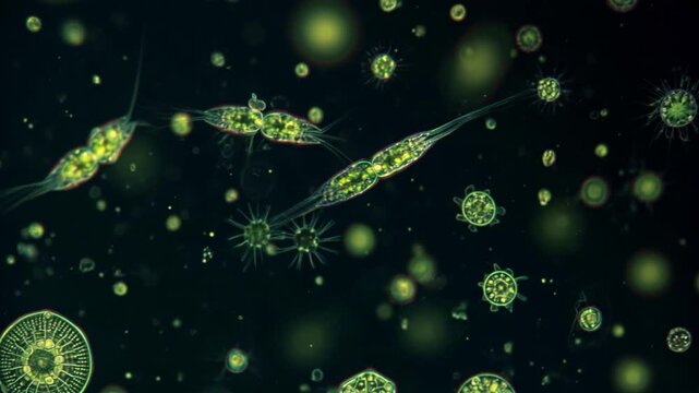
3D animation: Green glowing plankton, algae, and diatoms float in dark water. Microscopic aquatic life, darkfield microscopy style. Science, biology visualization.
00:10
HD
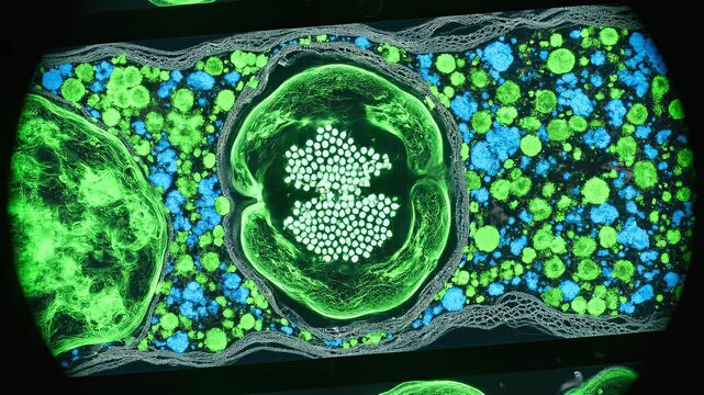
Exploring the Intricacies of Cellular Structures: Vibrant Microscopy Visuals Showcases Microscopic Cells Amidst Ethereal Backgrounds Blending Science and Art
00:05
HD
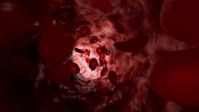
Red Blood Cells Moving Through Artery Interior Medical Visualization. 3D rendering
00:10
HD
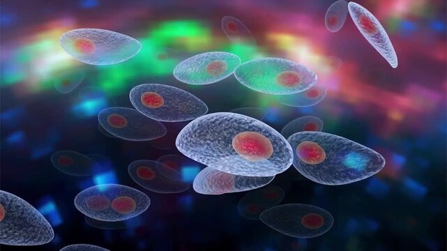
Toxoplasma gondii tachyzoites, 3D animation of crescent-shaped intracellular protozoan parasites with apical complex, causative agent of toxoplasmosis in humans and animals.
00:14
HD
Asian Female Scientist Works at a Multi-monitor Computer in a Clinical Lab, Reviewing AI Microscopy, CT Scans and Cell Maps to Accelerate Diagnosis, Drug Targeting and Patient Care. Medium Side Shot.
00:13
HD
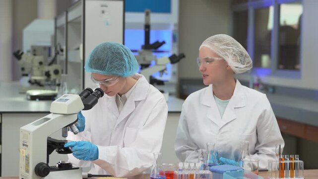
Two women scientists in white coats, protective eyewear and sterile hairnets straightening from the eyepiece and turning to an approaching colleague at a professional laboratory workstation.
00:11
HD
Laboratory scientist takes a prepared sample slide and places it under a microscope for examination. Scientific research, microscopy and laboratory analysis process.
00:10
HD
Glowing Blue Biological Cellular Structures Under Microscope.
00:06
HD
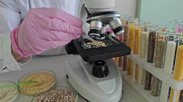
Microscope examination of grains and seeds in a laboratory setting with various samples displayed on the table
00:10
HD
Examining Microscope View of Cell Sample in Laboratory Imaging
00:10
HD
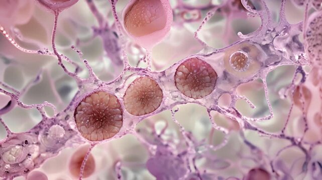
Nomarski Differential Interference Contrast Microscopy Cell Relief Seamless Loop in rich rose tones
00:10
HD
Cancer Cell Invasion and Metastasis Developing Across Human Tissue Surface 3D Rendering
00:10
HD
Glycogen granules and branched glucose polymers in cell microscopy
00:08
HD
Active nematode roundworms swimming around organic detritus under 40x darkfield microscopy 4K
00:22
HD
Fluorescence UV 365nm microscopy of marine collagen and urea mixture with moving ultraviolet light source showing dynamic glowing neon blue texture changes for futuristic science background
00:11
HD
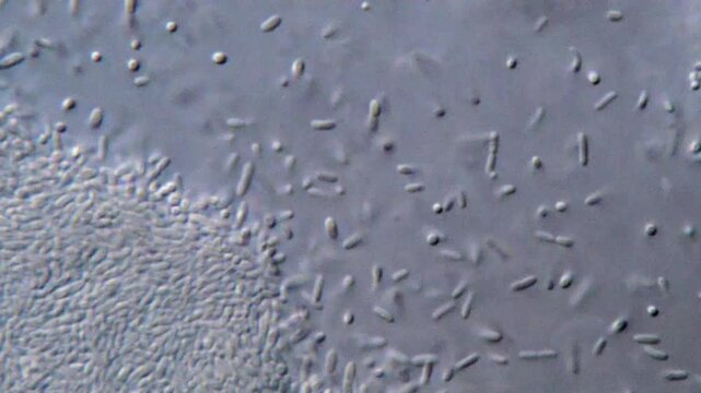
High resolution microscopy of bacteria colonies
00:44
HD
Urinalysis under microscopy showing plenty Pus cells, Pyuria
00:12
HD
Scientist analyzes a biological sample with automated laboratory equipment while viewing DNA data, illustrating genetics, biotechnology, diagnostics, pharmaceutical research and advanced scientific te
00:05
HD
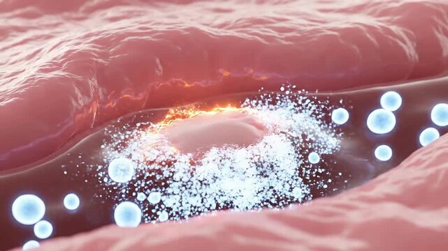
Detailed Animation Of Cellular Response During Wound Healing Process With Dynamic Particle Interaction And Realistic Textures On Solid-Color Background
00:08
HD
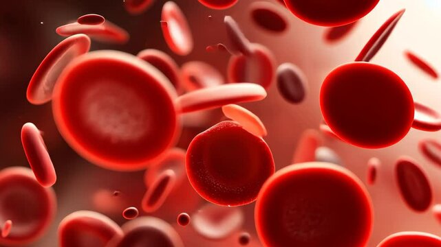
This video captures red blood cells coursing through a blood vessel, showcasing the vital process of oxygen transport in the human body, Dynamic depiction of oxygenated red blood cells
00:10
HD
View through microscope at in vitro fertilization process
00:07
HD
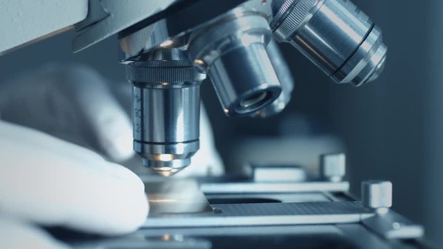
Close-up shot of microscope at laboratory.
00:25
HD
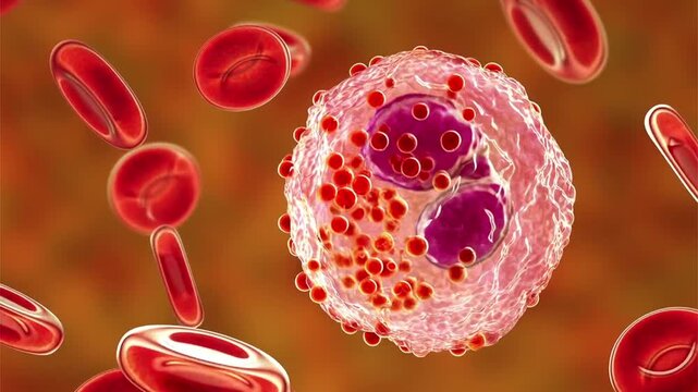
Eosinophil, 3D animation of activated white blood cell releasing granules in blood among red blood cells during allergic or antiparasitic immune response.
00:16
HD
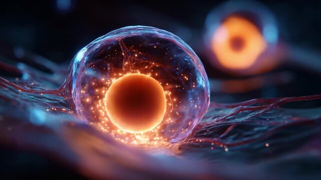
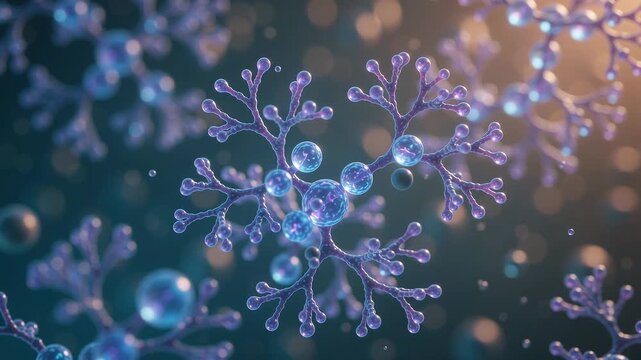
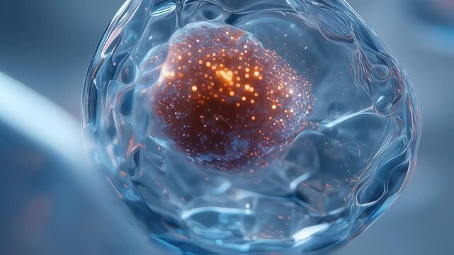
Detailed closeup image featuring exosomes encased in translucent spheres radiating subtle glows in a serene light atmosphere. A compelling visualization that embodies the forefront
00:06
HD
Blood erythrocyte movement in blood vessels capillaries pigment cells microscopy in tadpole tail
00:14
HD
Pertussis bacteria under the microscope
00:08
HD
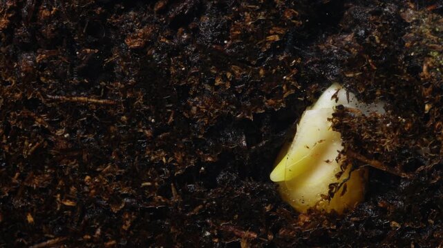
Young corn plant pushing upward from moist earth. Seedling with swollen cotyledon emerging through rich dark humus. Tiny sprout with green tip and delicate roots breaking through moist soil
00:14
HD
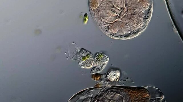
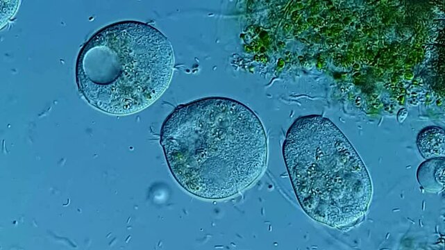
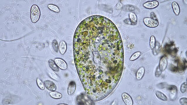
fresh water ciliate, Paramecium bursaria, with ingested green algae circulating in its cytosol. 450x magnification, phase contrast microscopy
00:57
HD
Electron Microscopy Enables Precise Scientific Examination of Glowing Biological Cellular Material
00:10
HD
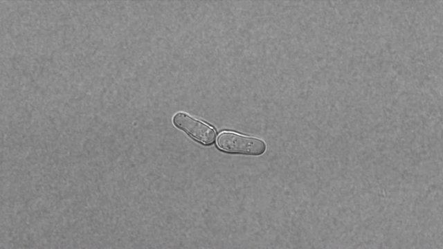
Fission yeast (Schizosaccharomyces pombe) reproduction under microscope
00:29
HD
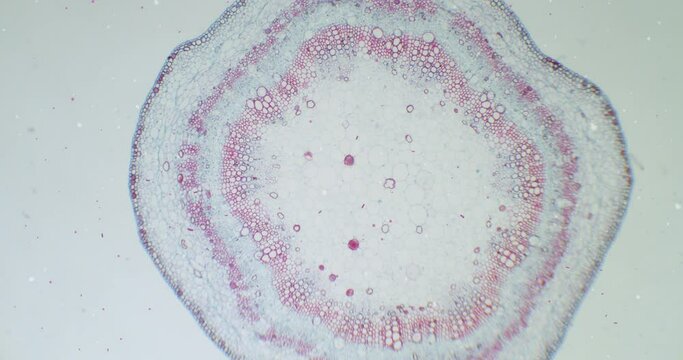
Cotton stem under a microscope with moving germs, microbes and bacteria. Infected view. Microscopic view of plant cells for botanic education. Cotton under a scope. A plant stem microscopic wideshot.
00:26
HD
3D Animation of Microscopic View of Stem Cells. Medical Illustration.
00:05
HD
Wide view of Demodex brevis skin mite on human epidermis background under 40x brightfield microscopy
00:13
HD
Real time crystal growth under polarized light microscopy showing unique bronze and silver metallic luster of chemical structures with cinematic soft focus effect around edges for abstract technology
00:20
HD
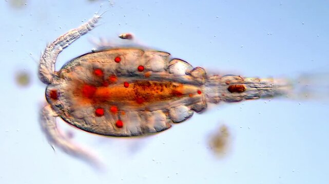
Copepoda microorganism under microscopy view, Copepod larvae
00:33
HD
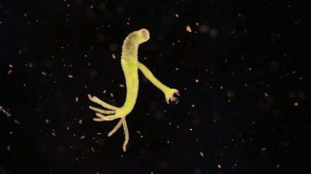
Hydra is a genus of small, fresh-water animals of the phylum Cnidaria and class Hydrozoa under the microscope for education.
00:20
HD
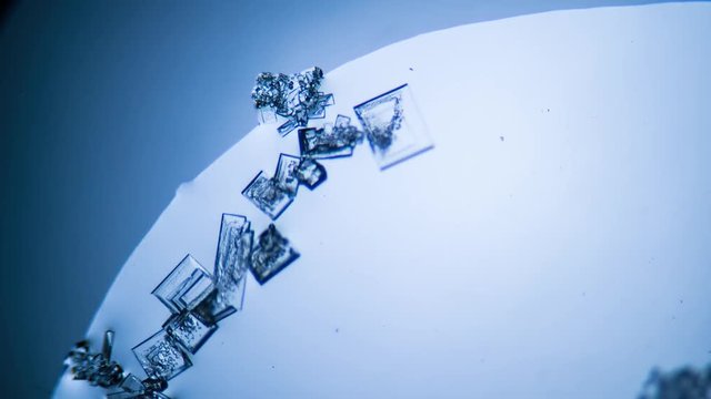
Crystal growth, microscope photography using a microscope
00:18
HD
Analytical scientist reviews microscopic tissue slides carefully
00:10
HD
3D Illustration of Ebola Virus Disease
00:05
HD
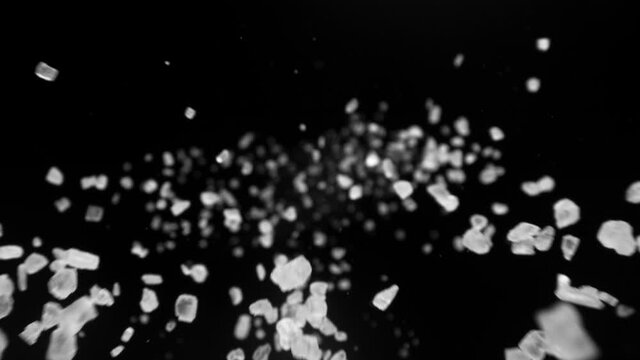
Super slow motion of falling whole grained salt pieces on black background. Filmed on high speed camera, 1000 fps
00:23
HD
Elegant scientific visualization of smooth translucent vesicles housing vibrant blue exosome particles, surrounded by a soft-focus cellular matrix, highlighting their role in regen
00:10
HD
Scientist using microscopy glass setup in laboratory
00:07
HD
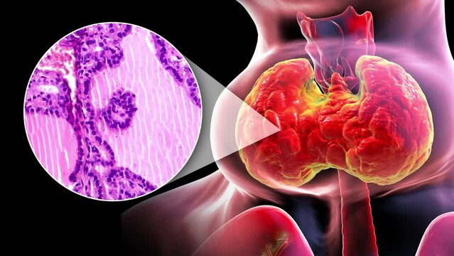
A 3D animation of a person with enlarged thyroid gland, alongside with a light micrograph footage of thyroid tissue affected by toxic goiter.
00:17
HD
Medium shot of an engineer adjusting the electron microscope settings to capture highresolution images of metal grain boundaries and inclusions
00:06
HD
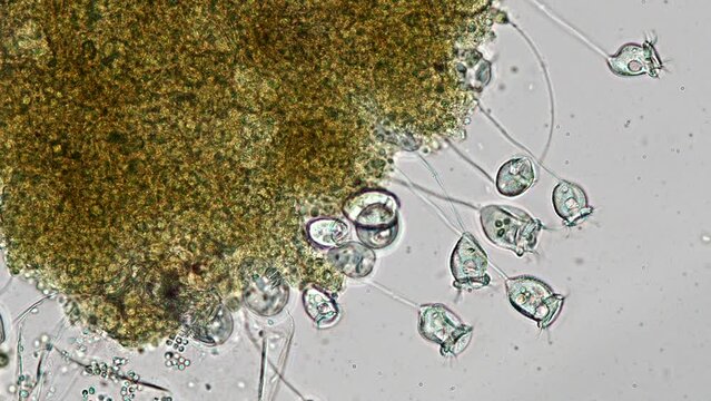
vorticella in aquarium water under the microscope - genus of bell-shaped ciliates (protozoa)
00:45
HD
Microscopy of parasite worm nematode (Strongyloides stercoralis). Movement of active parasite infection form in magnification 150x.
00:11
HD
scientist working with microscope
00:11
HD
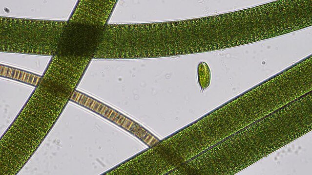
Euglena phacus trapped between a forest of filamenous cyanobacteria. Phacus swims with a beautiful corkscrew motion. Shot in real time under the microscope at 200x magnification.
00:47
HD
Lassa virus microscopy video showing cellular degeneration vacuoles and membrane rupture
00:10
HD
Glowing green particles drift above red intestinal villi in a microscopic view of gut absorption and microbiome
00:10
HD
Scientist performing microscopic analysis in a modern laboratory setting for medical research study
00:28
HD
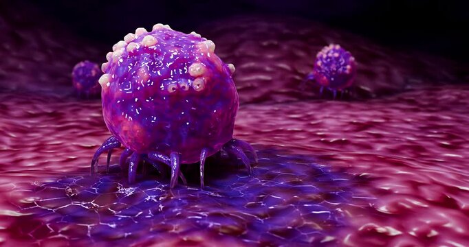
In a captivating illustration, a cancer cell is depicted interacting within its surrounding environment.
00:10
HD