암호 설정을 위한 링크가 다음으로 전송됨: 나중에 구매 기능을 이용하려면 암호가 필요합니다.
무제한 다운로드와 강력한 AI 편집 기능으로 제약 없이 창작해 보세요
새 필터 추가됨
정렬 기준

비디오에서 acetabulum에 관한 226개 결과

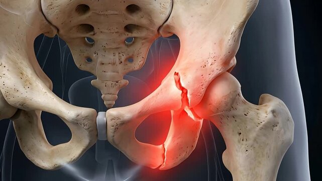
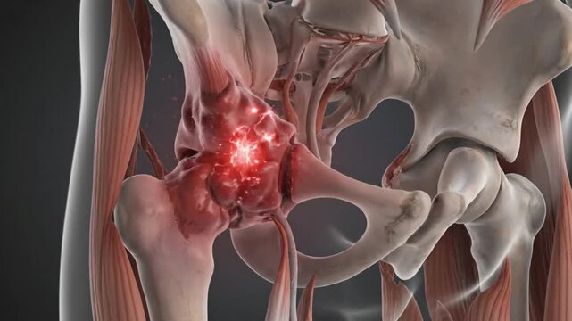
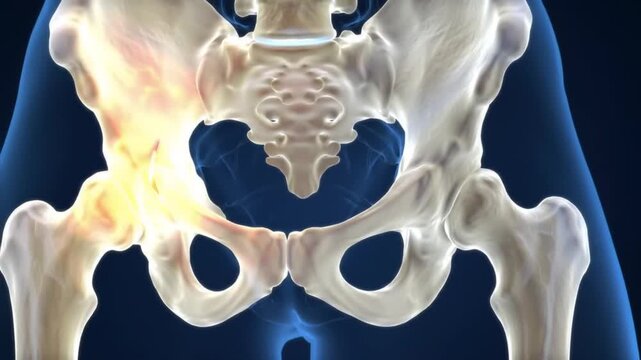
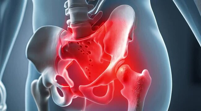
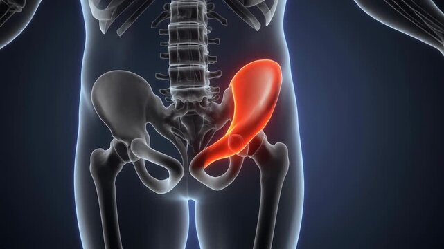
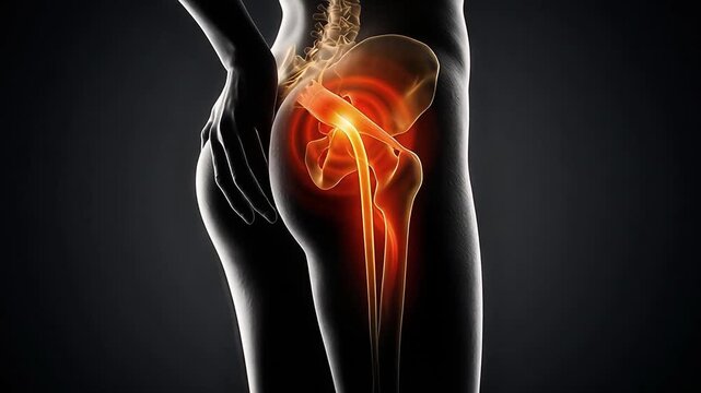
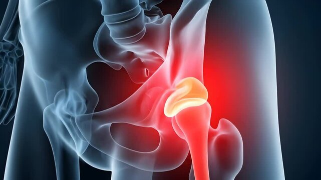
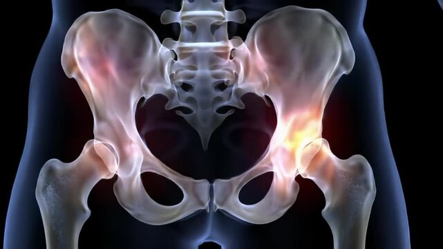
Medical Illustration of Human Pelvis Showing a Hip Joint Fracture With Red Glow Indicating Pain and Damage Against a Dark Transparent Human Body

00:05
HD
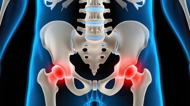
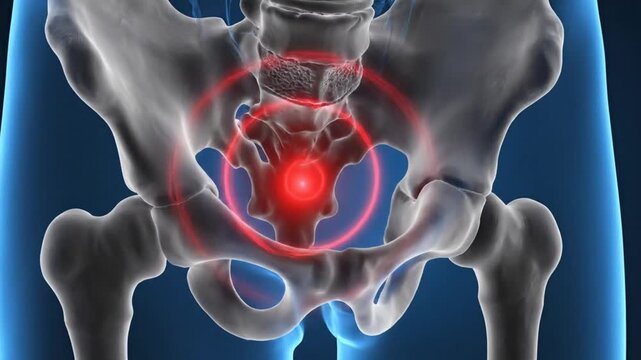
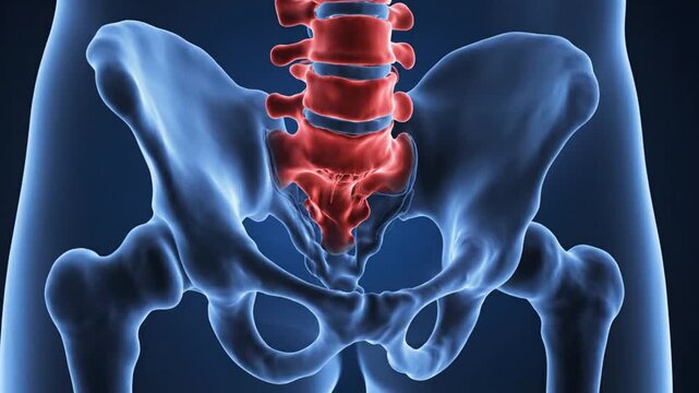
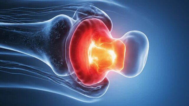
Blue X-Ray Animation of Pelvic Bone Joint Inflammation for Medical Illustration and Healthcare Promotion
00:08
HD
Understanding hip pain causes symptoms and treatment options for joint inflammation relief. Human pelvis illustration shows hip pain. Doctor guideline for hip joint health
00:10
HD
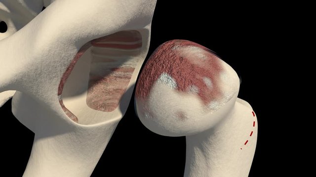
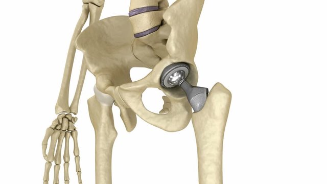
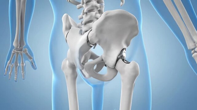
Hip Joint Replacement Anatomy Medical Illustration, Healthcare, Surgery
00:08
HD
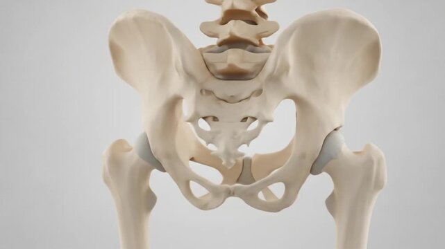
Human pelvis bone structure anatomy on white background.
00:08
HD
Closep-up on colony of unicellular green algae Mermaid's wine-glass or Mermaid's cup (Acetabularia acetabulum) Adriatic Sea, Montenegro, Europe
00:15
HD
Total Hip Replacement
00:16
HD
Mature doctor demonstrating hip joint
00:13
HD
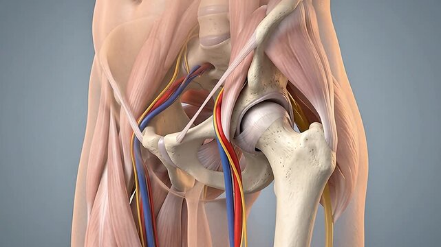
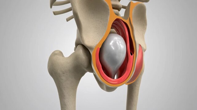
3D Medical Animation of Human Hip Joint Anatomy with Femoral Head, Acetabulum, Muscles, Blood Vessels and Nerves on Gray Background
00:05
HD
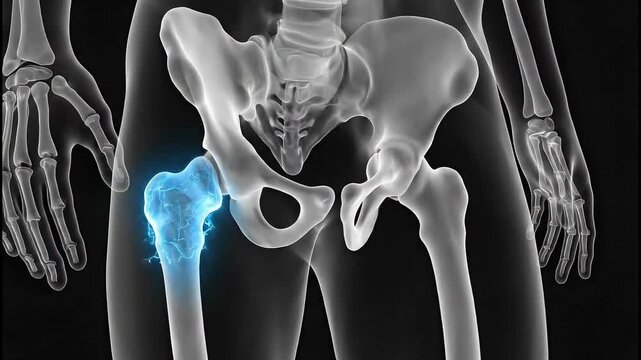
Detailed X-ray View of Human Hip Joint and Pelvis with Highlighted Femur Head.
00:07
HD
Medical X-Ray Style of Male Pelvis – Frontal View with Realistic Pelvic Bones Anatomy
00:05
HD
Anatomical Pelvic Bones: Front and Back View of Human Hip Structure for Study.
00:05
HD
3D medical animation of a total hip replacement (endoprosthesis)
00:15
HD
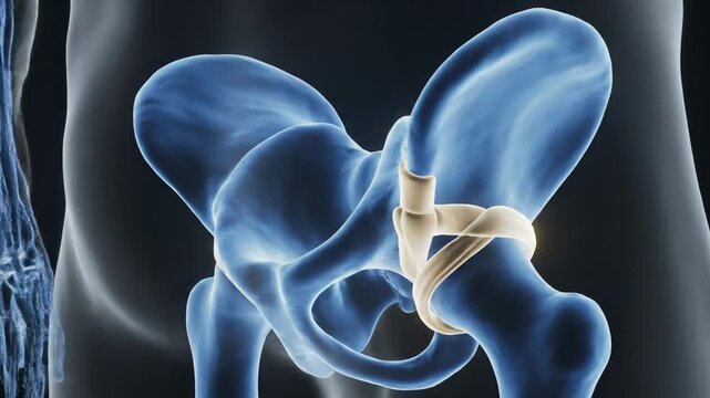
Ligamentum Teres Vascular Ribbons Shown in Transparent Hip Anatomy for Medical Education
00:05
HD
Hip replacement implant installed in the pelvis bone. Medically accurate  3D animation
00:13
HD
Human anatomy pelvis and sacrum background
00:15
HD
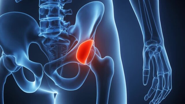
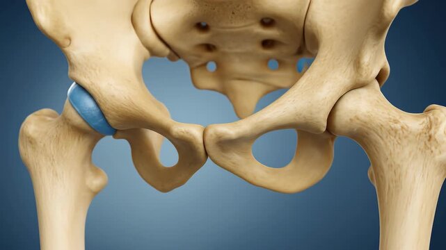
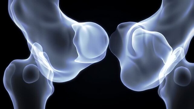
Human hip joint anatomy in blue with highlighted acetabulum for medical visualization
00:05
HD
3D rendering of human hip joint with visible bone structure and osteoporosis, medical illustration.
00:08
HD
Human pelvis anatomy illustration skeleton bones health medical glowing scan render
00:08
HD
Human skeleton with a metal hip prosthesis concept arthroplasty 3d render animation X-ray image
00:13
HD
The hip bone structure highlights the pelvis and spine, with the spine aligning with the pelvis. Muscles attach to the hip, emphasizing how muscles connect with the hip bone.
00:08
HD
Anatomical illustration of a human pelvis with a highlighted area indicating pain or inflammation on the right side.
00:10
HD
Anatomical 3D Render Illustrating Hip Joint Damage Showing Red Highlighted Pain and Inflammation
00:08
HD
Anatomy of the Human Pelvis: A detailed anatomical illustration showcasing the pelvic region with an emphasis on the skeletal structure and internal organs.
00:10
HD
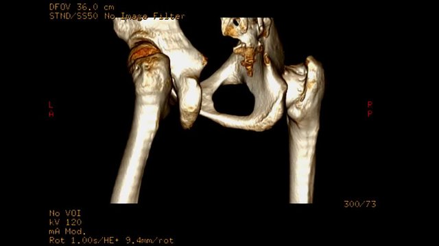
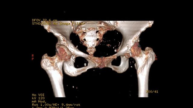
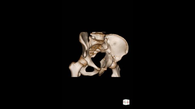
CT SCAN image of pelvic bone / Hip bone 3D rendering image show fracture left hip joint Rotating on monitor .
00:28
HD
3D Human Pelvis and Hip Joint Anatomy with Highlighted Pain Area.
00:08
HD
Hip Replacement Anatomy Animation Medical Visualization
00:08
HD
Detailed Medical Illustration of Hip Joint Anatomy with Pain Indication.
00:08
HD
A detailed anatomical model of the human pelvic girdle, hip joint, and lower spine against a neutral background.
00:08
HD
Pelvis with Skeleton Body
00:12
HD
Total hip replacement implant positioned within femur and acetabulum, camera slowly rotating around prosthesis on white background, detailed orthopaedic 3D animation.
00:12
HD
Human Pelvic Bone Illustration Showing the Os Coxae, Sacrum, Coccyx, and Pelvic Girdle Anatomy
00:07
HD
Understanding Shoulder Pain Anatomy and Causes of Discomfort Affecting the Joint
00:08
HD
Animation of a man's hip joint
00:13
HD
Hip replacement implant installed in the pelvis bone. Medically accurate  3D animation
00:16
HD
Human painful hip medical concept
00:10
HD
Animated Hip Fracture Video Detailed View of Pelvic Bone Injury and Inflammation
00:08
HD
Human skeleton model with extended arms
00:05
HD
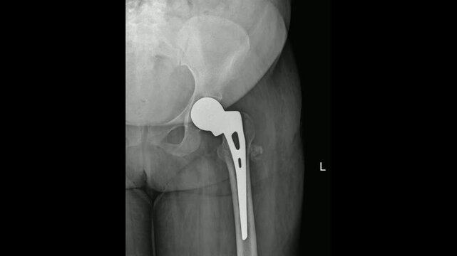
Hip Prosthetic X-Ray Showing Artificial Joint Replacement
00:15
HD
3D Animation of Femoral Head Fitting into the Hip Socket
00:10
HD
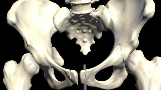
Fractured Human Pelvis Anatomical Illustration Skeletal Structure Medical Visualization Bone Injury Study in Black Background
00:08
HD
Animation Displaying Hip Dysplasia Diagnosis and Pain Pointed Out on Multiple Human Hip X-Ray Images Highlighting Medical Conditions and Bone Health
00:07
HD
Vertical video: Increasing magenta glow marking left hip in medical 3D view showing pelvic joint
00:05
HD
Illustration of the human hip joint highlighting pain, representing arthritis, injury, or inflammation in the pelvic area and surrounding bone
00:08
HD
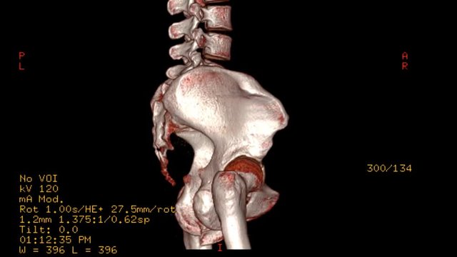
CT SCAN image of pelvic bone 3D rendering image demonstrate fracture pelvic bone . Rotating on monitor .
00:15
HD
Human Pelvis Anatomy A Detailed 3D Visualization of the Skeletal Structure.
00:08
HD
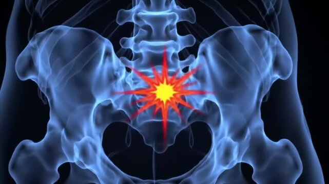
3D Animation of Human Hip and Sacroiliac Joint Pain
00:08
HD
Human Hip Joint Anatomy and Potential Implant Scenario
00:08
HD
A human hip joint showing a red area indicating inflammation. The image resembles a medical illustration, with a blue and white color scheme.
00:05
HD
Detailed 3D animation of the human hip and pelvic bones, showcasing anatomical structure and movement for medical education.
00:08
HD
Detailed illustration of a human body pelvis and lower spine against a clean white background with subtle gradient shading.
00:08
HD
High-End 3D CGI Octane Render of the Human Hip Joint
00:07
HD
Medical X-Ray Style of Male Pelvis – Frontal View with Realistic Pelvic Bones Anatomy
00:05
HD
3D Rendered Anatomy: Hip and Pelvis Structure Visualization
00:07
HD
Hip replacement implant installed in the pelvis bone. Medically accurate 3D animation
00:13
HD
3d medical figure with hip bone pain
00:14
HD
Bones and Ligaments of the Hip Joint
00:31
HD
Child Pelvis AP X-Ray Showing Hip and Sacral Anatomy
00:15
HD
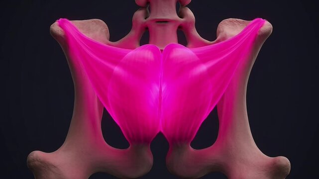
Detailed 3D Rendering Of The Human Pelvis Bone Structure With Highlighted Pink Gluteal Muscles On A Dark Gradient Background Anatomical Medical Scientific Illustration
00:06
HD
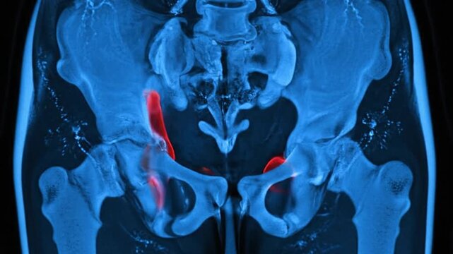
MRI Scan of Hip Joint.
00:08
HD
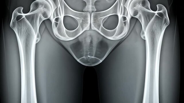
Detailed X-ray of Human Pelvis and Hip Joints Showing Bone Structure
00:05
HD
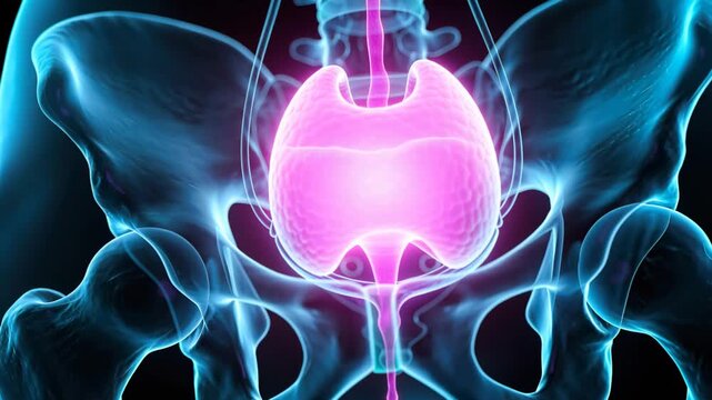
Filling magenta bladder against pelvic bones, receiving ureter flow and releasing via urethra
00:05
HD
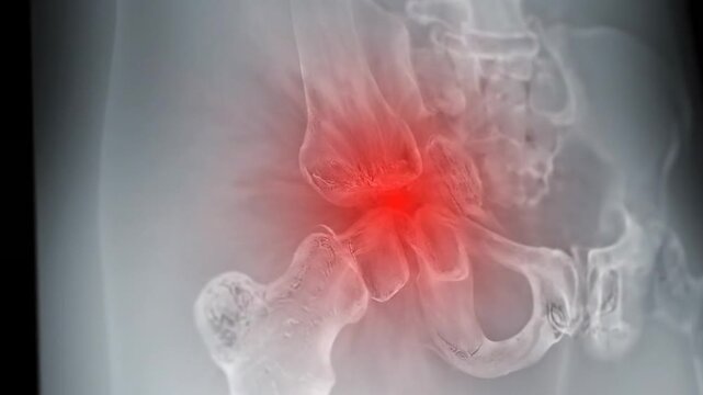
X-ray of the human hip joint on a gray background, with the painful areas highlighted in red. The image shows osteoarthritis in the hip joints of a woman, with the affected regions marked in red
00:08
HD
Anatomical illustration showing hip joint pain and skeletal structure with blue background and black space
00:10
HD
Human Pelvis Anatomy XRay Visualization for Medical Education.
00:08
HD
3D Medical Animation of Human Pelvis and Sacroiliac Joint Anatomy with Hip Bones, Nerves, Femur and Inflammation Pain Highlight on Gray Background
00:05
HD
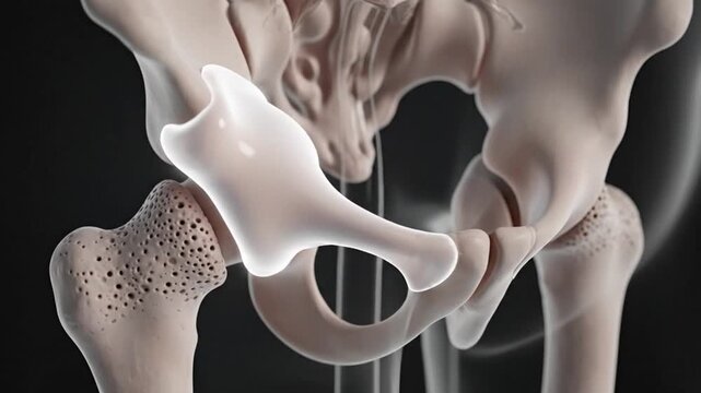
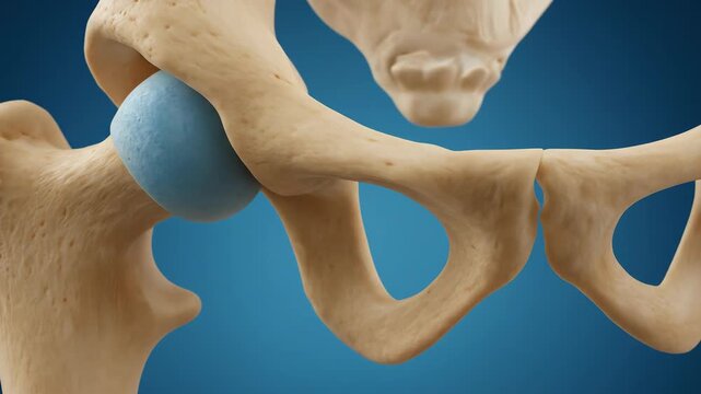
Detailed 3D Anatomy of the Human Hip Joint: Acetabulum and Cotyloid Notch
00:07
HD
Detailed 3D CGI Medical Animation of the Human Pelvis and Hip Anatomy with Blue Highlighted Joints Against a Dark Background for Healthcare Education
00:08
HD
Bipolar hemiarthroplasty, 3D animation showing prosthetic femoral head replacement with dual mobility articulation within the native acetabulum of the hip joint.
00:12
HD
Minimalist illustration of human pelvic bone anatomy on white
00:06
HD
Pelvic Anatomy 3D Animation Visualizing the Human Skeleton Bone Structure and Joint Movement with Detailed Medical
00:08
HD
Close-up perspective of hip joint model highlighting femoral head, acetabulum, and surrounding muscle fibers, warm directional lighting enhancing anatomical textures
00:10
HD
Human Pelvis Anatomy: A Detailed 3D Model of Bones, Muscles, and Ligaments
00:07
HD
A human hip joint illustration with highlighted pain area, showing bone structure and potential injury.
00:08
HD
Pain in the hip joint medical background
00:14
HD
Understanding Shoulder Joint Pain and Inflammation in the Human Anatomy
00:08
HD
Medical X-Ray Style of Male Pelvis – Frontal View with Realistic Pelvic Bones Anatomy
00:05
HD
Hip Fracture X-Ray Demonstrating Femoral Neck Break
00:15
HD
3D Medical Animation: Pelvic Bone Anatomy and Hip Joint, Acetabular Notch Highlighted
00:10
HD
Hip Joint Pain Animation 3D Medical Visualization of Pelvis and Femur with Highlighted Acetabulum
00:05
HD
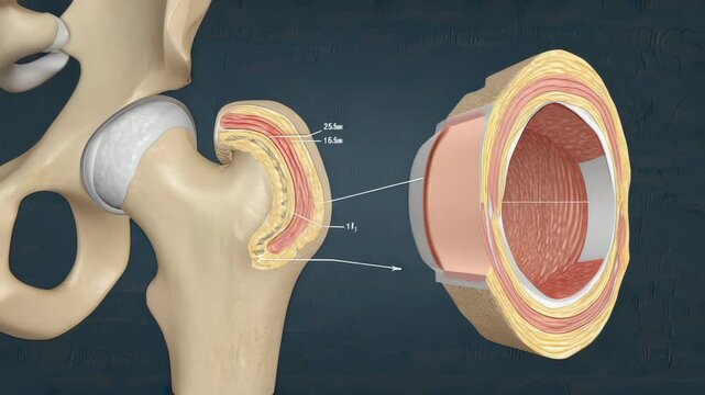
Hip Joint Anatomy Illustration with Cross-Sectional View, Femur Head, Acetabulum, Cartilage Layers, and Bone Structure for Medical Education
00:10
HD
Human Hip Joint XRay Anatomy.
00:08
HD
Human pelvis anatomy highlighted with green glow on transparent body.
00:08
HD
Human Hip Joint XRay Anatomy.
00:08
HD
Medical Illustration of Hip Joint Anatomy and Inflammation.
00:08
HD
Hip Joint Pain Inflammation 3D Medical Animation Human Pelvis Femur Anatomy X-Ray Glowing Red Dark Background
00:07
HD
Human Hip Joint Anatomy Illustration
00:08
HD
Detailed anatomical illustration of the human pelvis and hip joint on a white background.
00:08
HD
Hip pain animation. Medical visualization of hip joint inflammation, arthritis, bursitis or injury. Healthcare promo, educational content, orthopedic campaigns.
00:07
HD
Human hip joint anatomy illustration with highlighted red area
00:05
HD
CT SCAN  of  Both hip joint 3D rendering image Rotating on the screen  from patient case trauma .
00:18
HD
CT Scan pelvis 3D render Impression:Transverse fracture of acetabulum. Fracture Lt ishiopubic rami.Posterosuperior dislocation of Left femoral head. Medical technology concept.
00:09
HD
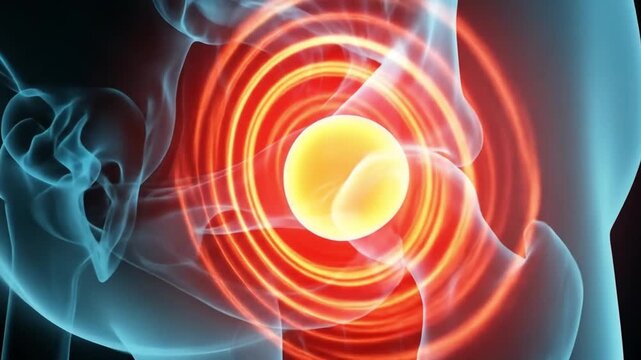
Hyper-realistic 3D rendered human skeletal hip and pelvis video, translucent blue x-ray view, pulsating red/orange glow, against black background. Concept of medical pain and inflammation
00:08
HD
3D Animation of Human Hip Joint Anatomy with Femoral Head Focus
00:07
HD
Pelvic Pain Animation XRay View of Hip Bones with Red and Yellow Pain Indicator
00:08
HD
Doctor holding in hand Hip Dysplasia
00:15
HD
Medical X-Ray Style of Male Pelvis – Frontal View with Realistic Pelvic Bones Anatomy
00:05
HD
Detailed Anatomical Diagram of Human Hip Joint and Pelvis Structure
00:08
HD
Medical Analysis of the Pelvic Bone Structure and Examination Process with Surgical Gloves
00:10
HD