Il collegamento per impostare la password è stato inviato a: La password sarà necessaria per accedere in futuro ai tuoi acquisti.
Versione di prova gratuita: ottieni 10 immagini stock, più strumenti di modifica AI
Aggiunti nuovi filtri
Ordina per

238 risultati per pupillometry in tutto

Visualizza pupillometry in video (137)
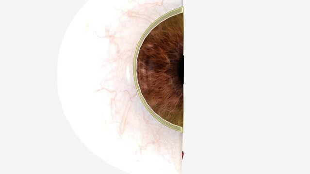
The cornea is the clear outer layer at the front of the eye .
00:10
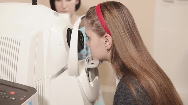
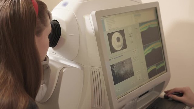
Side view shot of young girl having her eyes examed on eye tomography machine in hospital. Ophthalmologist's office.
00:09
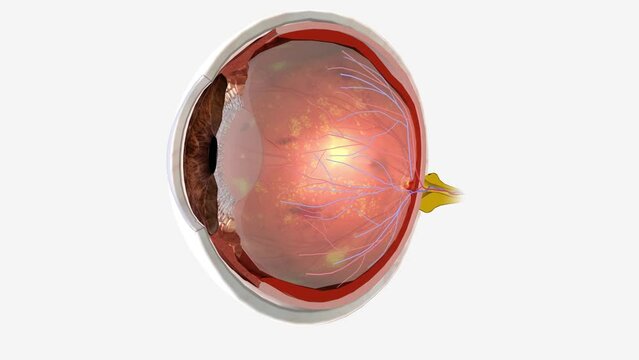
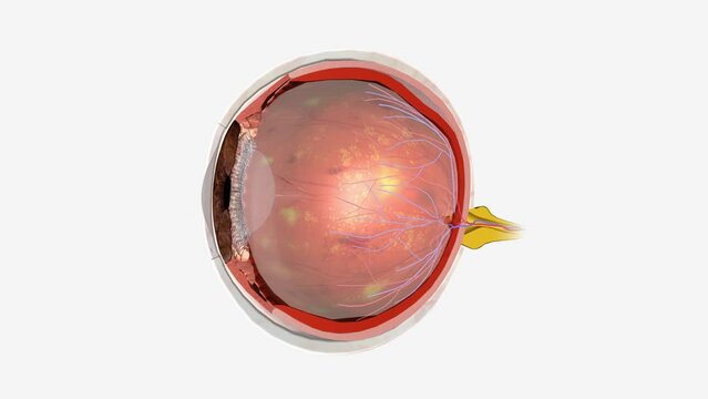
Diabetic retinopathy is an eye condition that can cause vision loss and blindness in people who have diabetes
00:08
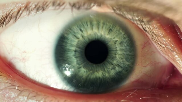
Macro footage of blue iris reacting to light, showcasing intricate details of the pupil's dilation and contraction
00:23
Eye icon with a clock symbol representing time monitoring or scheduling. Ideal for productivity, surveillance, and time management concepts.
00:06
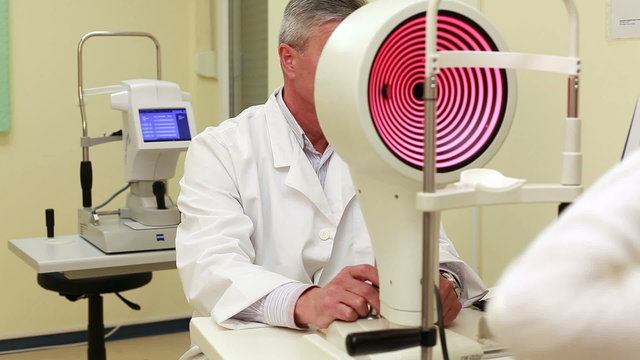
Close up shot of doctor checking on eyesight of patient. Tomoraghy of a cornea. Results and graphics on the monitor.
00:21
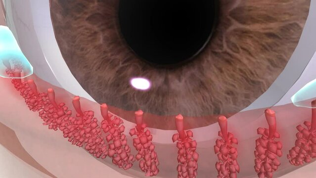
Blepharitis and Meibomian Gland Dysfunction (MGD) is when hardened oils trapped in the gland lead to inflammation along the eyelids
00:10
The cornea is the clear outer layer at the front of the eye .
00:12
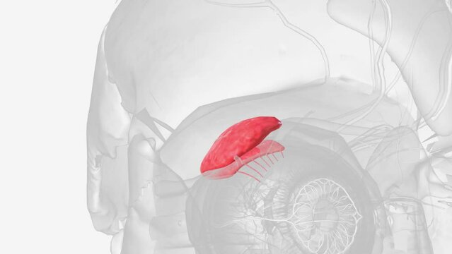
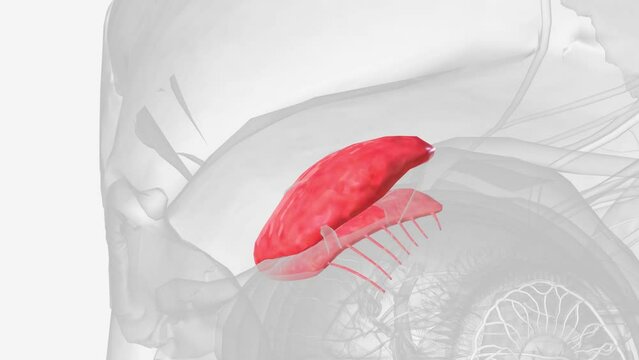
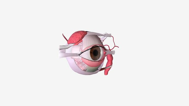
The lacrimal gland (tear gland) is an exocrine gland located above the eyeball, in the anterior part of the upper outer aspect of each orbit .
00:08
The conjunctiva is a thin, clear membrane that protects your eye
00:06
The lacrimal glands are serous type exocrine glands that secrete lacrimal fluid onto the surfaces of the conjunctiva and cornea of the eye
00:14
Dry Macular Degeneration
00:06
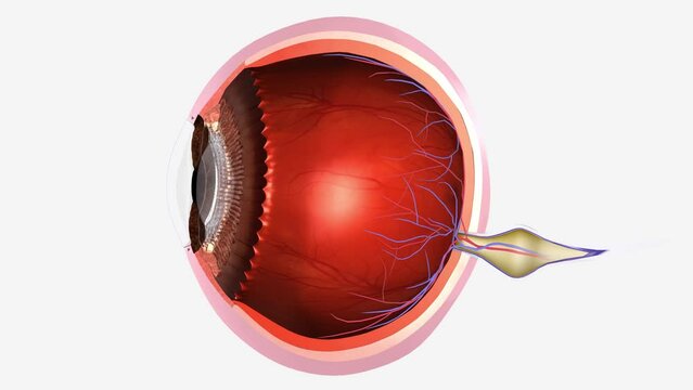
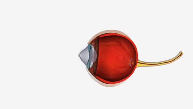
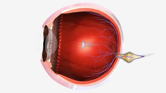
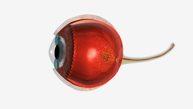
The eye is made up of three coats, which enclose the optically clear aqueous humour, lens, and vitreous body
00:07
The cornea is the transparent front part of the eye that covers the iris, pupil, and anterior chamber
00:09
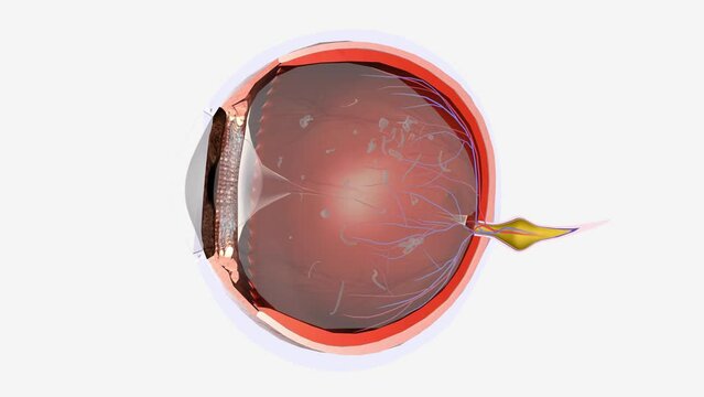
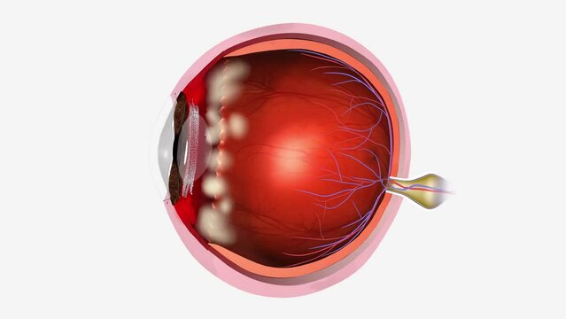
Floaters are small dark shapes that float across your vision
00:09
Blepharitis is an inflammation along the edges of the eyelids
00:09
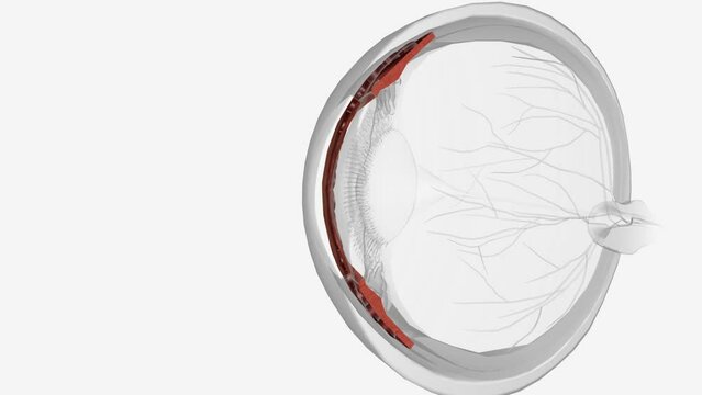
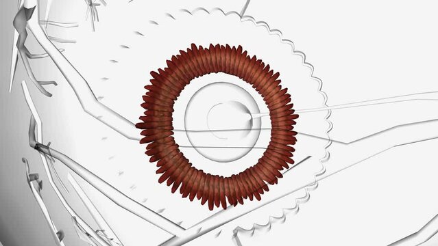
The ciliary body is found behind the iris and includes the ring-shaped muscle that changes the shape of the lens when the eye focuses
00:06
The lacrimal gland (tear gland) is an exocrine gland located above the eyeball, in the anterior part of the upper outer aspect of each orbit .
00:06
Diabetic retinopathy is an eye condition that can cause vision loss and blindness in people who have diabetes
00:07
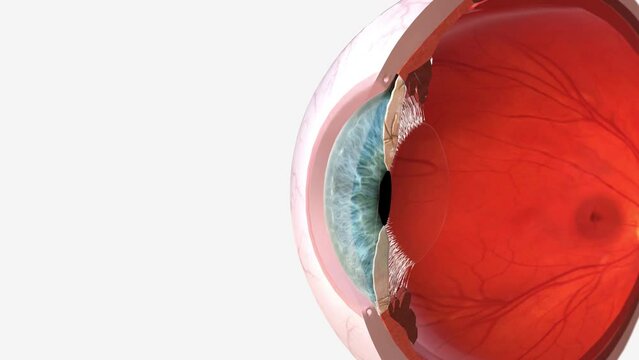
From the cornea, the light passes through the pupil
00:09
Young woman having her eyes examined by an eye handsome elderly
00:27
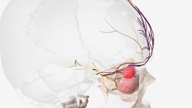
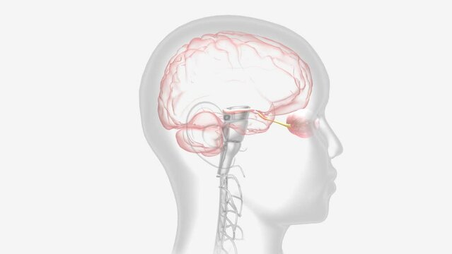
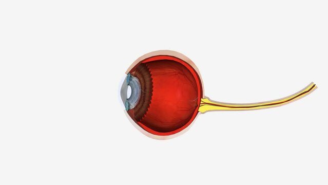
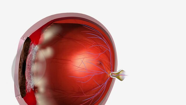
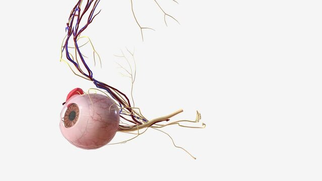
Nervous and vascular system in the eye .
00:21
Optic Neuritis Impaired Vision
00:08
The iris is the colored part of the eye that controls the amount of light that enters into the eye .
00:11
Structure forming the posterior ciliary arteries in the eye .
00:09
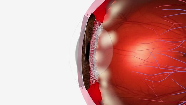
The zonules are the tiny thread-like fibers that hold the eye's lens firmly in place .
00:13
Optic neuritis occurs when swelling (inflammation) damages the optic nerve
00:07
Interstitial keratitis is chronic, nonulcerative inflammation of the mid-stroma that is sometimes associated with uveitis
00:14
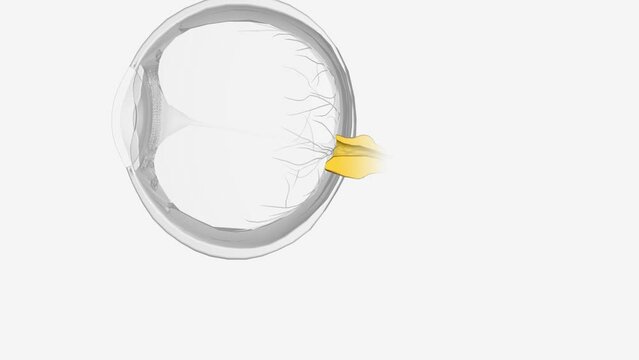
Eye Anatomy Lens
00:11
The pars plana is a narrow section of the ciliary body, inflammation of which is known as pars planitis
00:09
The pars plana is a narrow section of the ciliary body, inflammation of which is known as pars planitis
00:07
Keratoconus is a vision disorder that occurs when the normally round cornea becomes thin and irregular (cone) shaped.
00:12
The sclera is the white outer coating of the eye .
00:23
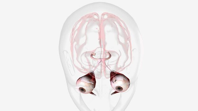
Six cranial nerves innervate motor, sensory, and autonomic structures in the eyes .
00:07
Nervous and vascular system in the eye .
00:10
Nearsightedness (myopia) is when close-up objects look clear but distant objects are blurry
00:10
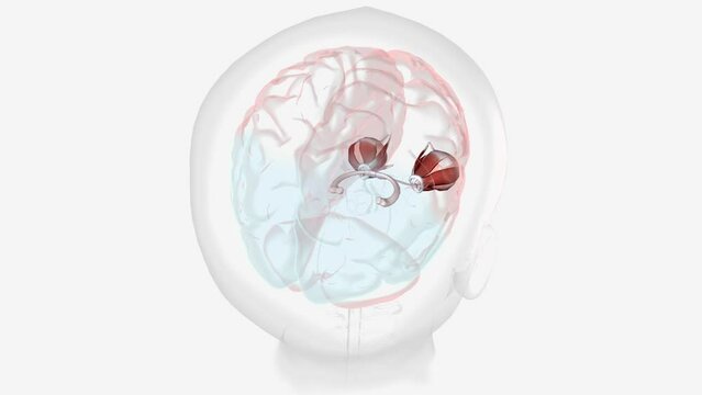
Normal Visual Pathway with Eye Movement
00:13
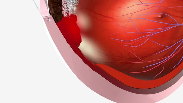
Pars Planitis Snowbank exudates
00:08
Normal Visual Pathway with Eye Movement
00:12
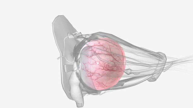
The veins that bring blood to the vessels in your eyes are called periorbital veins .
00:10
Tenon's capsule contains both collagen, elastin, or smooth muscle/fat tissue.
00:20
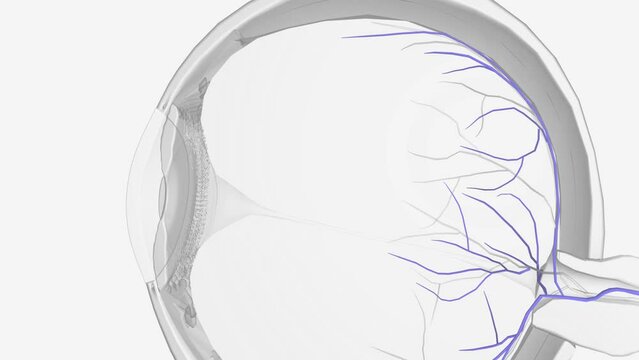
Similarly, ophthalmic veins (vortex veins) and a central retinal vein drain blood from the eye .
00:09
Dry Macular Degeneration 3d
00:09
he lacrimal canaliculi, are the small channels in each eyelid that drain lacrimal fluid, from the lacrimal puncta to the lacrimal sac
00:11
The sclera is the white outer coating of the eye. It is tough, fibrous tissue that extends from the cornea
00:12
The pars plana is a narrow section of the ciliary body, inflammation of which is known as pars planitis
00:12
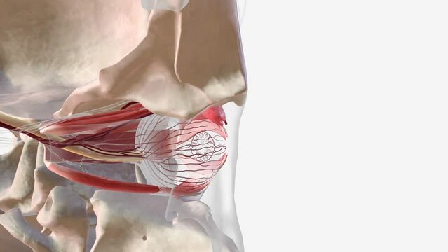
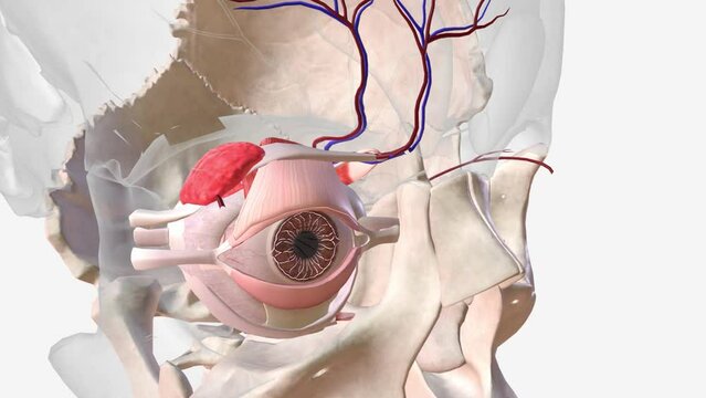
Eye Regional Anatomy
00:08
The orbicularis oculi muscle is a muscle located in the eyelids
00:13