Un lien pour définir votre mot de passe a été envoyé à : Pour accéder à vos achats à l’avenir, vous aurez besoin d’un mot de passe.
Obtenez 3 vidéos ou 25 images avec un essai gratuit
Nouveaux filtres ajoutés
Trier par

947 résultats pour parasitology dans vidéos

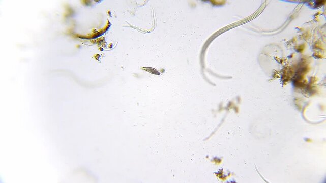
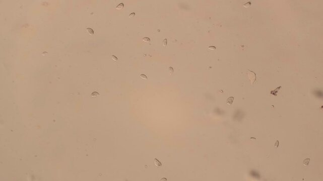
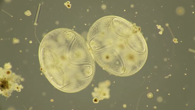
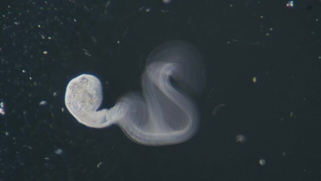
Ciliate swimming near nematodes under 100x magnification brightfield microscopy. 4K scientific microscopy footage.
00:26
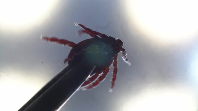
Closeup of researcher removing tick specimen. Scientific imaging showing tick with disease risk indicators. Educational depiction of tick removal highlighting danger of encephalitis and borrelia
00:34
Anaplasma bacteria infecting neutrophils, 3D animation. Anaplasma phagocytophilum causes human granulocytic anaplasmosis transmitted by the black-legged tick and the sheep tick.
00:10
Macro of a tick laying hundreds of eggs in moist sandy soil under a plant, then departing. High-detail parasitology scene highlighting vector reproduction, useful for medical, veterinary, pest control
00:35
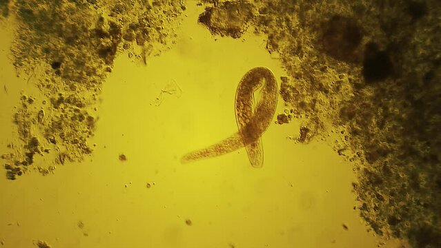
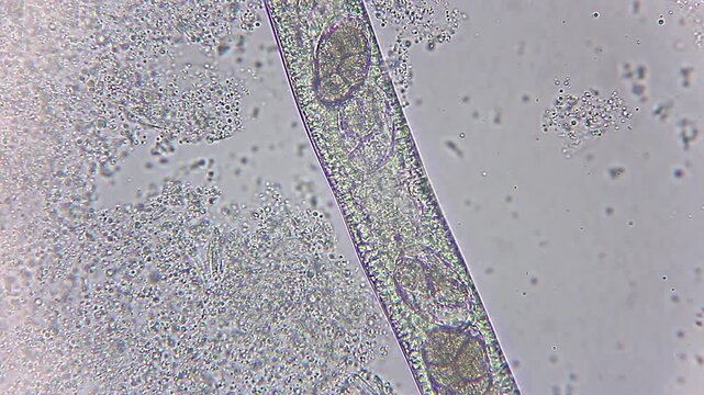
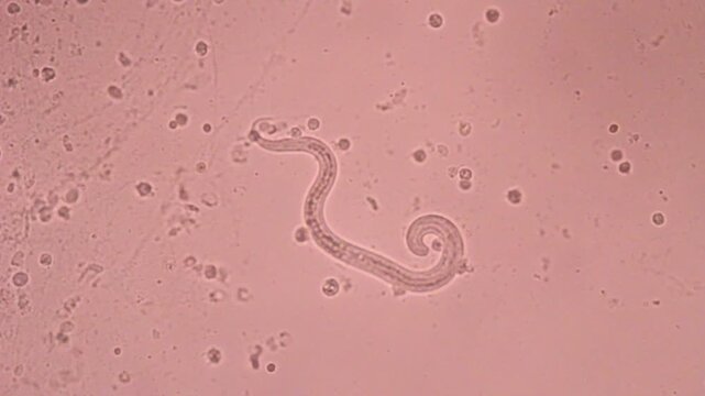
View in microscopic Strongyloides stercoralis or threadworm in human stool.Parasite or fluke infection.Medical background analyze by microscope, original magnification 400x
00:21
ROUNDWORM INFECTION treatment tablets used to eliminate nematode parasites from the intestine
00:26
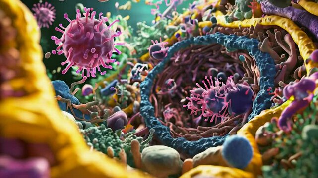
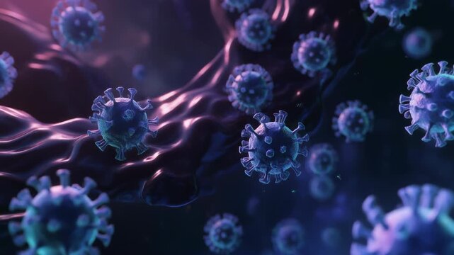
Viral particles floating in a dark, abstract environment, a microscopic view of virus-20.mp4
00:05
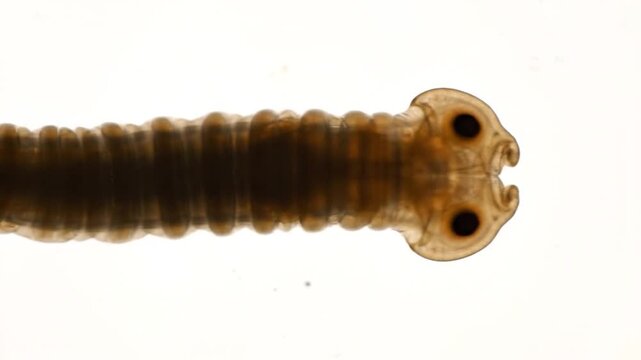
Parasitic worm close-up helminth parasite head.
00:08
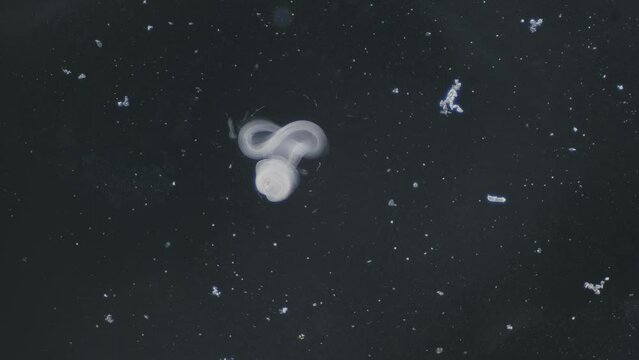
A roundworm on a dark background, viewed under a microscope, slowly wriggles sideways.
00:18
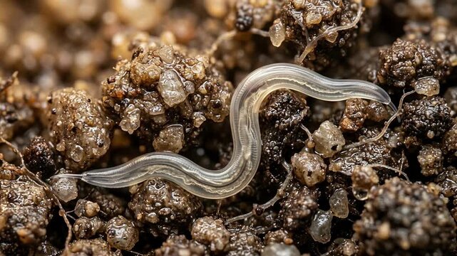
Translucent nematode roundworm moving through soil, microbiology
00:10
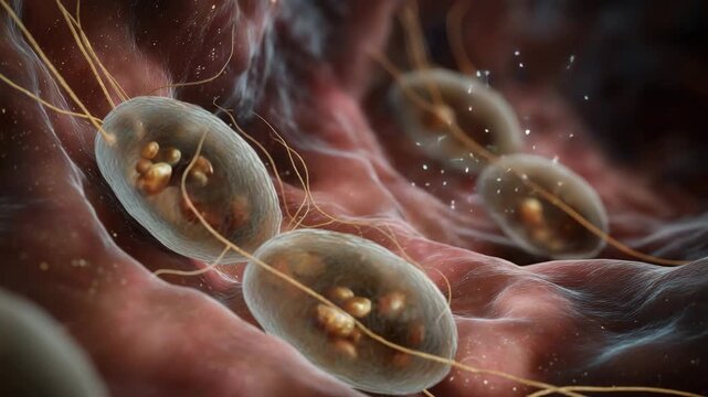
Microscopic view of Giardia lamblia parasites.
00:08
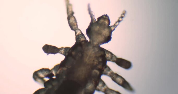
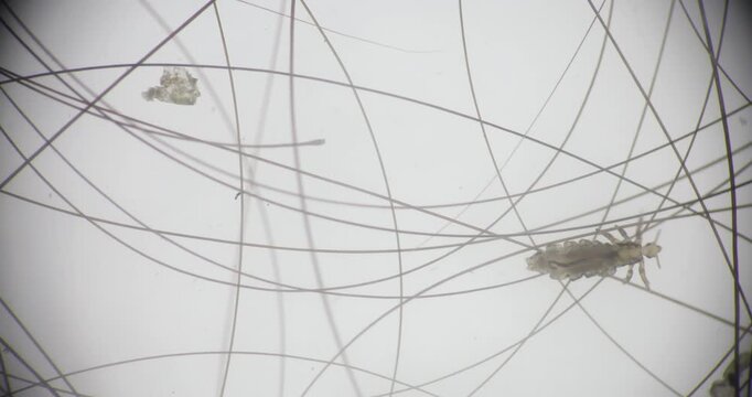
Microscopic head louse parasite moving isolated on white background
00:28
selective focus premium medical visual of parasitology
00:05
Macro shot of a human louse and a nit on a metallic surface. Pediculosis parasite and egg close-up
00:05
Close up macro view representing movement of intestinal parasites inside human body
.
00:06
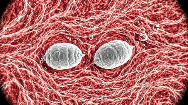
Microscopic View of Malaria Parasites Infecting Red Blood Cells, Detailed 3D Rendering
00:08
A microscopic view of a worm specimen on a glass slide, captured through a microscope lens in a laboratory setting.
00:05
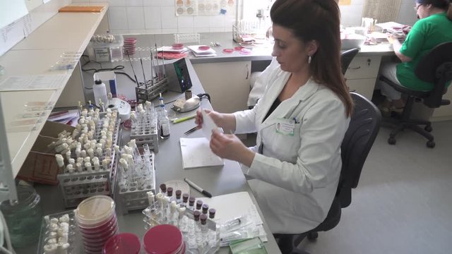
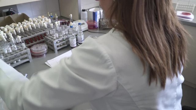
Microbiology or bacteriology analysis, two female doctors specialist microbiologist are testing samples at the desk with glassware in laboratory, crane shot, tracking, high angle view, room interior
00:26
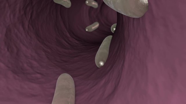
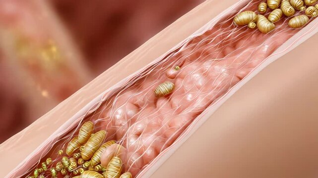
3D animation of parasitic worms in an intestines of human or animal.
00:12
Nematode worm
00:10
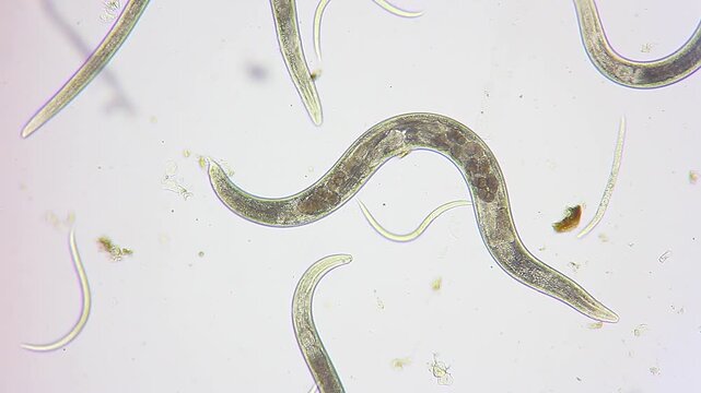
Nematodes moving slowly in blue water droplet under 400x magnification brightfield microscopy. 4K scientific microscopy footage.
00:17
Microscope Pine Pollen Whole Mount x1200
00:50
Leishmania promastigotes, 3D animation showing elongated flagellated protozoa in the promastigote stage, transmitted by sandflies and responsible for leishmaniasis in humans.
00:10
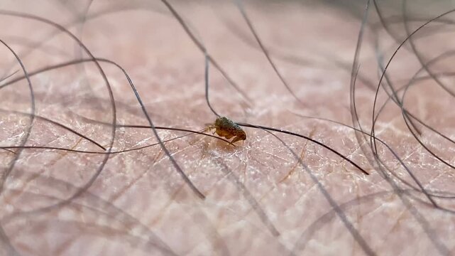
Macro stubborn flea biting human skin and sucking blood, refusing to detach when pushed by a finger. Powerful visual for zoonotic diseases, flea-borne infections, parasitology, dermatology and health.
00:14
View in microscopic Strongyloides stercoralis or threadworm in human stool.Parasite or fluke infection.Medical background analyze by microscope, original magnification 400x
00:16
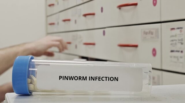
PINWORM INFECTION treatment tablets used to eliminate enterobius vermicularis parasites
00:26
Schistosoma is a genus of trematodes, commonly known as blood flukes for study under a microscope in lab.
00:30
Glistening Nematode Close Up View Under Microscopy Isolated on Black Background for Parasitology Research and Scientific Visualization of Microscopic Worms and Internal Parasites
00:08
Notoedres cati under the microscope. Notoedric mange, also referred to as Feline scabies, is a highly contagious skin infestation caused by an ectoparasitic and skin burrowing mite Notoedres cati.
00:13
 an antibiotic interacting with bacteria, focusing on how it disrupts bacterial cell walls, inhibits protein synthesis, or interferes with DNA replication.
00:07
Microscopic View of Colorful Hookworms and Microorganisms, Showcasing Intricate Movement and Anatomy in Vibrant Biological Environment
00:05
strongyloides stercoralis
00:09
selective focus stylized flat illustration of parasite life cycle stages
00:05
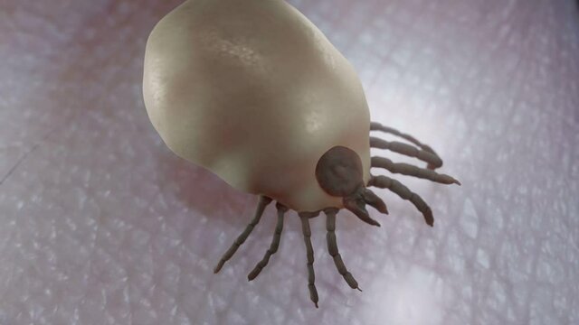
Tick full of blood
00:13
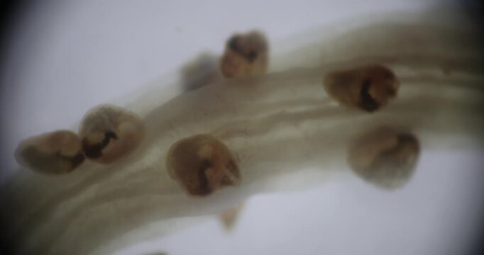
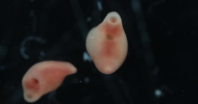
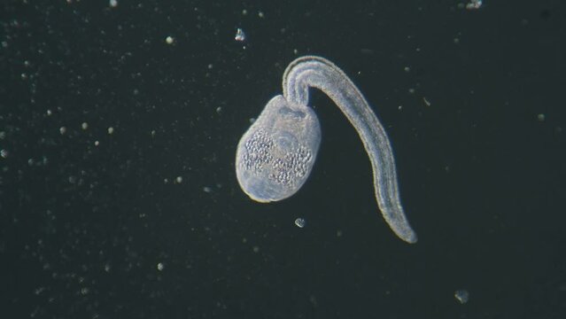
Trematode parasitic worm is escaping leaving parent body microscopical dark field view
00:31
Close up of serious young caucasian woman looking at sick plant which is suffering because of parasites. Death of indoor plants from dry air at home. Water or wrong temperature, gardening concept
00:12
Scientist Examining Dna Helix Structure In Test Tube Under Bright Laboratory Lights In Science Concept Presentation and
00:08
Plasmodium falciparium
00:30
3D animation of parasitic worms in an intestines of human or animal.
00:12
Parasite floating in the blood stream. 3D rendered animation.
00:20
Nematodes actively moving in blue water droplet under 400x magnification brightfield microscopy. 4K scientific microscopy footage.
00:20
Microscope Cerebellum Mammal Section x1200
00:50
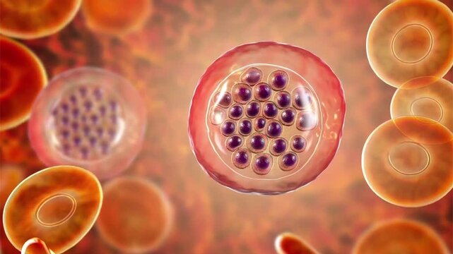
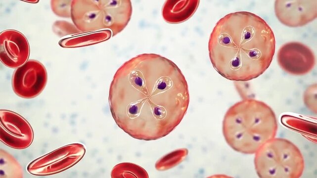
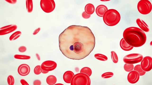
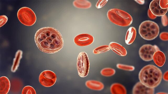
Plasmodium falciparum ring stage, 3D animation showing early intraerythrocytic trophozoites within red blood cells, characteristic of falciparum malaria in humans.
00:10
Educational veterinary footage showing sheep myiasis caused by fly larvae infestation, highlighting animal health, parasitic infection, and the importance of veterinary care.
00:19
View in microscopic Strongyloides stercoralis or threadworm in human stool.Parasite or fluke infection.Medical background analyze by microscope, original magnification 400x
00:09
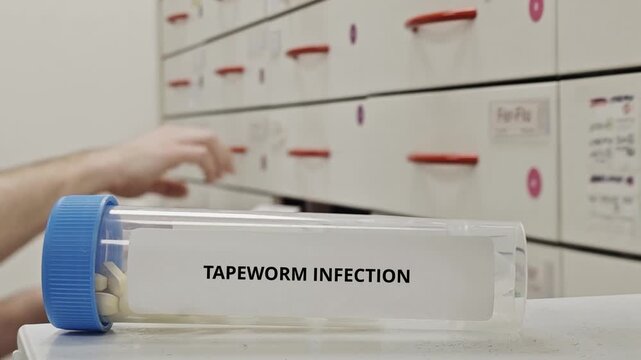
TAPEWORM INFECTION treatment tablets used to eliminate cestode parasites from the intestine
00:10
Schistosoma is a genus of trematodes, commonly known as blood flukes for study under a microscope in lab.
00:24
Close-Up of a Mosquito on Human Skin in the Wild, A Detailed Glimpse into Nature's Tiny Predator
00:06
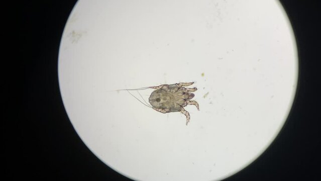
Otodectes cynotis, or ear mites under the microscope. This mites are found in cat's ear.
00:08
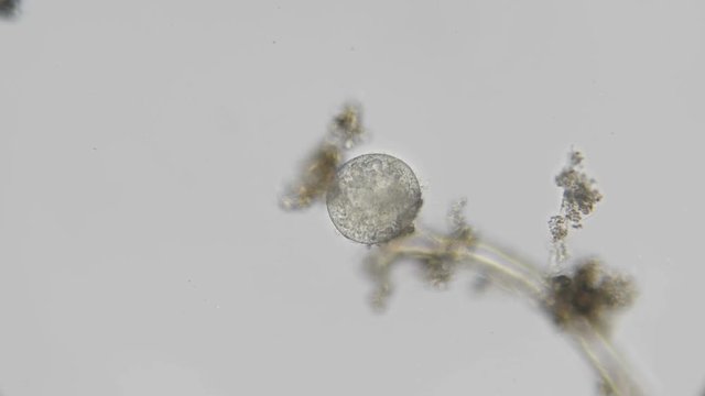
Nematode worm egg
00:10
Parasite attacking red blood cells in human bloodstream.
00:08
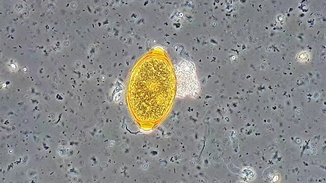
Stool sample under phase microscopy view, Trichuris egg
00:10
Malaria Merozoite Plasmodium Falciparum Invading Red Blood Cell With Membrane Deformation and Cellular Infection Process Visualization
00:10
red blood cells as they move through arteries, veins, and capillaries, carrying oxygen to organs and tissue
00:07
Trematode parasitic worm is escaping leaving parent body microscopical dark field view
00:19
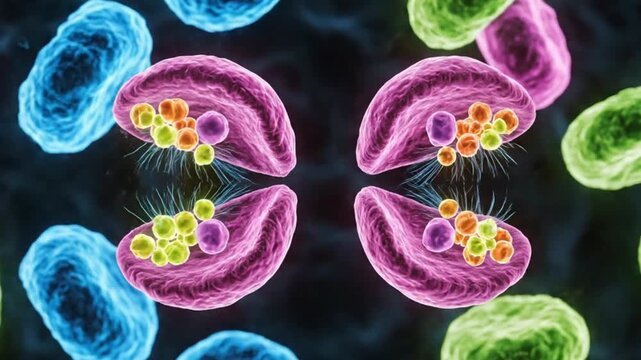
Microscopic World: Exploring the intricacies of Giardia Lamblia, a microscopic parasitic flagellate. Highlighted in vibrant detail.
00:10
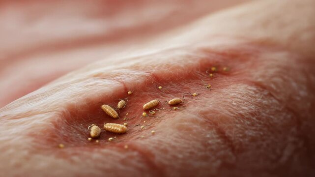
A person's arm has a rash with small bugs on it. Closeup realistic medical illustration of a myiasis wound showing small maggots inside human skin lesion, Myiasis clinical clean style
00:10
Microscopic visualization of plasmodium.
00:15
Schistosoma mansoni with medical digital technology concept
00:18
Veterinary laboratory technicians perform diagnostic testing on animal samples analyzing blood work and parasitology specimens for companion animal and livestock health blurred
00:10
Nematodes actively moving in blue water droplet under 40x magnification brightfield microscopy. 4K scientific microscopy footage.
00:53
Microscope Stem Collenchyma Cross Section 1200x
00:50
Plasmodium falciparum ring stage, 3D animation showing early intraerythrocytic trophozoites within red blood cells, characteristic of falciparum malaria in humans.
00:14
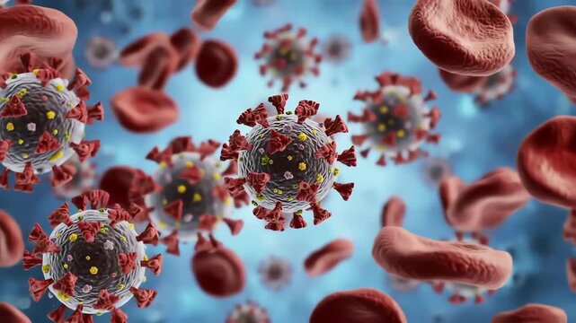
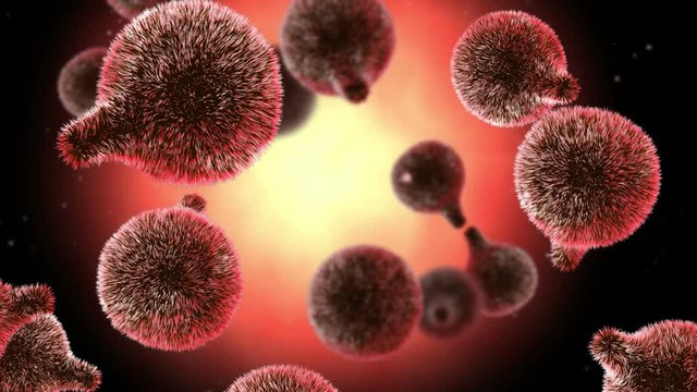
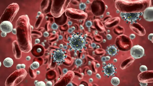
dengue virus particles in bloodstream, spherical with surface spikes
00:06
View in microscopic Strongyloides stercoralis or threadworm in human stool.Parasite or fluke infection.Medical background analyze by microscope, original magnification 400x
00:29
PINWORM INFECTION treatment tablets used to eliminate enterobius vermicularis parasites
00:15
Anatomical illustration of the trapezius muscle highlighted in orange.
00:05
Close-Up of a Mosquito on Human Skin in the Wild, A Detailed Glimpse into Nature's Tiny Predator
00:05
Otodectes cynotis, or ear mites under the microscope. This mites are found in cat's ear.
00:06
Bed bug, insect and pest on human skin for healthcare, medicine and hygiene. Parasite, biting and infestation with blood, health risk and dermatologist for treatment, prevention and sanitation.
00:06
Study of Parasitic helminths (Trematodes) of marine fishes under a microscope.
00:12
Two female doctors specialist microbiologist are looking through the loupe and analyzing samples at the desk with glassware in laboratory, microbiology or bacteriology analysis,  crane shot, tilt down
00:18
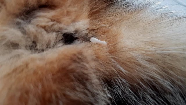
a tapeworm crawling slowly on the tail of a cat lying on the table. Dipylidium caninum is a dangerous animal and human parasite.
00:13
bacteriophages (viruses that infect bacteria) attacking a colony of bacteria. Visualize the phages binding to bacterial surfaces
00:07
4K footage highlighting Kissing Bug disease and Trypanosoma cruzi, ideal for medical education, parasitology awareness, and public health visuals.
00:10
Dipylidium caninum isolated on a white background, a euro cent coin for size comparison. Dipylidium caninum is a common parasite in domestic animals.
00:08
3D animation of parasitic worms in an intestines of human or animal.
00:12
Intestinal Parasites: Roundworms in Digestive System
00:08
Trematode parasitic worm is escaping leaving parent body microscopical dark field view
00:20
Young beautiful woman blogger florist sit on sofa at home,holding smartphone and take photo for social media to tell about plant diseases.Indoor plants diseases disorders Identification and treatment
00:18
Nematodes aggregation actively moving panoramic view under 40x magnification brightfield microscopy. 4K scientific microscopy footage.
00:10
Microscope Dog Colurmar Cilitd Epihelium Section x1200
00:50
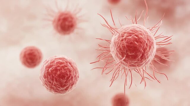
Anaplasma bacteria infecting neutrophils, 3D animation. Anaplasma phagocytophilum causes human granulocytic anaplasmosis transmitted by the black-legged tick and the sheep tick.
00:10
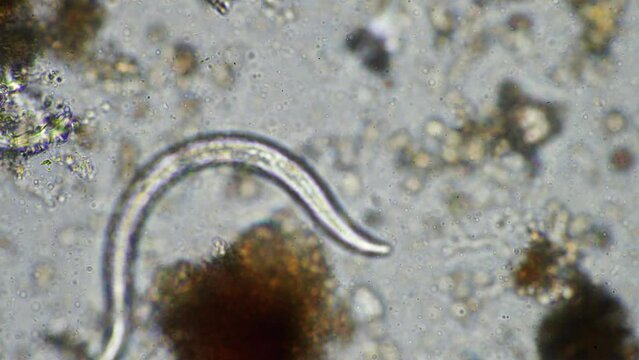
Toxoplasma gondii Tachyzoites: Microscopic view of Toxoplasma gondii tachyzoites, the rapidly multiplying stage of this common parasite.
00:05
View in microscopic Strongyloides stercoralis or threadworm in human stool.Parasite or fluke infection.Medical background analyze by microscope, original magnification 400x
00:15
TAPEWORM INFECTION treatment tablets used to eliminate cestode parasites from the intestine
00:15
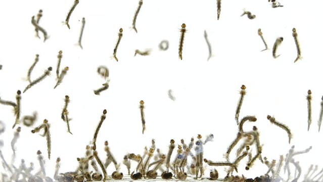
 mosquito's larva in water 4K
00:29
Veterinarians analyze data using tablets in barn with livestock in background, showing modern approach to animal health management, collaborative veterinary practice.
00:10
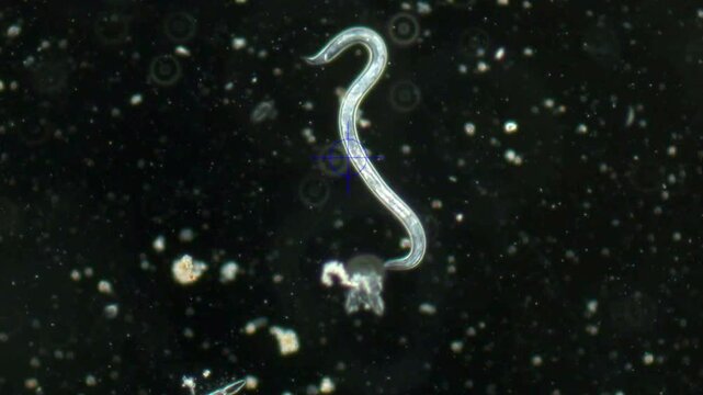
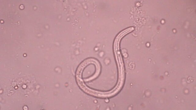
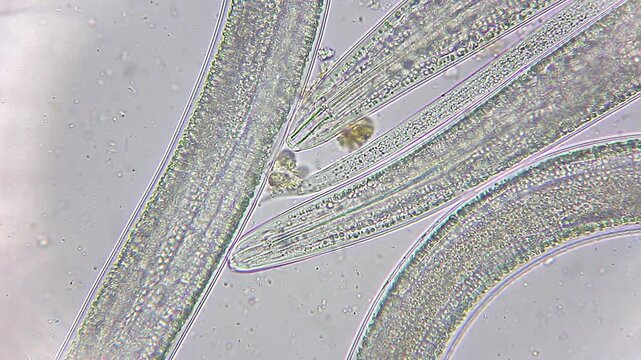
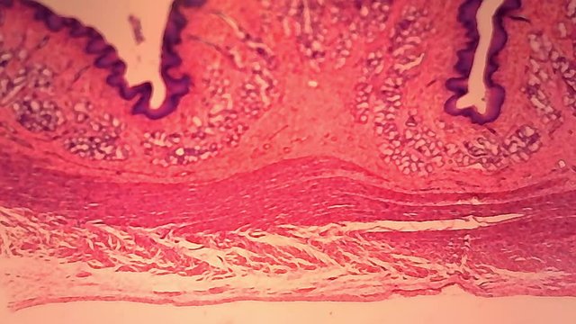
A large magnified nematode worm bends under microscope magnification. Studying parasites with a microscope. Diagnosing parasitic forms of infection.
00:37
Mosquito larvae are eating organic matter under water
Larva developed from eggs.
Breathe through siphon tube, fish food.
Life cycle of Mosquitoes, larva, gnats.
larva, wigglers.
Insects, bugs.
Animals
00:54
Study of Parasitic helminths (Trematodes) of marine fishes under a microscope.
00:11
closeup view of various human parasites affecting immune system depicted in sterile environment
00:10
multitude of mosquitos larvaes and pupa state moving quickly in water
00:12
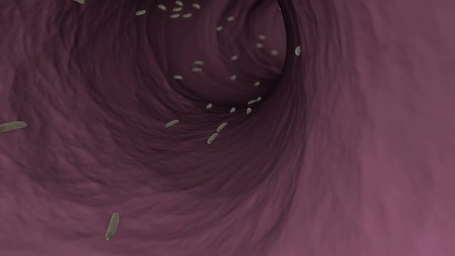
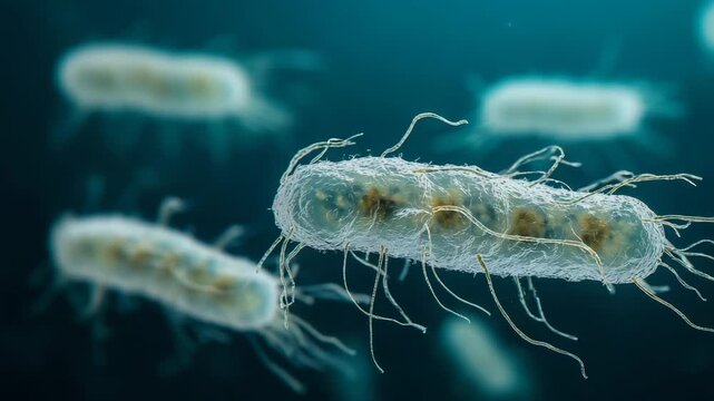
a single bacterium as it moves through the intricate environment of the human gut. Visualize it navigating a maze of villi and interacting with various other microbes,
00:07
Tick on human skin closeup
00:10
Hands of female technician during microbiology and bacteriology analysis, dolly macro shot  through the glassware, shallow depth of field, room interior
00:20
Nematode worm egg
00:10
A group of bugs are crawling on a red surface. There are at least 13 bugs in the image. Realistic illustration of fly larvae developing inside human tissue, Myiasis medical educational concept
00:06
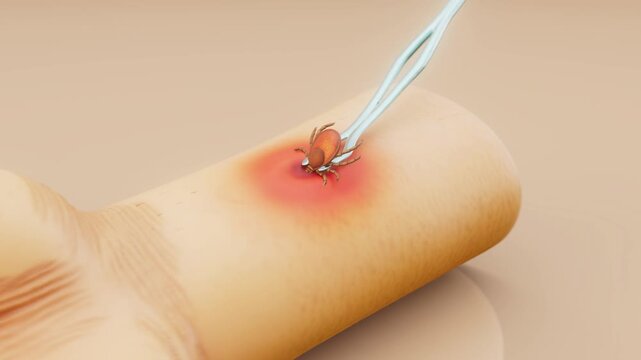
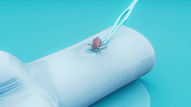
Conceptual 3D animation of the removal of a tick from human skin
00:11
Trematode parasitic worm is escaping leaving parent body microscopical dark field view
00:14