Un lien pour définir votre mot de passe a été envoyé à : Pour accéder à vos achats à l’avenir, vous aurez besoin d’un mot de passe.
Obtenez 3 vidéos ou 25 images avec un essai gratuit
Nouveaux filtres ajoutés
Trier par

2 585 résultats pour fibia dans vidéos

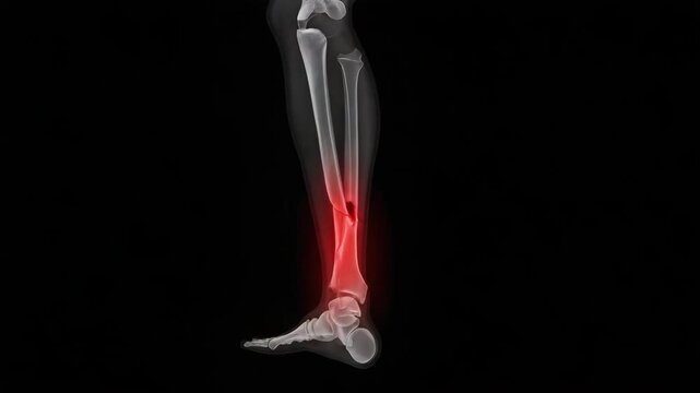
Medical x ray of a broken tibia
00:05
HD
Lateral knee xray human leg joint orthopedic medical scan skeletal injury diagnosis bone fracture anatomy healthcare hospital imaging radiology science research knee joint xray medical skeleton
00:08
HD
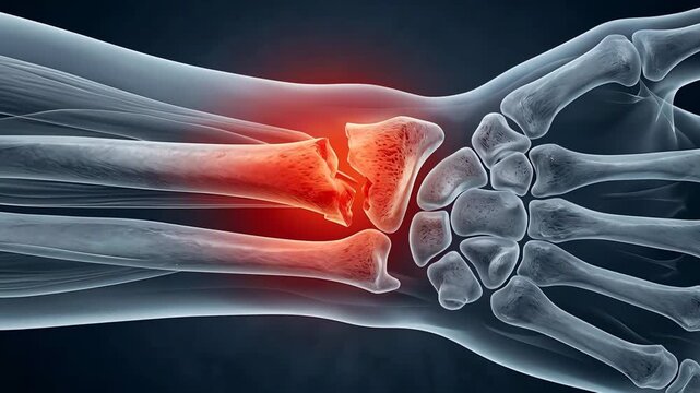
Close Up Medical Illustration Of A Broken Wrist Bone Showing Severe Pain And Inflammation On A Dark Blue Background
00:05
HD
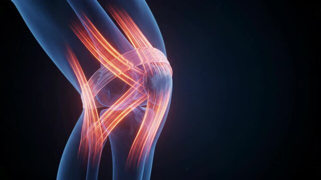
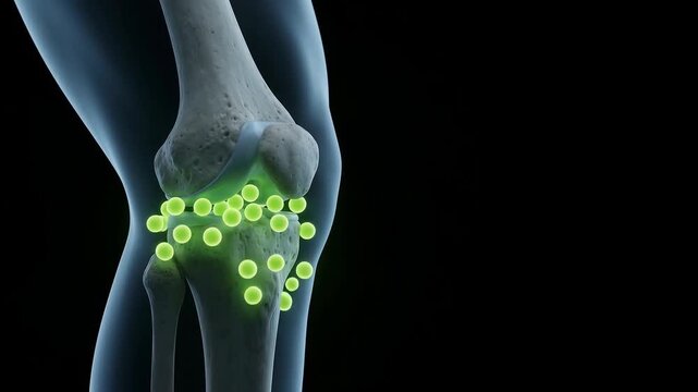
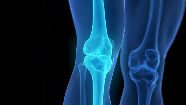
3D Rendered Human Knee Joint Anatomy Showing Femur Tibia Fibula and Glowing Particles
00:05
HD
Man Sitting Medical Exam Table Holding Ankle Fracture Pain Glowing X-Ray Bone Anatomy Overlay Crack Visible Clinic Hospital Background
00:07
HD
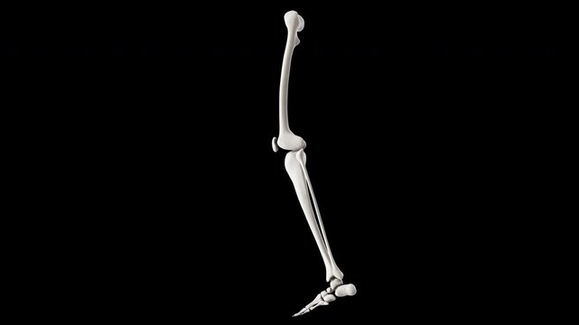
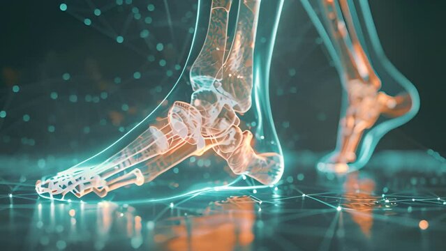
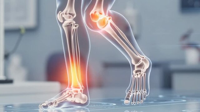
Human leg and foot bones in a skeletal representation during a walking motion
00:05
HD
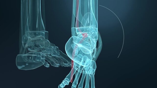
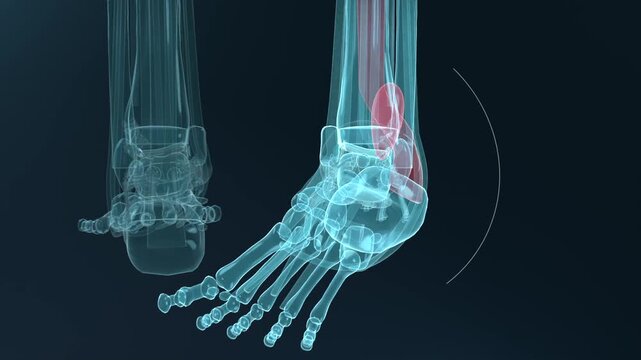
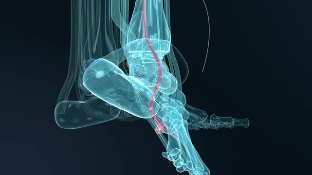
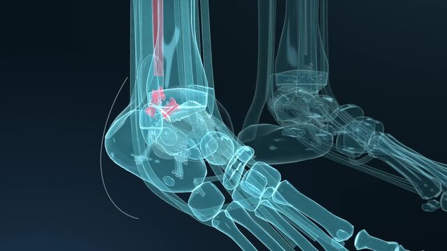
Animation of the bones of a man's right ankle
00:13
HD
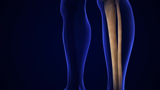
Anatomy of human lower leg skeletal structure with tibia and fibula bones in medical visualization skeleton bone orthopedic healthcare scientific
00:10
HD
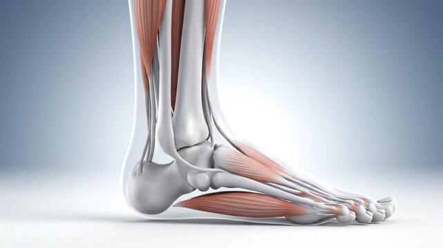
Detailed 3D Anatomy of the Human Foot and Ankle with Bones and Muscles.
00:08
HD
A detailed side view of a full human skeleton model sitting upright on a black stool against a plain grey background.
00:06
HD
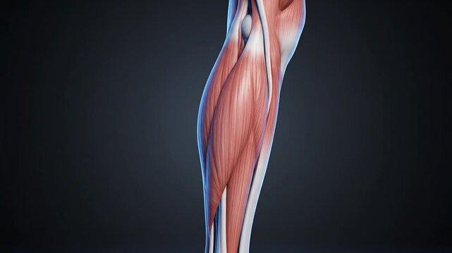
Detailed 3D anatomical rendering of the human calf muscles, showcasing the complex muscular and skeletal structures against a dark background, for human muscular system education.
00:05
HD
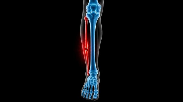
Broken Fibula Bone Pain Anatomy Medical Concept
00:12
HD
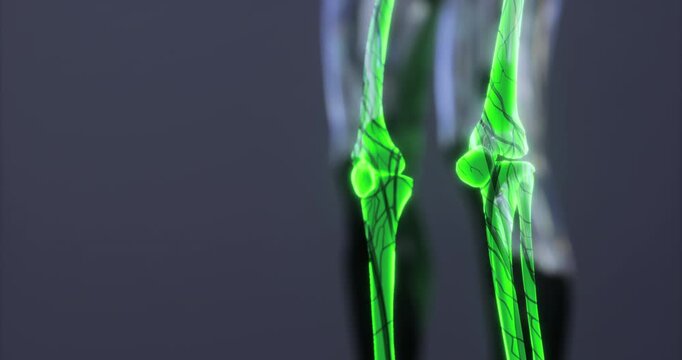
A vivid representation of human skeleton anatomy focuses on the knee joint, showcasing intricate details of the bones and surrounding structures in a striking green hue.
00:06
HD
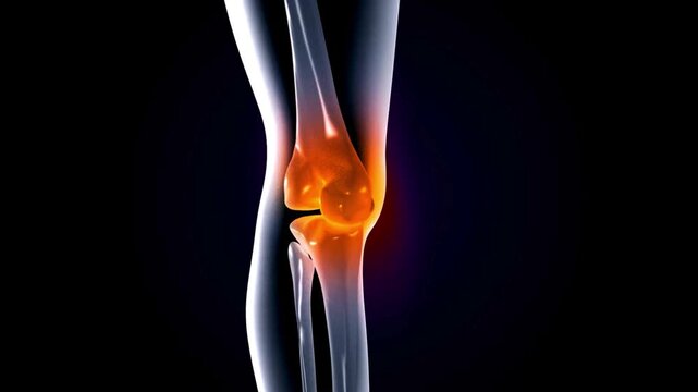
Knee Anatomy: Detailed medical illustration highlights the knee joint, emphasizing bone structure with a focus on inflammation.
00:10
HD
Anatomical 3D Render of Human Skeleton Highlighting Leg Bones and Pelvis.
00:05
HD
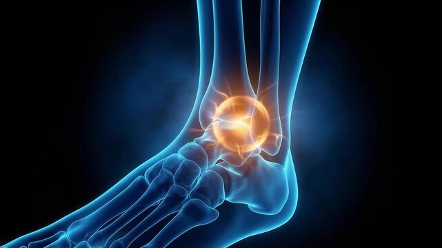
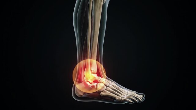
3D medical illustration of a human ankle and foot skeleton with a glowing orange point indicating joint pain or inflammation at the talus bone on a dark background.
00:08
HD
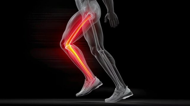
Semi transparent legs of an athlete running showing glowing tibia and fibula bones, highlighting a stress fracture, against a black background
00:05
HD
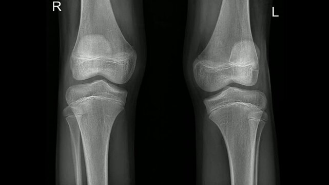
Pediatric Normal Both Knee X-ray Showing Healthy Bone Structure
00:15
HD
Anatomy of running legs showing bones while exercising on a treadmill in a gym.
00:08
HD
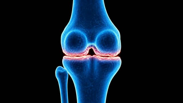
3D medical animation of human knee joint anatomy with blue x-ray style bones and highlighted meniscus cartilage on a black background.
00:06
HD
Pain and edema in the sprained ankle. Ankle Pain Affecting Mobility and Daily Activities. Ankle injury with dislocation and sprains.
00:10
HD
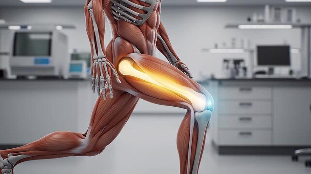
Anatomy of the Human Leg Performing a Lunge Exercise Showing Muscle Engagement and Bone Structure in a Medical Lab
00:06
HD
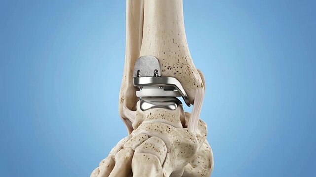
Close up 3D medical animation showcasing a metallic ankle joint prosthesis surgically implanted into a human tibia fibula and tarsal bones with
00:05
HD
Professional medical leg scan showcasing a cross-sectional analysis of the tibia and fibula for clinical evaluation.
00:56
HD
Detailed anatomical model of a human knee joint showing progressive angles and components, including femur, tibia, fibula, and metallic prosthetic elements in a dark background
00:08
HD
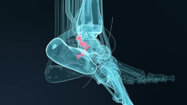
Ankle Joint Pain Injury 3D Medical Animation Foot Anatomy Achilles Tendon Bone X-Ray Glowing Dark Background
00:06
HD
Animation of an inflamed knee
00:20
HD
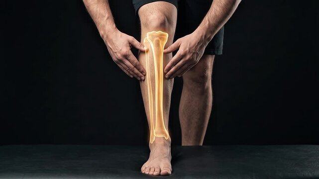
Detailed digital illustration of lower leg bones highlighting pain area in medical setting
00:05
HD
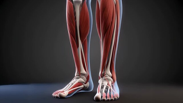
Detailed anatomical illustration of human leg muscles and skeletal structure.
00:07
HD
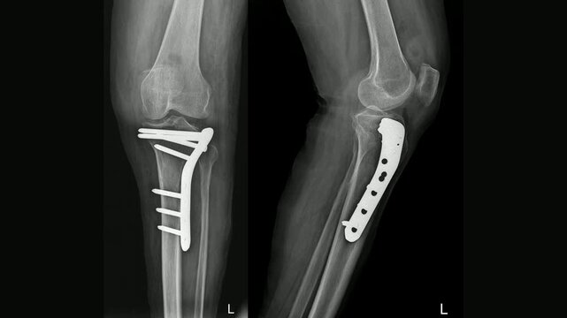
Anatomical knee joint x-ray examination for diagnostic analysis and bone structure, side view
00:10
HD
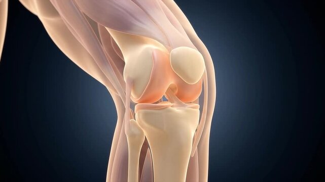
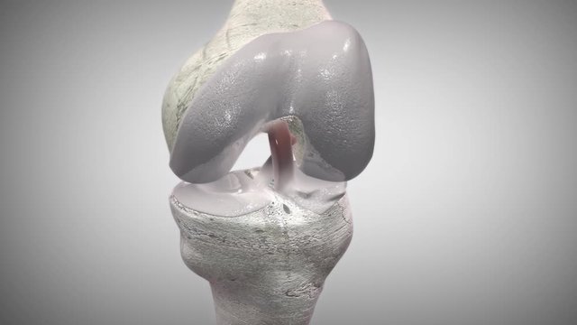
Detailed Human Knee Joint Anatomy Model
00:05
HD
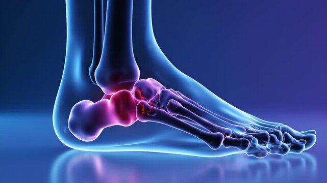
Animation displaying foot bones with inflamed calcaneus, showcasing anatomy and potential pain points related to bone injury and musculoskeletal disorders requiring medical attention.
00:10
HD
This visualization presents the human skeleton, showcasing the intricate details of the skeletal system. Joint structures are illuminated for better understanding and study of human anatomy.
00:06
HD
The Human Knee Joint in Pain: An abstract, artistic representation of a human knee joint glowing with intense red, highlighting areas of pain and inflammation.
00:10
HD
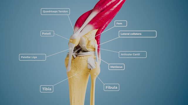
3D rendering of human raising knee, zooming into joint anatomy with labeled tendons, bones, ligaments, and cartilage. Ideal for medical, scientific, and educational use
00:08
HD
Innovative healthcare professional showcasing virtual skeletal structure technology with advanced
00:08
HD
Glowing Bone Held by Gloved Hands.
00:08
HD
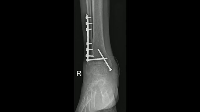
Ankle Joint Fracture Fixation X-Ray Post-Surgical Assessment,
00:15
HD
Medical illustration highlighting pain and inflammation in the human lower leg bones with a glowing effect
00:08
HD
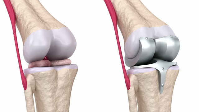
Anatomical illustration of human knee joint.
00:08
HD
Human Leg Anatomy Knee Joint Medical Visualization
00:05
HD
Anatomical illustration of a human foot and ankle showcasing inflamed bones and joints, highlighting pain and discomfort associated with foot conditions and injuries
00:10
HD
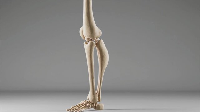
3D model of human tibia and fibula leg bones, lower leg anatomy
00:10
HD
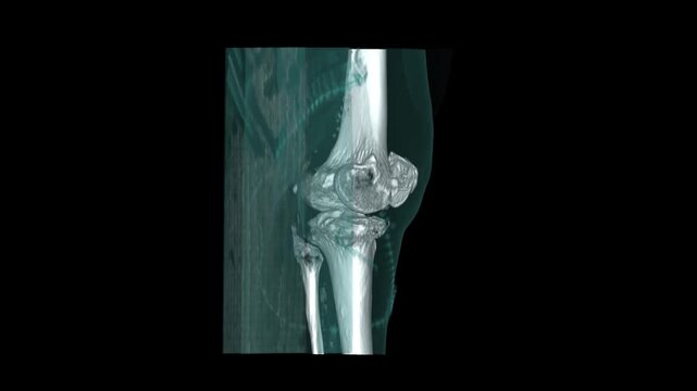
3D volume rendered CT scan reconstruction of a left knee joint showing femur patella tibia and fibula bones
00:16
HD
Human Skeleton Model Rotating on Black Background.
00:10
HD
Bone Cancer Illustration - This video depicts an illustration of bone cancer in a knee joint. The cancerous growth is shown in red and yellow, highlighting the affected area on the bone.
00:08
HD
Radiating Pain in the Shoulder A Visual Exploration of Bone Tumors and Upper Arm Discomfort with X Ray Style Body
00:08
HD
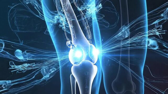
Detailed 3D anatomical illustration showcasing the human knee joint experiencing significant pain and inflammation, emphasizing nerve pathways and bone structure for medical and educational purposes
00:05
HD
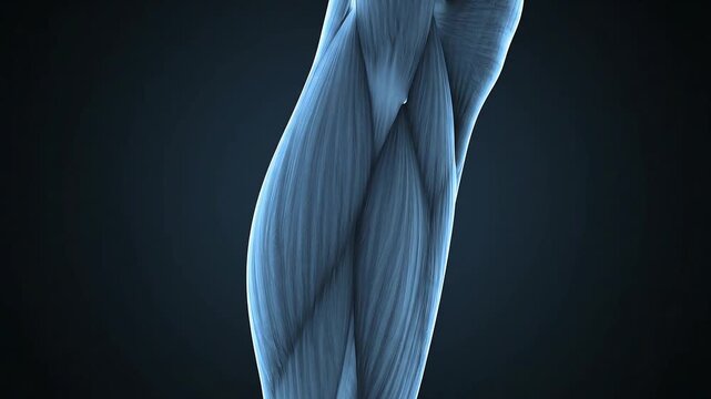
Detailed anatomical rendering of human leg muscles in a blue-toned scientific illustration on a dark background.
00:05
HD
Medical 3D animation showing a fibula bone fracture in the human lower leg
00:06
HD
Human knee joint anatomy with highlighted cartilage and synovial fluid.
00:08
HD
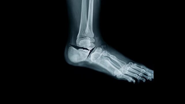
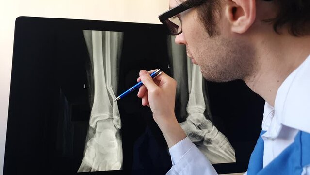
X-ray of a broken ankle showing a fracture in the fibula and surrounding bones taken in a medical setting
00:09
HD
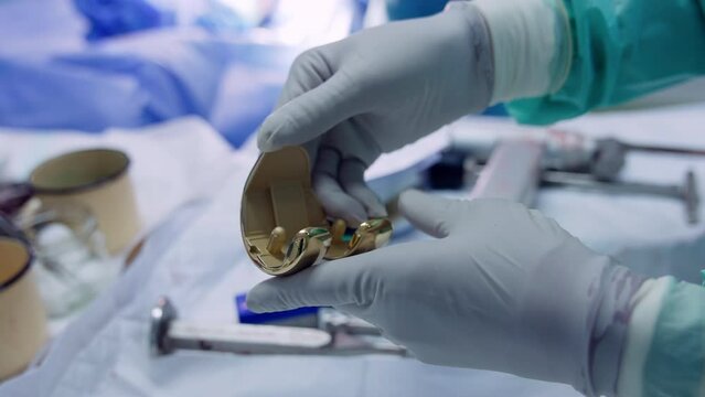
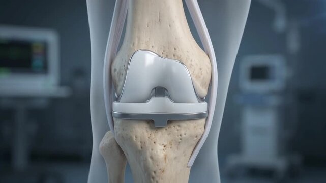
Hands of doctor in latex gloves holding a knee prosthesis. Materials for knee joint replacement. Close up.
00:12
HD
Lumbar Disc Herniation and Sciatic Nerve Pain Visualization - This 3D medical illustration depicts a herniated lumbar disc compressing a nerve root, causing inflammation and radiating pain.
00:08
HD
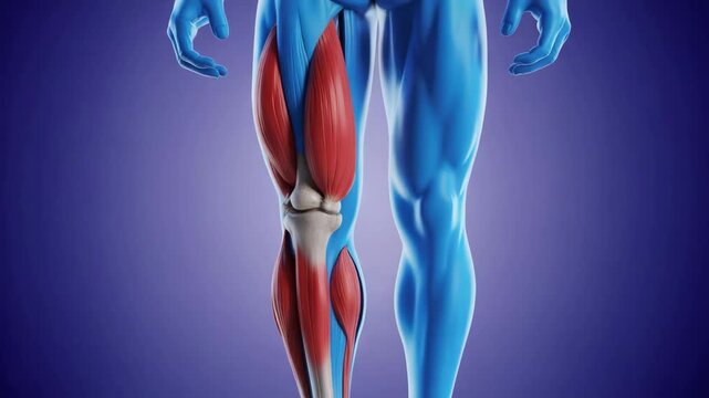
3d rendering of human leg showing hamstring and calf muscle anatomy with knee joint bone structure highlighted against a blue background
00:05
HD
Anatomical View of a Woman in Downward Facing Dog Yoga Pose.
00:08
HD
Bubbles and metallic objects submerged in green liquid, concept of innovation
00:08
HD
Anatomical Study of Human Leg Muscles and Bones in Detailed 3D Render Against
00:05
HD
Detailed 3D animation of a male human anatomy model highlighting the knee joint with glowing orange pain or inflammation, showing anatomical structures and muscle tendons
00:15
HD
X-ray image of an ankle joint, showcasing bones and structure for medical analysis.
00:04
HD
Human Skeletal System and Nerves: Anatomical Illustration
00:07
HD
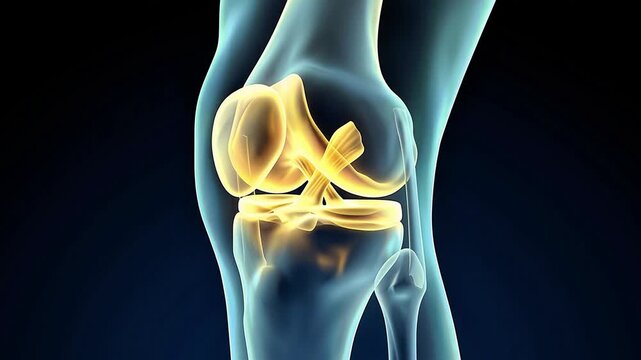
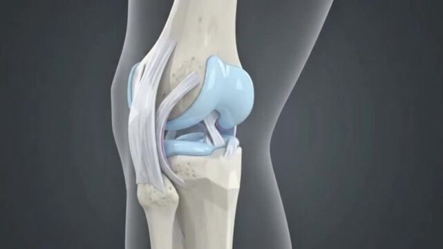
3D Medical Animation Human Knee Joint Anatomy Showing Bones Ligaments Cartilage Meniscus Tendon Orthopedic Healthcare Concept Blue Background
00:05
HD
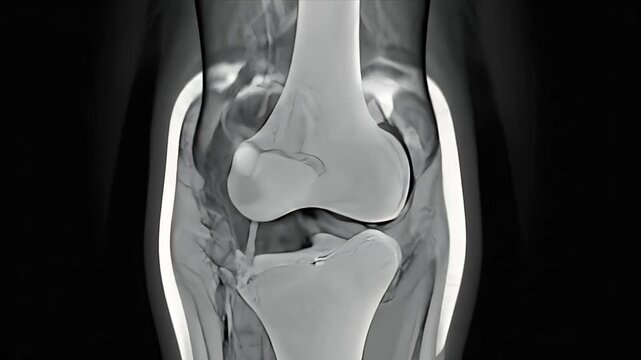
hospital x ray image of knee joint
00:06
HD
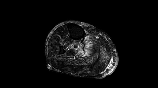
Academic 3D MRI Visualization of Lower Leg Cross-Section and Tibia Fibula Skeletal Architecture
00:11
HD
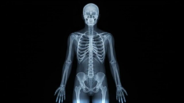
Human Skeleton 3D Rendering Rotation Animation.
00:10
HD
Human having pain in feet
00:08
HD
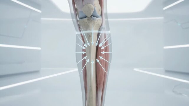
Shin Splints Pain Relief Lower Leg and Knee Injury Recovery Medical Animation Bone Health Inflammation Anatomy Relief
00:08
HD
minimalist knee joint icons with depicting human anatomy, orthopedic health, and Healthcare Biology Outline for Medical Video Presentation motion graphics, graphic design.
00:10
HD
3D Medical Animation of Knee Anatomy, Patella Structure, and Lower Body Skeleton
00:08
HD
Digital X-ray visualization of human knee joint. Orthopedic anatomy, injury diagnosis and joint health, copy space for text
00:05
HD
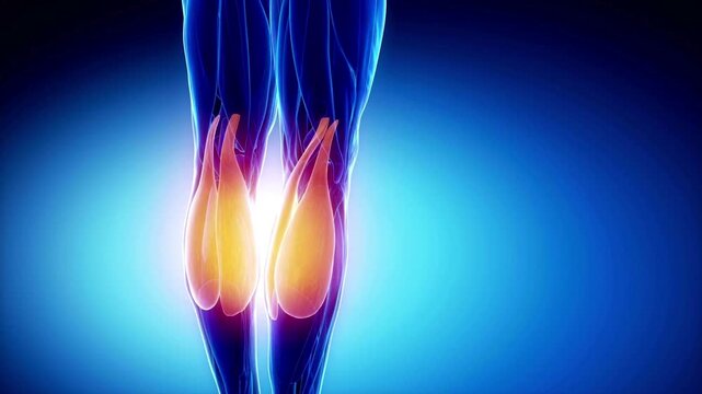
Human leg anatomy illustration showing glowing calf muscles and bones
00:10
HD
A detailed illustration showcasing the intricate mechanics of a knee joint, highlighting the bones, ligaments, and tendons essential for movement.
00:05
HD
Anatomical Visualisation of Foot Sprain with Highlighted Ligament Injury
00:16
HD
Professional Workspace Modern Office Setup for Virtual InPerson Meetings
00:05
HD
Human skeleton model for medical education.
00:08
HD
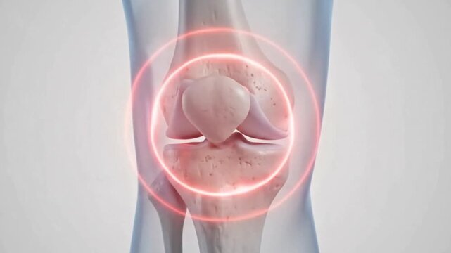
Detailed anatomical illustration of a human knee joint with a red glow indicating pain or inflammation.
00:06
HD
Exploring the human skeletal system in detail with advanced digital imaging technology
00:10
HD
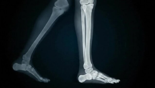
Lateral X-ray view of human legs and feet in a walking motion, showing skeletal bone structure including the tibia, fibula, ankle joint, and metatarsals against a black background.
00:08
HD
Runner with leg pain demonstrating muscle inflammation and knee joint discomfort during exercise showcasing potential sports injury
00:10
HD
Animated Foot Anatomy Detailed 3D Visualization of Bones and Structure on Blue Background
00:08
HD
Rotating 3D Human Skeleton Animation with Detailed Bone Structure and Transparent Body Outline on Dark Blue Background, symbolizing medical education and anatomical study
00:05
HD
Knee Joint Pain Inflammation 3D Medical Animation Human Leg Anatomy Bone X-Ray Glowing Red Dark Background
00:06
HD
Wireframe runner legs highlighting a stress fracture injury during running, providing a visualization of the impact and stress on bones during physical activity
00:05
HD
3D volume rendered CT scan reconstruction of human left ankle and foot bones showing tibia fibula tarsals and metatarsals
00:08
HD
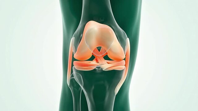
A close-up view of a human knee joint, showing the bones, muscles, and ligaments.
00:08
HD
3d rendering of human foot and ankle bones, highlighting the skeletal structure and anatomy
00:08
HD
Detailed Human Skeleton Leg Anatomy Articulated Bones and Joints Displayed in a Studio Setting with Dramatic Lighting and Dark Background for Medical Educational Purposes
00:06
HD
minimalist knee joint icons with depicting human anatomy, orthopedic health, and Healthcare Biology Outline for Medical Video Presentation motion graphics, graphic design.
00:10
HD
Medical illustration of a knee joint highlighting cartilage, bones, and inflammation.
00:08
HD
Close-up view of a human foot skeleton on a dark background with detailed bones exposed.
00:05
HD
Detailed medical illustration of human leg bones showing tibia and fibula anatomy in high resolution
00:05
HD
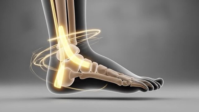
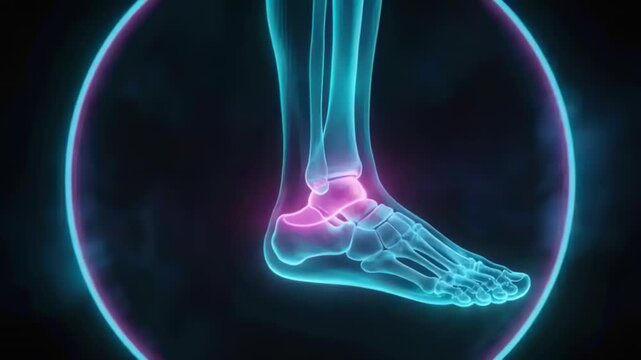
Animating cyan foot bones pulsing, tilting on cue to highlight ankle with pink overlay, neon ring
00:05
HD
Anatomy of the human calcaneus
00:14
HD
Knee Pain Anatomy Animation in Hospital Setting
00:07
HD
Knee Anatomy: A close-up view highlights the intricate structure of a knee joint. The image, illuminated with a cool blue glow.
00:10
HD
X ray of a human leg and lower torso with highlighted knee joint
00:08
HD
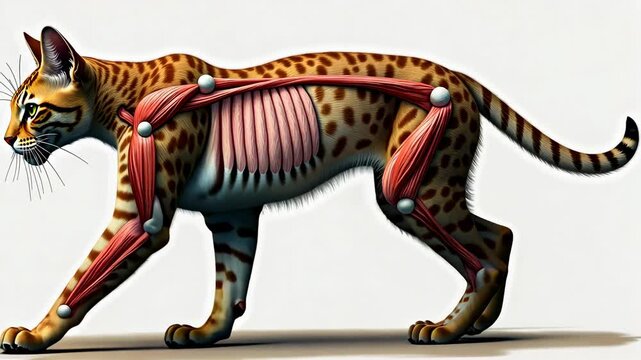
Cat anatomy, muscle structure, skeletal system. Animal biology. Muscles expand during movement. Joints enable flexible motion. 3D medical illustration enhances learning.
00:10
HD
Time Lapse video animation of R.M.N. Nuclear magnetic resonance, of human foot, with cross-section and sagittal section.
00:06
HD
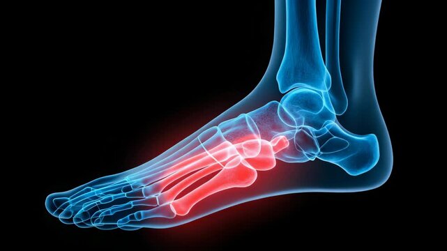
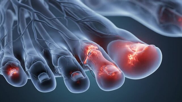
Foot Anatomy Illustration Showing Pain and Inflammation in Toes.
00:08
HD
Anatomical visualization of a human knee joint highlighting inflammation and pain with a glowing red spot on the femur and tibia bones against a dark blue scientific
00:08
HD